SLHS 566 Exam 1
 Call Kai
 Learn
Learn Practice Test
 Spaced Repetition
 Match
Match Flashcards
 Knowt Play
1/200

There's no tags or description
Looks like no tags are added yet.
Name | Mastery | Learn | Test | Matching | Spaced | Call with Kai | Chat |
|---|
No analytics yet
Send a link to your students to track their progress
201 Terms
What are non-speech functions of the airway system?
transport something into/out of the body
air
liquids
solids
What is anatomy?
study of structures of organisms and relation to their parts
What is physiology?
science dealing with functions of living organisms or their parts
Describe the following anatomical position: Anterior
closer to the front of the body
Describe the following anatomical position: Posterior
closer to the back of the body
Describe the following anatomical position: Rostral
closer to the head of the body
Describe the following anatomical position: Caudal
closer to the tail/feet, away from head
Describe the following anatomical position: superior
upper (NOT superficial)
Describe the following anatomical position: inferior
lower (NOT deep)
Describe the following anatomical position: superficial
closer to the surface of the body
Describe the following anatomical position: deep
away from the surface of the body
Describe the following anatomical position: medial
closer to the middle of the body
Describe the following anatomical position: lateral
closer to the side of the body
Describe the following anatomical position: proximal
closer to the trunk/start of an extremity
Describe the following anatomical position: distal
away from the trunk/away from the start of an extremity
Describe the following anatomical position: central
related to the center
Describe the following anatomical position: Caudal
related to the periphery

What anatomical direction describes A?
Posterior

What anatomical direction describes B?
Anterior

What anatomical direction describes A?
rostral

What anatomical direction describes B?
caudal
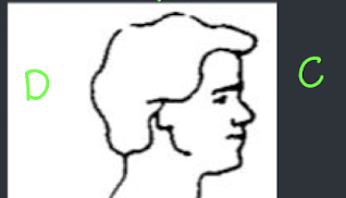
What anatomical direction describes C? ***This is referring to the brain!
rostral

What anatomical direction describes D? This is referring to the brain!
caudal
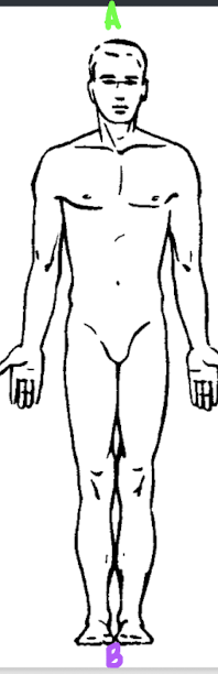
What anatomical direction describes A?
Superior
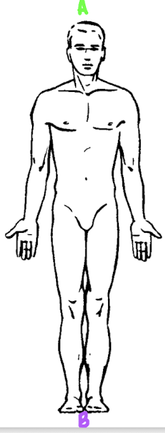
What anatomical direction describes B?
inferior

What anatomical direction describes A?
medial

What anatomical direction describes B?
lateral

What anatomical direction describes A?
proximal
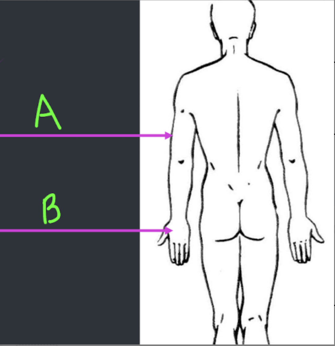
What anatomical direction describes B?
distal
What are the three anatomical planes? How do they divide the body and cortex?
sagittal
divides right from left
coronal
divides front from back
transverse
divides up from down

Which anatomical plane describes A?
Coronal

Which anatomical plane describes B?
transverse (horizontal)

Which anatomical plane describes C?
sagittal
What are the two main nervous systems in the body and what components make them up?
Central Nervous System
Brain and Spinal Cord
Peripheral Nervous System
Cranial Nerves and Spinal Nerves
What two main nervous systems make up the peripheral nervous system? What functions do they govern?
Somatic Nervous System
voluntary body movements
conscious perception/integration of sensory stimuli
Autonomic Nervous System
involuntary body control
breaks down into 3 divisions
The autonomic nervous system breaks down into 3 divisions, identify them and describe their individual functions.
enteric NS
everything related to the GI track
sympathetic NS
fight or flight responses
parasympathetic NS
rest and digest responses
What are the 6 types of glial cells? Where are they found and what is their main function?
Microglia
CNS
Clean up crew
Ependymal Cells
CNS
CSF production + balance
Oligodendrocytes
CNS
produce myelin
Astrocytes
CNS
maintain the blood brain barrier
Satellite Cells
PNS
Repair muscle and are the astrocytes of the PNS
Schwann Cells
PNS
produce Myelin
What are the two types of neurons?
motor neurons
sensory neurons
What are motor neurons?
neurons located in the brain/brain stem/spinal cord (Upper motor neurons)
axon extends to muscle fiber (lower motor neurons)
discharges electrochemical signal to cause muscle contraction
What are sensory neurons?
neurons located throughout the body that transmit info from body back to CNS
What are the two types of motor neurons?
upper motor neurons (UMN)
lower motor neurons (LMN)
Discuss how an upper motor neuron functions
axons of UMN are entirely contained in CNS and never exit the neuraxis (brain + spinal cord)
cell bodies of these neurons sit in the cerebral cortex
Wherever the UNM ends in the spinal cord is where a LMN originates — at the point of communication
Discuss how a lower motor neuron functions
LMN send motor axons outside of the neuraxis to a muscle
it is what ultimately moves a structure
The corticospinal tract is a major neuronal pathway for motor movement. Outline from the point where an upper motor neuron starts its way to where it connects with a lower motor neuron.
upper motor neurons start in the primary motor cortex. the cell bodies of these neurons reside in the PMC.
at the internal capsule, all of the axons from the UMN’s in the OMC meet and congregate, creating a bundle of axons called a tract
this tract travels down the cortex to the brainstem. It passes through the midbrain and the pons
at the level of the medulla, this tract made of pyramids (axons) crosses over to the other side of the medulla, moving contralaterally. This is known as the DECUSSATION OF PYRAMIDS. 90% of the fibers will do this but a small group will remain ipsilateral.
past the medulla the tract reaches the spinal cord where cell bodies of lower motor neurons (LMN) live. Axons from this tract will send electrical signals to LMNs causing the LMNs to send signals to our muscles to contract.
In general what are the two main functions of neurons?
send signals
to other neurons (UMN)
to muscles (LMN)
receive signals
from other neurons
from the environment → sensory neurons
What are the main parts of a neuron?
dendrites
soma/cell body
axon hillock
nodes of ranvier
myelin sheath
axon
axon terminal
What are dendrites and what is their function?
extend from the cell body (soma)
receive electrochemical signals from other neurons or sensory receptors
What is an axon and what is its function?
one long, thin process
sends electrochemical signals to other neurons (which will be received by dendrites!)
What is a process?
anything that hangs off the cell body
What is the difference between a unipolar, bipolar, pseudounipolar, and multipolar neuron? How can we determine this?
We can determine what kind of neuron it is by looking at how many processes are hanging off of the cell body!
unipolar
only one process
bipolar
two processes
pseudounipolar
only one process off of the cell body but that process bifurcates into two
multipolar
many processes!
we have one axon BUT dendrites are processes two which is what makes it multipolar
What is the myelin sheath?
myelin is a fatty (lipid), white-ish substance that wraps around portions of the axon. Insulates the axon and aids in the conduction of the electrical signal.
What are nodes of ranvier?
Gaps in the myelin sheath with a high concentration of ion channels that maintain the electrical signal throughout the axon.
What are the two types of conduction and what kind is used with myelinated axons?
continuous and saltatory
Neurons with myelinated axons function via saltatory conduction
this is much more efficient than continuous conduction because instead of all ion channels having to be open along the axon, only some of the ion channels open at a time at the nodes of ranvier.
What happens at the axon hillock?
decides if the input coming from the dendrites is enough to send an action potential down the axon
if not strong enough to reach threshold - an AP will not go off
What happens in the axon terminal that leads to the synapse between the presynaptic and postsynaptic neuron?
When the action potential arrives at the axon terminal, it reaches voltage-gated Ca2+ channels
The voltage of the action potential opens up these calcium ion channels
Ca2+ then enters the presynaptic neuron which sends a signal to vesicles filled with neurotransmitters
these neurotransmitter vesicles move to the membrane of the presynaptic neuron and dock on the membrane via a docking protein
Neurotransmitters are then released via exocytosis. Exocytosis is an active transport process where a cell moves large molecules, proteins, or waste out of the cell by fusing a small sac (vesicle) with the outer cell membrane.
Now these neurotransmitters are in the SYNAPTIC CLEFT where they will bind to receptors located on the dendrites of the postsynaptic neuron.
Once the NTs bind to the receptors, a signal is initiated in the postsynaptic cell and is sent to the axon hillock to determine if an action potential will be sent down the axon.
What happens to the NT in the synaptic cleft?
The NT doesn’t just disappear on its own. If there were no “clean up” mechanisms they would remain in the cleft and continuously stimulate the postsynaptic neuron.
Enzymes go to the NT and bring it back into the neuron where its recycled or repackaged
What is a refractory period?
period after an action potential before a neuron can fire again
during this period an action potential cannot go off
What are the four types of gated-channels? What opens and closes them?
Mechanical gated ion channels
opens and closes in response to touch, stretch, force
Leakage gated ion channels
always passively open and lets specific ions to move passively across the membrane → help maintain the balance
Voltage-gated ion channels
opens and closes in response to voltage
Ligand gated ion channels
opens and closes in response to chemical substances like Neurotransmitters
Where do neurons eventually connect to? Where does this occur?
Neurons eventually connect to the muscle at the neuromuscular junction
What are the three types of muscle?
Striated Muscles (aka skeletal muscle)
voluntary muscle
Smooth Muscles (non-striated)
involuntary muscle
organs like our stomach
Cardiac Muscle
no control
heart muscle
Cardiac muscle is also striated. What makes it different than skeletal muscle?
cardiac muscle is also striated but it is a different subset.
It is different because
cardiac muscle is involuntary and skeletal muscle is voluntary
cardiac muscle has 1 or 2 nuclei while skeletal muscle has many nuclei
cardiac muscle does not regenerate but skeletal muscle does

What kind of muscle is pictured here?
cardiac muscle

What kind of muscle is pictured here?
skeletal/striated muscle

What kind of muscle is pictured here?
smooth muscle
How many striated muscles do we have? Are they all paired?
we have ~329 striated muscles
all of them are pared except the procerus and diaphragm
Muscles have 4 properties. What are they?
excitability: respond to a stimuli
contractibility: can contract
extensibility: can be stretched without tearing
elasticity: return to normal shape
What is the outermost protective layer of a muscle called?
epimysium
Explain the Russian nesting dolls of a muscle.
Epimysium
Muscle
Perimysium
Fascicle
Endomysium
muscle fiber
Myofibril
filaments containing actin and myosin
In what part of the muscle does contraction actually occur?
contraction occurs at the level of the myofibrils in the muscle fiber. The actin and myosin are involved in contraction.

What part of the muscle is labeled A?
tendon

What part of the muscle is labeled B?
epimysium
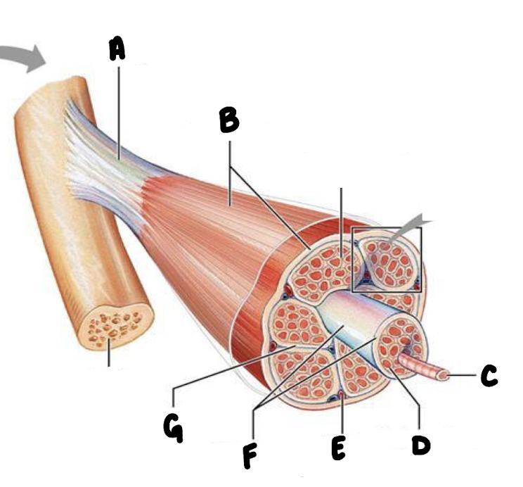
What part of the muscle is labeled C?
muscle fiber

What part of the muscle is labeled D?
endomysium (between fibers)

What part of the muscle is labeled E?
blood vessel

What part of the muscle is labeled F?
fascicle
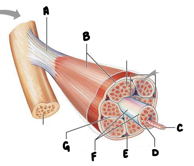
What part of the muscle is labeled G?
perimysium (wraps around each fascicle)

What part of the muscle fiber is labeled A?
striations

What part of the muscle fiber is labeled B?
nucleus

What part of the muscle fiber is labeled C?
myofibrils

What part of the muscle fiber is labeled D?
Filaments containing actin and myosin
What is the difference between the sarcolemma and the endomysium of the muscle fiber?
the sarcolemma is the cell membrane of a muscle fiber, while the endomysium is the layer of connective tissue that wraps around that membrane
What is the smallest functional unit of striated muscle?
the muscle fiber
What are properties of a muscle fiber?
multinucleated
made up of many myofibrils
endomysium separates muscle fivers from one another
Each myofibril contains:
filaments of MYOSIN protein molecules
these are thick!
filaments of ACTIN protein molecules
these are long and thin!
What two components are important for muscle contraction?
myosin and actin are important for muscle contraction
What is a sarcomere?
contractile unit
smallest unit in a muscle that can contract
a repeated sequence of actin and myosin along the length of the myofibril

What is represented between the Z lines?
the sarcomere is the unit represented between 2 Z-lines
What is the sliding filament theory of muscle contraction?
When an electrical signal is sent from a lower motor neuron to a muscle, Acetylcholine is released which causes calcium to be released.
The calcium is what allows myosin to bind to the actin filament. When the filaments bind together they form a crossbridge.
Energy being released from ATP allows myosin to then pull the actin filaments inward which shortens the sarcomere and leads to muscle contraction.
What is a motor unit?
Has 2 parts
a single lower motor neuron.
all of the muscle fibers innervated by LMN axon
What is the motor unit signal?
this is the action potential that travels down the neuron
Discuss the different types of motor unit size
gross motor unit
large motor unit
controls many muscle fibers
GROSS MOTOR MOVEMENTS
small motor unit
controls only few muscle fibers
FINE MOTOR MOVEMENTS
Explain motor unit spacing and why it is helpful to preserving function if damage occurs.
Motor units are spaced in an intermingled arrangement. This evenly spreads activation among muscle fibers.
If an injury or neurodegenerative disease damages a specific motor neuron, the muscle fibers connected to it will lose their stimulation. Because those fibers are intermingled rather than clustered, their loss won't leave a dead zone or a massive hole in the muscle's functional capacity.
Why do muscle fibers only attach to 1 neuron?
muscle fibers do not attach to more than one neuron because we want a clear signal. Receiving multiple different signals would confuse the muscle fiber.
How do motor units respond?
in an all or nothing fashion
either contract or dont
Grading force is characterize by what two properties?
the number of motor units active at one time → need a lot of force? activate A LOT of neurons!! need only a little force? activate only a FEW neurons.
frequency of firing of motor units → the more often the neuron fires the stronger the contraction. if you don’t need much force fire once or few times.
What is Henneman’s Size Principle?
This principle tells us the order by which motor units are activated
Activated first: motor units with smallest number of fibers
Activated last: motor units with biggest number of fibers
This translates to… SMALL MOTOR UNITS FIRST THEN GROSS MOTOR UNITS
What is the innervation ratio of motor units?
the innervation ratio is the number of fibers controlled by 1 neuron
low if ~5
small fine motor movements
high if ~1000
leg + thigh muscles
What is the motor neuron pool?
collection of motor neurons innervating a single muscle
all the LMNs that control one specific muscle → lots of muscle fibers innervated by individual neurons
What is a facet?
a small area on structure where it contacts another structure
often act like cup and saucers to create a joint
joints in the larynx are facet joints
one side is hollowed and the other is a little bowed
What is articulation in anatomy?
a joint or juncture of bones or cartilages
“articulated structures” = structures that come together at a joint