NPB 101: Lecture 8 - Action Potential II

0.0(0)
Studied by 2 people
call kaiCall Kai
learnLearn
examPractice Test
spaced repetitionSpaced Repetition
heart puzzleMatch
flashcardsFlashcards
GameKnowt Play
Card Sorting

1/10

flashcard set

Earn XP

Description and Tags

Learning Objectives: Distinguish the absolute and relative refractory periods in terms of sodium-channel availability, potassium conductance, and the ability to initiate another action potential. Explain how refractory periods limit firing frequency and help produce unidirectional action-potential propagation. Compare continuous and saltatory conduction and explain how myelin and nodes of Ranvier increase the speed and efficiency of signal propagation.

11 Terms

1
New cards

Refractory Period

A brief period of time after an action potential begins during which a neuron cannot fire another electrical impulse.

  • Limits the rate APs are generated and makes sure that APs only propagate in one direction.

  • Two Phases:

    • Absolute Refractory Period

    • Relative Refractory Period

  • Action Potentials can vary across cell types

    • Duration of APs can range

      • from 200 microseconds to many milliseconds

    • Rate of APs can vary

      • from a few/second to 1000/second

    • Differences are attributed to variations in depolarization and repolarization


<p>A brief period of time after an action potential begins during which a neuron <strong>cannot fire another electrical impulse</strong>.</p><ul><li><p><u>Limits the rate</u> APs are generated and makes sure that APs only propagate in <u>one direction</u>.</p></li></ul><ul><li><p>Two Phases:</p><ul><li><p><strong>Absolute Refractory Period</strong></p></li><li><p><strong>Relative Refractory Period</strong></p></li></ul></li><li><p>Action Potentials <u>can vary</u> across cell types</p><ul><li><p><em>Duration</em> of APs can range</p><ul><li><p>from 200 microseconds to many milliseconds</p></li></ul></li><li><p><em>Rate </em>of APs can vary </p><ul><li><p>from a few/second to 1000/second</p></li></ul></li><li><p>Differences are attributed to <u>variations in depolarization and repolarization</u></p></li></ul></li></ul><p></p>
2
New cards

Absolute Refractory Period

Period in which a new action potential is impossible to trigger, no matter how strong the stimulus is. This ensures each AP is separate and distinct.

  • Begins with the activation of voltage-gates Na+ channels

    • Na+ channels are either open or inactive from first AP

  • Ends when inactivation is removed (i.e. the voltage-gate closes and the time-gated ball is dislodged upon repolarization)


<p>Period in which a new action potential is <strong>impossible</strong> to trigger, no matter how strong the stimulus is. This ensures each AP is <strong>separate and distinct.</strong></p><ul><li><p><u>Begins</u> with the activation of voltage-gates Na<sup>+</sup> channels</p><ul><li><p>Na<sup>+</sup> channels are either open or inactive from first AP</p></li></ul></li><li><p><u>Ends</u> when inactivation is removed (i.e. the voltage-gate closes and the time-gated ball is dislodged upon <strong>repolarization</strong>)</p></li></ul><p></p>
3
New cards

Relative Refractory Period

A brief period after the Absolute Refractory Period in which a new action potential can be triggered, but only by a stronger-than-normal stimulus.

  • Begins when Na+ channel inactivation is removed

  • Ends when voltage-gated K+ channels deactivate

  • During this period:

    • Inactivation of the Na+ channel is maximal

      • Few if any channels are available to increase Na+ conductance

    • K+ conductance is very large

    • In order to initiate a regenerative depolarization, a very large increase in Na+ conductance is required to override the K+ conductance


<p>A brief period after the Absolute Refractory Period in which a new action potential <strong>can </strong>be triggered, but only by a <strong>stronger-than-normal</strong> stimulus.</p><ul><li><p><u>Begins</u> when Na<sup>+</sup> channel inactivation is removed</p></li><li><p><u>Ends</u> when voltage-gated K<sup>+</sup> channels deactivate</p></li><li><p>During this period:</p><ul><li><p>Inactivation of the Na<sup>+</sup> channel is maximal</p><ul><li><p>Few if any channels are available to increase Na<sup>+</sup> conductance</p></li></ul></li><li><p>K<sup>+</sup> conductance is very large</p></li><li><p>In order to initiate a <u>regenerative depolarization</u>, a <strong>very large increase</strong> in Na<sup>+</sup> conductance is required to <strong>override</strong> the K<sup>+ </sup>conductance</p></li></ul></li></ul><p></p>
4
New cards

Graded Potential

A localized, variable-magnitude change in a neuron’s membrane potential that grows stronger or weaker depending on the strength of the incoming stimulus.

  • Graded potentials code stimulus intensity through changes in amplitude

  • If a graded potential is strong enough, it will cause a cell to fire an action potential(s)


<p>A localized, variable-magnitude change in a neuron’s membrane potential that grows stronger or weaker <strong>depending on the strength</strong> of the incoming stimulus.</p><ul><li><p>Graded potentials code stimulus intensity through changes in <strong>amplitude</strong> </p></li><li><p>If a graded potential is strong enough, it will <strong>cause a cell to fire an action potential(s)</strong></p></li></ul><p></p>
5
New cards

Action Potential

A rapid, temporary reversal of voltage across a cell membrane that allows neurons and other excitable cells to transmit electrical signals.

  • Action potentials code stimulus intensity through changes in frequency


<p>A rapid, temporary reversal of voltage across a cell membrane that allows neurons and other excitable cells to transmit electrical signals.</p><ul><li><p>Action potentials code stimulus intensity through changes in <strong>frequency</strong></p></li></ul><p></p>
6
New cards

Action Potential Propagation

As the action potential flows along the nerve axon, some current is passively lost through the membrane.

  • Two strategies to improve conduction:

    • Increase diameter of axon

      • Decreases internal resistance

    • Myelination

      • Increases electrical insulation around the axon

      • Allows for rapid nerve impulse conduction

        • Faster propagation than nonmyelinated fibers of the same axon diameter

          • (10-120 m/second vs. 1 m/second respectively)


<p>As the action potential flows along the nerve axon, some current is <strong>passively lost</strong> through the membrane.</p><ul><li><p>Two strategies to improve conduction:</p><ul><li><p><strong><u>Increase diameter of axon</u></strong></p><ul><li><p>Decreases internal resistance</p></li></ul></li><li><p><strong><u>Myelination</u></strong></p><ul><li><p>Increases <u>electrical insulation</u> around the axon</p></li><li><p>Allows for <strong>rapid</strong> nerve impulse conduction</p><ul><li><p><u>Faster propagation than nonmyelinated</u><strong> </strong>fibers of the same axon diameter</p><ul><li><p>(10-120 m/second vs. 1 m/second respectively)</p></li></ul></li></ul></li></ul></li></ul></li></ul><p></p>
7
New cards

Glial Cells

Non-neuronal cells in the Central Nervous System (CNS) and Peripheral Nervous System (PNS) that support, protect, and nourish neurons rather than generating electrical impulses.

  • Glial cells outnumber neurons

  • Provides nutrients to neurons (astrocytes) and helps remove damaged neurons and infectious agents (microglia)

  • Myelin sheaths are made from glia:

    • CNS: Oligodendrocytes

    • PNS: Schwann cells

  • Participates in the uptake and metabolism of the neurotransmitters and intercellular signaling


8
New cards

Oligodendrocytes (Myelination)

Myelin-forming glial cells in the Central Nervous System (brain and spinal cord).

  • A single oligodendrocyte uses multiple branch-like extensions to myelinate multiple separate axons simultaneously (up to 60)


<p>Myelin-forming glial cells in the Central Nervous System (brain and spinal cord).</p><ul><li><p>A single oligodendrocyte uses multiple branch-like extensions to <strong>myelinate multiple separate axons simultaneously</strong> (up to 60)</p></li></ul><p></p>
9
New cards

Schwann Cells (Myelination)

Myelin-forming glial cells in the Peripheral Nervous System (afferent and efferent divisions).

  • A single Schwann cell forms myelin around just one axon segment


<p>Myelin-forming glial cells in the Peripheral Nervous System (afferent and efferent divisions).</p><ul><li><p>A single Schwann cell forms myelin around <strong>just one axon segment</strong></p></li></ul><p></p>
10
New cards

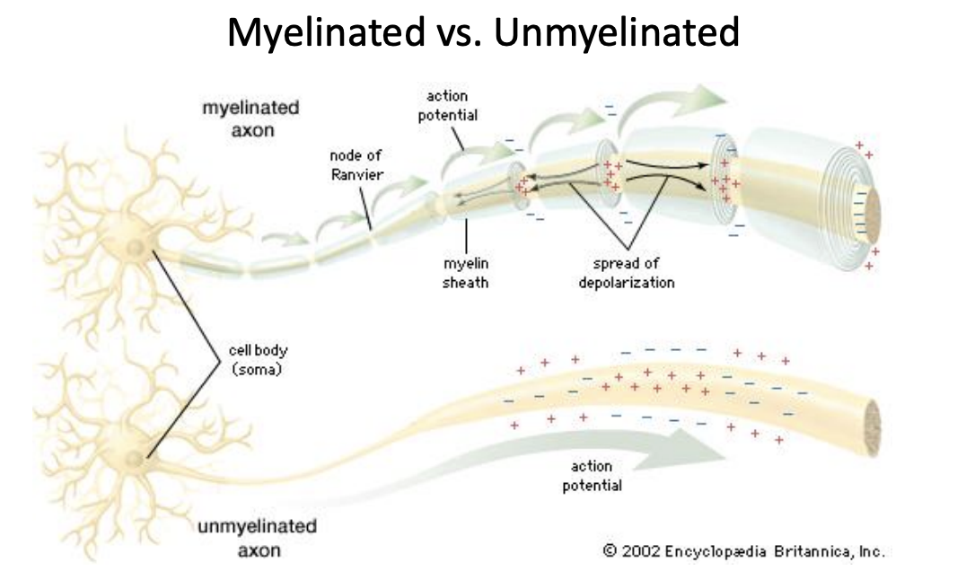
Myelinated Axons

Nerve fibers wrapped in multiple layers of myelin membrane by Oligodendrocytes in the CNS and Schwann cells in the PNS.

  • Myelin prevents current flow through the membrane

  • Nodes of Ranvier:

    • Periodic gaps in the myelin sheath

    • Saltatory conduction

      • Current appears to “jump” from node to node


<p>Nerve fibers wrapped in multiple layers of myelin membrane by <u>Oligodendrocytes</u><strong> </strong>in the CNS and <u>Schwann cells</u><strong> </strong>in the PNS.</p><ul><li><p>Myelin prevents current flow through the membrane</p></li></ul><ul><li><p><strong><u>Nodes of Ranvier:</u></strong></p><ul><li><p>Periodic gaps in the myelin sheath</p></li><li><p><strong>Saltatory conduction</strong></p><ul><li><p>Current appears to “jump” from node to node</p></li></ul></li></ul></li></ul><p></p>
11
New cards

Unmyelinated Axons

Nerve fibers that lack a multi-layer myelin wrapping.

  • Direct conduction

    • Channels must open sequentially all the way down the axon membrane

      • This is to maintain the amplitude of the action potential

    • Slow propagation

  • Helpful to have when neurons are in close proximity

<p>Nerve fibers that <strong>lack</strong> a multi-layer myelin wrapping.</p><ul><li><p><strong>Direct conduction</strong></p><ul><li><p>Channels must open sequentially <u>all the way down the axon membrane</u></p><ul><li><p>This is to maintain the amplitude of the action potential</p></li></ul></li><li><p><strong>Slow propagation</strong></p></li></ul></li><li><p>Helpful to have when neurons are in close proximity</p></li></ul><p></p>



