anatomy lab - muscles physiology
 Call Kai
 Learn
 Practice Test
 Spaced Repetition
 Match
 Flashcards
 Knowt Play
1/83

There's no tags or description
Looks like no tags are added yet.
84 Terms
A skeletal muscle may be supplied by hundreds
Cell bodies in the ventral horn
Axons leave the spinal cord in the ventral root
Skeletal muscle fiber
Multinucleate cell
striated
voluntary
Receives synaptic input from only one motor neuron
That synapse is the only source of excitation for the fiber

Motor unit
A motor axon/neuron plus the muscle fibers it connects with
Action potential in the motor neuron → action potential in all its fibers → single twitch
NO - the gap is the synaptic cleft of the neuromuscular junction
Events at the neuromuscular junction
Action potential arrives at synaptic knob
Calcium enters synaptic knob
ACh released
ACh binds ACh receptors
End-plate potential forms
Muscle action potential
ACh removed from synaptic cleft
What starts the events at the neuromuscular junction?
Action poential travels down the motor neuron axon
Reaches the axon terminal (synaptic knob)
After the action potential reaches the synaptic knob, what happens?
Voltage-gated calcium channels in the synaptic knob open
Calcium diffuses into the terminal
After calcium enters the synaptic knob, what happens?
Synaptic vesicles release ACh by exocytosis
ACh enters the synaptic cleft
After ACh is released into the synaptic cleft, what happens?
ACh diffuses across the synaptic cleft
Binds to ACh receptors on the motor end plate
Ligand-gated (chemically gated) sodium channels open
After the ligand-gated sodium channels open, what happens?
Na⁺ enters and K⁺ leaves the muscle fiber
More Na⁺ in than K⁺ out → membrane potential becomes less negative
This graded potential is the end-plate potential
How do the events at the neuromuscular junction end?
Signal continues until ACh is removed from the synaptic cleft
By diffusion or breakdown by acetylcholinesterase
Enzyme that breaks down ACh into acetic acid and choline
Choline is taken back into the axon terminal to make new ACh
Blocks exocytosis (release) of ACh at the neuromuscular junction
No end-plate potential can form → no muscle contraction
Action potential still reaches the synaptic knob and calcium still enters
Fundamental life process
Carried out by filamentous proteins: actin, myosin, and tubulin
Muscle uses it for locomotion and transport of fluids through tubes of digestive, circulatory, and glandular systems
Smooth
Cardiac
Skeletal
Differ in function, structure, and distribution
Can be described as voluntary/involuntary, striated/nonstriated, uninucleate/multinucleate
Smooth muscle
Sheets of cells, each with one nucleus
Involuntary
Nonstriated
Found in walls of blood vessels, digestive tract, and reproductive system
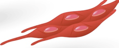
Cardiac muscle
Found only in the heart
Uninucleate cells bound together at dense plaques called intercalated disks
Striated
Due to parallel arrangement of bundles of contractile fibers
Involuntary

Only voluntary muscle type
Generally attached to the skeleton
Cells fused into multinucleated fibers
Gives maximum strong, directional contraction
Prominent striations visible in light microscope
Due to extreme regularity of arrangement of contractile proteins
is a complex tissue that is comprised of several levels of organization
composed of fibers (muscle cells), which are made of myofibrils
muscle cells which are made of myofibrils
made of sarcomeres, which are made of myofilaments
muscle → fiber → myofibril → sarcomere → myofilament
since they are the smallest units of contraction within the muscle fiber, they are considered to be functional units of skeletal muscle
made of thick myosin and thin actin - both are contractile proteins arranged in alternating pattern
z disc to z disc
reach from one side of the A band to the next side of the A band
the myosin heads project to the binding sites on the thin filaments to form cross bridges
fascicle
A small bundle of muscle fibers within a muscle
Layer of connective tissue that surrounds an individual muscle fiber
endomysium
one end to the Z line and one end not entirely over to the A band
since it does not reach all the way over there is an area in the middle of the A band that is lighter because there is only thick filament - H zone
area in A zone where thin filament does not reach all the way over so it is only thick filament - lighter looking

I band

Center of the sarcomere
Anchors thick filaments in place

Protein that makes up elastic filaments
Runs through the core of thick filaments and attaches them to Z discs at either end
Resists overstretching and recoils like a spring after the muscle is stretched
troponin and tropomyosin
troponin and tropomyosin are found on top of actin thin filaments
Protein that covers myosin binding sites on actin in a relaxed cell
Bound to troponin
Protein bound to tropomyosin
Calcium binds to it to start contraction
Where thick and thin filaments overlap and cross-bridges form
More overlap (up to optimal) → more cross-bridges → more tension
1. Excitation of the muscle cell
ACh released at neuromuscular junction → end-plate potential
2. Excitation-contraction coupling
Action potential travels along sarcolemma and down T tubules → calcium released from SR
3. Cross-bridge cycling
Calcium binds troponin → tropomyosin moves → cross-bridges form and power stroke causes contraction
Physiological process that converts an action potential in a muscle fiber into a mechanical muscle contraction
Stimulated by a motor neuron
Stimulates several muscle fibers when it generates action potentials
Motor neuron + the muscle fibers it stimulates = motor unit
Action potential travels from neuromuscular junction (end plate) along sarcolemma
T tubules conduct it into the interior of the muscle fiber
Activates voltage-sensitive dihydropyridine (DHP) receptors
DHP receptors open calcium channels in sarcoplasmic reticulum membrane
Calcium flows into the sarcoplasm and triggers contraction
infoldings of the sarcolemma

Terminal cisternae
Linked together by calcium-regulating proteins
Store calcium that is released into the sarcoplasm during excitation-contraction coupling
Ca²⁺
Stores calcium in resting muscle
Releases calcium into cytoplasm when an action potential arrives
Takes calcium back up during relaxation
Na⁺: enters the cell to create the action potential
Ca²⁺: binds troponin to expose binding sites on actin
ATP: powers the cross-bridge cycle and the return of calcium to the SR
Calcium attaches to troponin
Troponin-tropomyosin complex shifts so tropomyosin moves
Uncovers myosin binding sites on actin
Myosin heads can then bind
Binding sites stay exposed as long as calcium stays high in the sarcoplasm
a high concentration of calcium is present in the sarcoplasm
Link formed when an activated myosin head binds to actin
Repeatedly form and break during contraction, creating tension
During contraction, portions of the sarcomere decrease in length, increase in length or stay the same, which ones decrease?
I band and H zone
Before it begins: myosin head is activated (cocked) by ATP
Step 1: cross-bridge formation
Activated myosin head binds to actin to form a cross-bridge
Inorganic phosphate is released
Bond between myosin and actin becomes stronger
Step 2: power stroke
ADP is released
Activated myosin head pivots, sliding the thin filament toward the center of the sarcomere
Step 3: cross-bridge detachment
Another ATP binds to the myosin head
Link between myosin head and actin weakens and the head detaches
Step 4: reactivation of the myosin head
ATP is hydrolyzed into ADP and inorganic phosphate
Energy released energizes the myosin head and returns it to the cocked position
Cycle repeats as long as calcium and ATP are present
Cross-bridges pull thin filaments toward the center of the sarcomere
Changes the filaments' position, NOT their length
Z discs are pulled along → sarcomere shortens
Shortens the muscle fiber and ultimately the entire muscle
Detaches the cross-bridge
ATP binding weakens the myosin-actin link so the head lets go (cycle step 3)
Without ATP, heads stay stuck to actin (rigor mortis)
Reactivates the myosin head
ATP hydrolysis energizes the head and returns it to the cocked position (cycle step 4)
Returns calcium to the SR
Powers active transport of calcium back into the SR, ending contraction
Rigor mortis
Example of ATP's role in breaking actin-myosin bonds
After death, ATP declines
Calcium pumps stop working without ATP, so calcium stays high and the binding sites stay exposed
Actin and myosin can still bind, but bonds can't break
Filaments stay bound → rigid muscle
Temporary; goes away as decomposition breaks down contractile proteins
Calcium ions are actively transported back into the SR
Cross-bridge formation ends
Troponin returns to its original shape
Tropomyosin glides over and covers the myosin binding sites on actin
Filaments return to their original position
Makes membranes permeable
Low-molecular-weight material is extracted
Contractile proteins remain
Side effect: troponin-tropomyosin disrupted
Myosin heads bind actin without Ca²⁺
Lets us add different solutions to find requirements for contraction
ATP and Mg²⁺
Ca²⁺ not needed because troponin-tropomyosin is disrupted
Mg²⁺ alone: no contraction
ATP alone: some contraction
ATP and Mg²⁺: most contraction
Binds with ATP to form Mg-ATP
Mg-ATP is the form of ATP myosin uses
Transducer: measures the force of muscle contraction
Electrode: electrically stimulates the muscle
Simulates nerve impulses normally coming from the brain
Smallest stimulus that produces a contraction
Below threshold, no force is generated
In lab: 0.2 V produced 0 g of force
Smallest stimulus that produces the maximum response (all motor units recruited)
Any stronger stimulus produces the same maximal response
In lab: 1.6 V and above all produced 235 g of force
Recruitment
Nervous system controls the number of motor axons firing
Controls number of active motor units → number of contracting fibers
higher voltage → more motor units recruited → stronger contraction, up to a maximum
motor unit recruitment
the successive activation of additional motor units. The higher the recruitment the stronger the force of the muscle contraction will be
Stimulate more fibers (recruitment)
Increase frequency of stimulation
Stimuli close together produce stronger contractions
Muscle length just before stimulation
Closest to optimal length → most tension
Tension depends on number of cross-bridges between thick and thin filaments
Stretching a muscle stretches its sarcomeres and changes filament overlap
Already contracted (too short)
Thick filaments butt up against Z discs and stop
Contraction is brief and weak
Optimal length (resting length)
Overlap is maximized → most tension
Overstretched
Little overlap, so myosin can't grip the thin filaments
Contraction is weak
Total tension = active + passive; both vary with muscle length
Active tension: force from thick filaments pulling on thin filaments (cross-bridge cycling)
Passive tension: occurs during stretch beyond the normal range of motion
Resisted by noncontractile proteins, especially titin
Collagen and elastin in connective tissue also contribute
At optimum length, active tension makes up most of total tension
When overstretched (little or no overlap), passive tension contributes significantly and can exceed active tension
About 28 mm
Tension increased to a maximum, then decreased as stretch increased
In lab, data peaked at 28.5 mm
Since the tension produced by a contracting muscle fiber depends upon the number of cross-bridges formed between the thick and thin myofilaments, then stretching the muscle beyond 28.5 mm will lead to what?
decreases the amount of overlap and decreases the amount of tension produced by a single contraction
Bones and joints
Prevent excessive shortening and lengthening so maximum tension can be achieved
Brief (a few milliseconds)
Lets the muscle be stimulated in quick succession
Makes summation and tetanus possible
Contraction from a single action potential
One cycle of contraction followed by relaxation
Weak and usually not observed
Produces jerky movement; only a fraction of the tension fibers can produce
Example: spontaneous, involuntary, rhythmic movements of fingers or eyelids
Latent period
Short delay after the stimulus before tension begins
Action potential spreads across sarcolemma and down T tubules; calcium is released from SR
Contraction phase
Calcium binds troponin and cross-bridges cycle
Tension rises to its peak
Relaxation phase
Calcium is pumped back into the SR and cross-bridges stop forming
Tension falls and the muscle returns to resting length
Returns to resting length before contracting again
Calcium channels close and SR quickly takes calcium back up
Elevated intracellular calcium is brief
Thick and thin filaments can't slide to maximum capacity
"Staircase effect"
Muscle is stimulated repeatedly and fully relaxes between stimuli
Each twitch is slightly stronger than the one before for the first several twitches, then levels off
Thought to be due to calcium building up in the sarcoplasm and the muscle warming up
Different from summation because the muscle fully relaxes between twitches

Summation
Second action potential occurs before relaxation from the first is complete
Second contraction builds on the first → greater strength
Muscle does not return to resting length before contracting again

Incomplete tetanus
Happens as stimulation frequency increases
Fluttering, sustained contraction
Single contractions still visible
Creates more tension than single twitches
Due to intracellular calcium accumulation

Frequency increases further so no relaxation occurs
Smooth, sustained, maximal (fused) contraction
Single contractions can't be seen
Only reached at unnaturally high frequencies produced in the lab
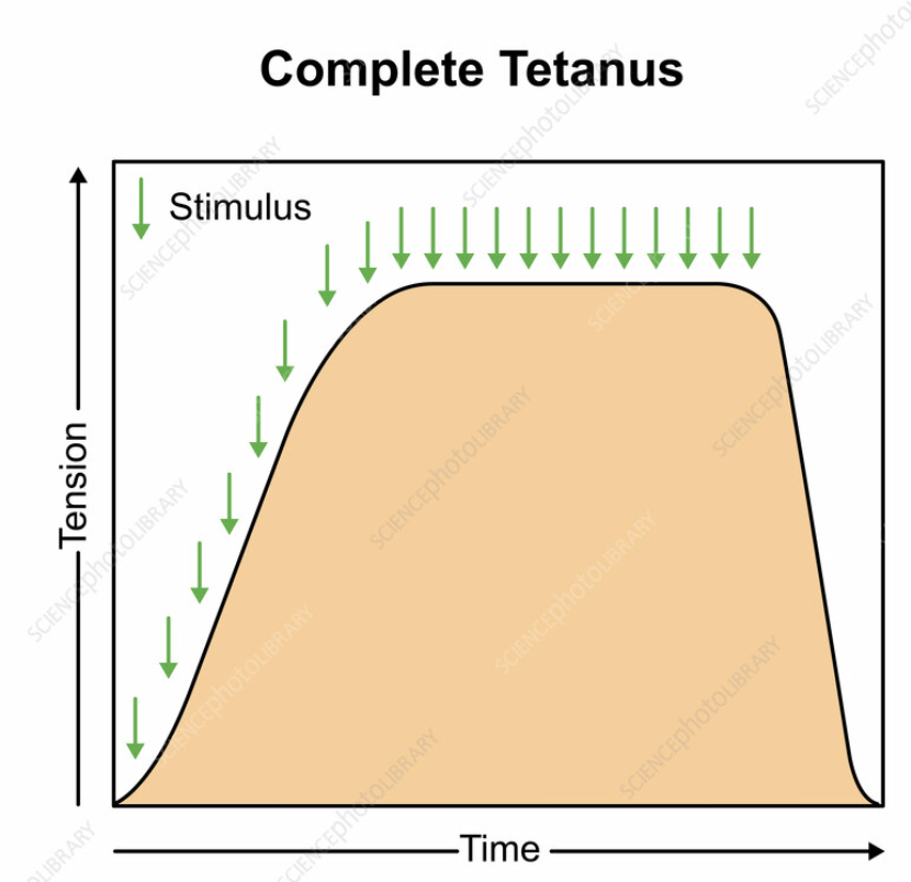
How tetanus reaches maximum tension?
Elevated intracellular calcium is maintained
Filaments have time to slide to fullest capacity
Tested in Exercise C with pairs of shocks at different intervals on frog gastrocnemius
Why relaxation is slower after tetanus?
Large amount of calcium in cytoplasm takes time for SR to take back up
What was the time interval between stimuli when single twitches were observed?
greater than 250 ms
Why would the rate of muscle relaxation be slower after tetanus than after a single twitch?
It takes time for the large amount of calcium in the sarcoplasm to be taken up by the sarcoplasmic reticulum
if there is only a single twitch induced at a low stimulus rate, that is because…
the stimuli were not frequent enough to cause summation
Longer intervals → muscle relaxes between stimuli; shorter intervals → contractions combine
250 ms (4 stimuli/sec): single twitches
Muscle completely relaxed before each stimulus; each twitch had the same force
120 ms (about 8 stimuli/sec): summation
Muscle only partially relaxed; contractions had more force than single twitches
70 ms (about 14 stimuli/sec): complete tetanus
Maximal, sustained contraction