Lab 7- the skull
 Call Kai
 Learn
 Practice Test
 Spaced Repetition
 Match
 Flashcards
 Knowt Play
1/53

There's no tags or description
Looks like no tags are added yet.
54 Terms
the skull
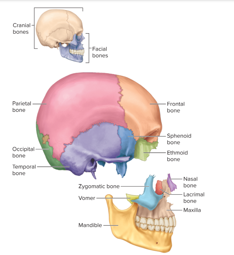
Notice the skull appears to be one bone. It is, however, made up of 22 bones, 21 of which are fused together. We can study these bones based on whether they are part of the neurocranium or the viscerocranium
Neurocranium
We will begin by looking at the neurocranium, or braincase. This is the part of the skull that contains the brain and is made up of eight bones. Notice the zigzag lines on the skull. These are sutures, where two bones join together. They define the edges of the skull bones.
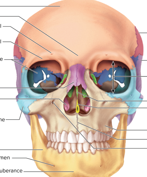
1. Find the frontal bone. This forms the anterior, superior part of the skull and forehead.
2. Find the two parietal bones, which form the superior and lateral sides of the skull.
3. Find the two temporal bones, which form the lateral skull near the temple. Temporal comes from the Latin tempus, which means "time." Since people start to go gray at the temples, this region of the head is so named because it marks the passage of one's life.
4. Find the occipital bone, which forms the inferior, posterior part of the skull.


Ethmoid bone
Frontal bone
Inferior nasal concha
✓ Lacrimal bone
V Mandible
Maxilla
✓ Nasal bones
Occipital bone
Palatine bone
Parietal bone
Temporal bone
Sphenoid bone
Vomer
Zygomatic bone


Ethmoid bone
Frontal bone
Inferior nasal concha
✓ Lacrimal bone
V Mandible
Maxilla
✓ Nasal bones
VOccipital bone
Palatine bone
Parietal bone
VTemporal bone
Sphenoid bone
Vomer
✓Zygomatic bone


Viscerocranium
Paired Bones (12 total / 6 pairs)
Maxillae (Maxillary bones) [2]: Form the upper jaw, the anterior hard palate, and a large portion of the midface.
Zygomatic bones [2]: Form the prominent cheeks and the lateral walls of the eye sockets (orbits).
Nasal bones [2]: Join together to form the bridge of the nose.
Lacrimal bones [2]: Small, thin bones located at the medial (inner) walls of the orbits that house part of the tear-drainage system.
Palatine bones [2]: Form the posterior part of the hard palate, part of the nasal cavity floor, and the floor of the orbits.
Inferior nasal conchae [2]: Scroll-like bones attached to the lateral walls of the nasal cavity that help filter, warm, and humidify inhaled air. [1]
Unpaired Bones (2 total)
Mandible [1]: The lower jaw bone, which forms the chin and lower dental arch; it is the only movable bone of the skull.
Vomer [1]: A thin, plow-shaped bone located on the midline that forms the inferior and posterior portion of the nasal septum. [1]

viscerocranium
Maxillae (Maxillary bones) [2]: Form the upper jaw, the anterior hard palate, and a large portion of the midface.
Zygomatic bones [2]: Form the prominent cheeks and the lateral walls of the eye sockets (orbits).
Nasal bones [2]: Join together to form the bridge of the nose.
Lacrimal bones [2]: Small, thin bones located at the medial (inner) walls of the orbits that house part of the tear-drainage system.
Palatine bones [2]: Form the posterior part of the hard palate, part of the nasal cavity floor, and the floor of the orbits.
Inferior nasal conchae [2]: Scroll-like bones attached to the lateral walls of the nasal cavity that help filter, warm, and humidify inhaled air. [1]
Unpaired Bones (2 total)
Mandible [1]: The lower jaw bone, which forms the chin and lower dental arch; it is the only movable bone of the skull.
Vomer [1]: A thin, plow-shaped bone located on the midline that forms the inferior and posterior portion of the nasal septum. [1]

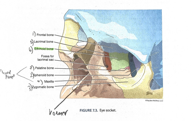
orbit of the eye
1. Frontal bone (forms part of roof)
2. Sphenoid bone (roof and lateral and medial walls)
3. Zygomatic bone (floor and lateral wall)
4. Maxilla bone (floor and medial wall)
5. Lacrimal bone (medial wall)
6. Ethmoid bone (medial wall)
7. Palatine bone (floor and medial wall)
Hard Palate
Bones of the hard palate form the roof of the mouth and the floor of the nasal cavity.
1. The maxillae and palatine bones form the hard palate.
2. The function of the hard palate is to separate the mouth from the nasal cavity. The soft pal- ate is made up of soft tissue and extends the hard palate posteriorly. Use your tongue to feel the roof of your mouth and notice where the hard and soft palates are.
Sutures


Sutures
are the synarthroses (non-moving junctions) between skull bones. 1. Find the coronal suture between the frontal bone and the parietal bones.
2. The sagittal suture connects the two parietal bones.
3. The lambdoid suture is between the parietals and the occipital bone.
4. The squamous suture is between the parietal and temporal bone.
5. Label sutures 1-3 on Figure 7.4. The two squamous sutures cannot be seen in this view.
6. Label the frontal, parietal, and occipital bones on Figure 7.4.

hyoid bone
The hyoid bone is found in the area of the throat and does not articulate with any other bone. It
looks like vampire teeth and is attached to muscles of the tongue and larynx.


Fetal Skull
The fontanelles are areas where the bone of the skull has not yet formed. The space is cov-
ered by a fibrous connective tissue and allows the skull bones to shift to allow the fetus to fit
through the birth canal more easily.
a. The anterior fontanelle is posterior to the frontal bone and medial to the parietal bones.
This suture closes between 7 months and 2 years.
b. The posterior fontanelle is anterior to the occipital bone and will be closed by the lamb-
doid suture. This fontanelle closes in the first few months of life.
2. Label these structures on Figure 7.6A and 7.6B. Also label the coronal suture, the sagittal su-
ture, and the lambdoid suture.


B: Vertebral Column and
Thoracic Cage
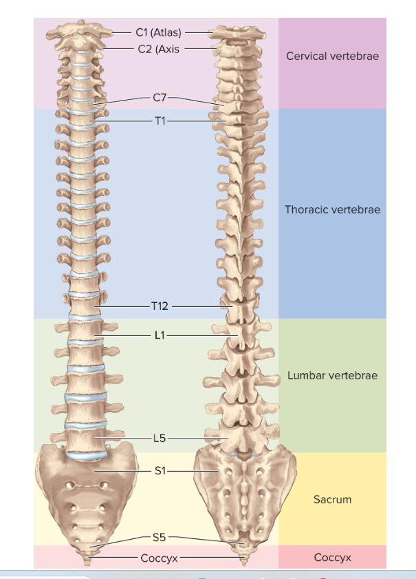
The vertebral column has 26 bones.
· There are seven cervical vertebrae (remember this be-
cause you get up at seven o'clock).
. The first vertebra (Cl) is called the atlas. In Greek myth,
Atlas was condemned to hold up the sky.
. The second vertebra (C2) is called the axis. It is named
this because its odontoid process forms the axis of rota-
tion for the atlas.

cervical vertebrae


label the vertebrae

the sacrum is fused from how many bones?
5
label the parts of the spine:

different vertebrae types
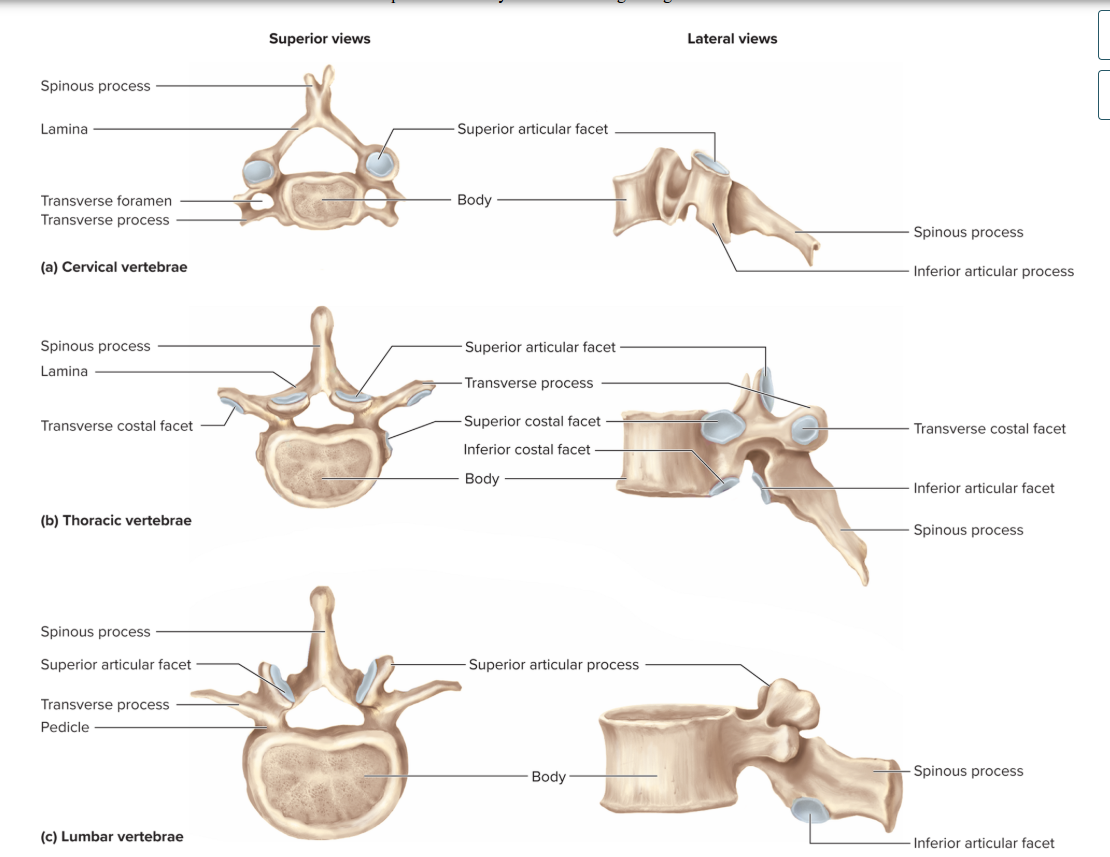
vertebral coloumn
thoracic cage:
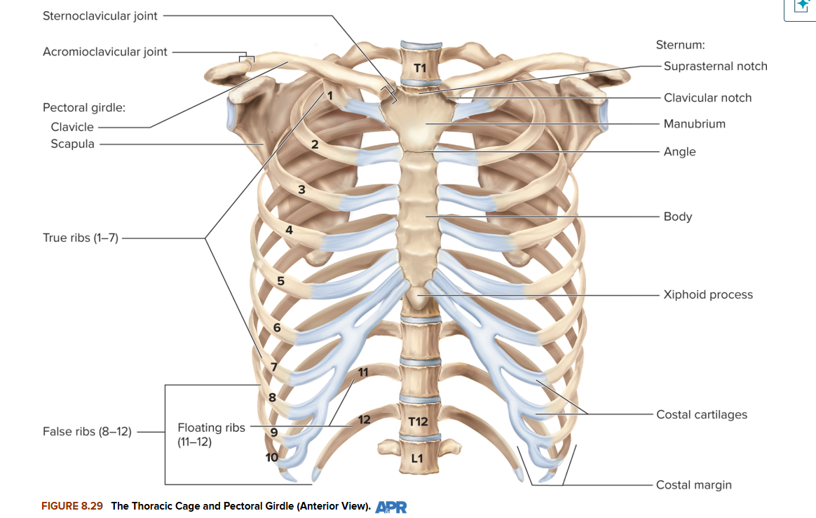
This cage protects your heart and lungs.
1. Find the sternum.
a. The sternum is made of three fused bones.
b. The superior portion is called the manubrium.
c. The middle portion is called the body,
d. The inferior portion is called the xiphoid process.
2. You have 12 pairs of ribs, numbered 1-12.
a. Ribs 1-7 are called true ribs because their costal cartilage attaches directly to the
sternum.
b. Ribs 8-12 are called false ribs because they do not attach directly to the sternum.
c. Ribs 8-10 are vertebrochondral ribs because they attach to the vertebrae posteriorly
and to the cartilage of the 7th rib anteriorly.
d. The last two ribs are floating ribs because they do not attach to the sternum at all.

C: Bone Tissue
Bone is a type of connective tissue characterized
by a hard extracellular matrix made up of a min-
eral compound called hydroxyapatite. The bone
cells (osteocytes) are found in lacunae. What
other connective tissue has cells in lacunae?

Label the following section through a long
bone (Figure 7.11). Be sure to use the follow-
ing labels:
Articular cartilage
Blood vessel
Compact bone
Diaphysis
Epiphyseal line
Epiphysis
Medullary cavity
Periosteum
Spongy bone
Yellow bone marrow


label this bone tissue
this, be sure to use the following labels on your diagram: osteon, lamel-
lae, central canal, lacunae, perforating canal, canaliculi, periosteum, and blood vessels.

articulations
Articulations are joints. A joint is the location where two bones come into contact-some allow
movement, while others are fixed.'
Suture-
An immovable fibrous joint that forms between bones, as seen in the sutures of the
skull.
Gomphosis-
A fibrous joint of collagen fibers that holds a tooth in the jaw, like a peg in a
socket.
Syndesmosis
-A fibrous joint where two structures are held together by longer connective
tissue fibers. This type of articulation is found between the tibia and fibula, for example.
Symphysis
-A joint formed when two structures are held together by fibrocartilage. The
symphysis pubis between the os coxae is an example.
Synchondrosis
-A cartilaginous joint formed when two structures are held together by hya-
line cartilage. The main example in your body is the joint between the epiphysis and diaphy-
sis in a long bone, in growing children.
Synovial --
A freely moving joint between two bones.
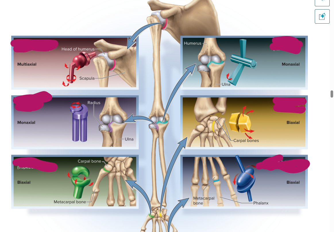
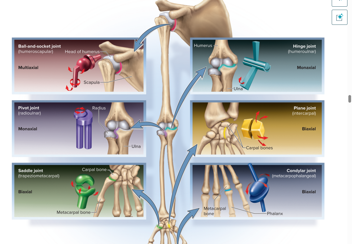
types of synovial joints
Ball and socket joint-The round head of one bone fits into the hemispherical socket of
another bone. Movement is in all three planes. (gleyotumerul)
Condylar joint-The oval shape of one bone fits into a matching depression of another
bone. You get movement in two planes. Rq o iotafpol 5oint,
Hinge joint-A convex articular surface of one bone fits into the concave articular sur-
face of another bone. Allows movement in only one plane.
. Pivot joint-The movement of the atlas turning on the axis or the head of the radius
turning in the radial notch of the ulna.
. Plane joint-The bones of a plane joint have flat surfaces and can glide past each other.
. Saddle joint-Both bones have a saddle shape. Movement in two planes but more move-
ment than with condylar joints.


types of synovial joints
1. Ball-and-socket joints. These are the shoulder and hip joints-the only multiaxial joints in the body. In both cases, one bone (the
humerus or femur) has a smooth hemispherical head that fits into a cuplike socket on the other (the glenoid cavity of the scapula or
the acetabulum of the hip bone).
2. Condylar (ellipsoid) joints. These joints exhibit an oval convex surface on one bone that fits into a complementary-shaped depression
on the other. The radiocarpal joint of the wrist and metacarpophalangeal (MET-uh-CAR-po-fah-LAN-jee-ul) joints at the bases of the
fingers are examples. They are biaxial joints, capable of movement in two planes. To demonstrate this, hold your hand with the palm
facing you. Make a fist, and these joints flex in the sagittal plane. Fan your fingers apart, and they move in the frontal plane.
3. Saddle joints. Here, both bones have a saddle-shaped surface-concave in one direction (like the front-to-rear curvature of a horse's
saddle) and convex in the other (like the left-to-right curvature of a saddle). The clearest example of this is the trapeziometacarpal joint
between the trapezium of the wrist and metacarpal I at the base of the thumb. Saddle joints are biaxial. The thumb, for example, moves
in a frontal plane when you spread the fingers apart, and in a sagittal plane when you move it as if to grasp a tool such as a hammer.
This range of motion gives us and other primates that invaluable anatomical hallmark, the opposable thumb. Another saddle joint is
the sternoclavicular joint, where the clavicle articulates with the sternum. The clavicle moves vertically in the frontal plane at this joint
when you lift a suitcase, and moves horizontally in the transverse plane when you reach forward to push open a door.
4. Plane (gliding) joints. Here the bone surfaces are flat or only slightly concave and convex. The adjacent bones slide over each other and
have relatively limited movement. Plane joints are found between the carpal bones of the wrist, the tarsal bones of the ankle, and the
articular processes of the vertebrae. Their movements, although slight, are complex. They are usually biaxial. For example, when the
head is tilted forward and back, the articular facets of the vertebrae slide anteriorly and posteriorly; when the head is tilted from side to
side, the facets slide laterally. Although any one joint moves only slightly, the combined action of the many joints in the wrist, ankle,
and spine allows for a significant amount of overall movement.


joints contd
5. Hinge joints. These are essentially monaxial joints, moving freely in one plane with very little movement in any other, like a door hinge.
Some examples are the elbow, knee, and interphalangeal (finger and toe) joints. In these cases, one bone has a convex (but not
hemispherical) surface, such as the trochlea of the humerus and the condyles of the femur. This fits into a concave depression on the
other bone, such as the trochlear notch of the ulna and the condyles of the tibia.
6. Pivot joints. These are monaxial joints in which a bone spins on its longitudinal axis like a bicycle wheel spinning on its axle. There are
two principal examples: the atlantoaxial joint between the first two vertebrae, and the radioulnar joint at the elbow. At the atlantoaxial
joint, the dens of the axis projects into the vertebral foramen of the atlas and is held against the anterior arch of the atlas by the
transverse ligament (see L fig. 8.26c). As the head rotates left and right, the skull and atlas pivot around the dens. At the radioulnar
joint, the anular ligament of the ulna wraps around the neck of the radius. During pronation and supination of the forearm, the disclike
radial head pivots like a wheel turning on its axle. The edge of the wheel spins against the radial notch of the ulna like a car tire
spinning in snow.

name the joint type

name the joint type
How are flexion and extension different?
Flexion
Definition: A movement that decreases the angle between two body parts, essentially "closing" or bending a joint. [1, 2]
Memory Trick: "F" stands for Folding or Forward motion
Examples:
Bending the elbow during a bicep curl
Extension
Definition: A movement that increases the angle between two body parts, "opening" or straightening a joint.
Straightening the elbow during a tricep extension.

How are abduction and adduction different?
Abduction
Definition: A movement that pulls a body part away from the midline of the body or a limb's central axis.
Adduction
Definition: A movement that pulls a body part toward the midline of the body or a limb's central axis.

What is circumduction?
Circumduction is a complex, circular anatomical movement that combines flexion, extension, abduction, and adduction in sequence. Instead of moving in just one straight line, the limb moves in a way that its far end draws a circle while the joint itself stays in place, creating a cone-like shape in space.
Key Characteristics
The Shape: The movement traces a cone. The joint (like your shoulder) forms the sharp point of the cone, and your hand or foot traces the flat, circular base.
The Components: To complete one full circle of circumduction, a joint must smoothly transition through four separate movements:
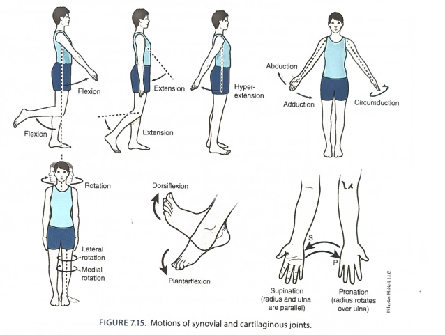
Describe the two ways you can move your foot.
Dorsiflexion (Foot Up):
What it is: Moving the top of your foot (the dorsum) closer to your shin.
Example: Lifting your toes off the ground while keeping your heels planted, walking on your heels, or pulling your toes back when stretching your calves.
Plantarflexion (Foot Down):
What it is: Pointing your toes downward, away from your shin.
Example: Pressing down on the gas pedal in a car, standing up on your tiptoes, or a ballet dancer pointing their feet.

nodding the head yes:
Main Bones Involved
Occipital Bone: The lower back and base of the skull, which features two smooth, rounded bumps called occipital condyles.
Atlas (C1): The very first cervical vertebra of the spine, shaped like a ring to support the weight of the head.
Joints Involved
Atlanto-Occipital Joint: This is the specific joint formed where the occipital condyles of the skull rest on top of the Atlas (C1) vertebra. You have one on the left and one on the right.
Type of Movement
Anatomical Classification: Flexion and Extension. Bending your head forward to lower your chin is flexion, and lifting your head back up or tilting it slightly upward is extension.
Joint Type: This is structurally a synovial condyloid (ellipsoid) joint
thumbs up
Main Bones Involved
Trapezium: A wrist bone (carpal bone) located right at the base of your thumb.
1st Metacarpal: The long bone inside the meaty palm section of your thumb.
Proximal Phalanx: The lower segment of your visible thumb.
Distal Phalanx: The top segment of your thumb (where your thumbnail sits).
Joints Involved
Interphalangeal (IP) Joint: The hinge joint right below your thumbnail.
Metacarpophalangeal (MCP) Joint: The middle joint connecting the thumb to the palm.
Carpometacarpal (CMC) Joint: The highly mobile saddle joint at the very base of your thumb near the wrist.
Type of Movement
To form a full thumbs-up, the thumb coordinates three synchronized actions across its different joints:
Extension: Straightening out the thumb. This occurs at the IP and MCP joints to uncurl the thumb, pointing the tip upward.
Abduction: Moving the thumb out and away from the palm. This happens at the CMC joint to clear the thumb away from your curled pointer finger.
Retraction / Lateral Rotation: Pulling the thumb backward into a hitchhiking position (also controlled at the CMC joint).
kick
Main Bones Involved
Pelvis (Coxal Bone): Serves as the stable anchor and platform for hip rotation and power generation.
Femur: The thigh bone, which is the longest and strongest bone in the body.
Tibia & Fibula: The shin bones, which make up the lower leg segment.
Patella: The kneecap, acting as a fulcrum to increase the leverage of your thigh muscles.
Tarsals & Metatarsals: The ankle and foot bones that create a rigid structure to impact the ball.
Joint Mechanics and Movement Types
The kinetic chain relies on three major joint complexes firing in rapid succession:
1. The Hip Joint
Joint Type: Synovial ball-and-socket joint.
Backswing Phase: Hyperextension and external rotation. The femur is pulled backward to wind up for the kick.
Strike Phase: Flexion and adduction. The thigh accelerates forward and slightly inward across the body to drive toward the ball.
2. The Knee Joint
Joint Type: Synovial hinge joint.
Backswing Phase: Flexion. The knee bends deeply, pulling the heel up toward the glutes to shorten the lever arm so the leg can swing forward faster.
Strike Phase: Extension. Just before contact, the knee violently snaps straight. This rapid extension acts as the primary whip mechanism that transfers maximum speed to the ball.
3. The Ankle Joint
Joint Type: Synovial hinge (talocrural) joint.
Strike Phase: Plantarflexion.
How many bones are there in the vertebral column?
adult - 26 babies 33
Why is the Cl vertebra also known as the atlas?
The C1 vertebra is named the atlas after the Titan from Greek mythology who was condemned to hold up the celestial heavens on his shoulders for eternity
.
Just like the mythical figure, this ring-shaped bone sits at the very top of the spine and is responsible for supporting the entire weight of the skull (head).
What feature of the C2 vertebra allows you to rotate your head laterally?
The feature of the C2 vertebra (axis) that allows lateral head rotation is a distinct, upward-projecting bony peg called the dens (also known as the odontoid process).
How it Works
The Pivot Point: The dens sticks straight up out of the body of C2 and inserts into the ring-shaped C1 vertebra (atlas) above it.
The Movement: The atlas loops around this peg, creating a specialized pivot joint called the atlanto-axial joint. When you shake your head "no," the atlas rotates laterally by spinning around the static dens of C2.
4. What might happen to you if you break your C3 or C4 vertebrae?
Breaking your C3 or C4 vertebrae is a life-threatening medical emergency that can result in permanent paralysis of all four limbs (quadriplegia) and the immediate loss of your ability to breathe on your own.
Disrupting the Phrenic Nerve ("The Breathing Nerve")
The C3, C4, and C5 nerve roots merge together to form the phrenic nerve, which sends the vital electrical signals that command your diaphragm to contract and pull air into your lungs.
A famous medical memory phrase taught to doctors is: "C3, C4, C5 keep the diaphragm alive."
If the spinal cord is severely damaged or severed at the C3 or C4 level, the brain can no longer send signals to the diaphragm. This causes immediate respiratory failure, meaning a person will stop breathing instantly and requires a mechanical ventilator to survive.
You looked at bones that started out as multiple bones but fused into one. Name two of these
fused bones.
The Sacrum: In a developing child, the sacrum consists of five separate sacral vertebrae (S1 through S5) separated by cartilage. As a person grows—typically between the ages of 16 and 25—these five bones completely fuse together into a single, wedge-shaped bone that anchors the pelvis to the spine.
The Pelvis (Coxal / Hip Bone): During childhood, each side of the pelvis is composed of three distinct bones: the ilium (the broad upper blade), the ischium (the lower back portion you sit on), and the pubis (the lower front portion). They meet inside the hip socket (acetabulum) and firmly fuse into a single, solid hip bone by the time a person reaches their early 20s.
Name two bones that might get broken if someone punches you in the eye.
The Maxilla (Maxillary Bone): This bone forms the floor of the eye socket (as well as your upper jaw and cheek). The orbital floor is paper-thin, making it the single most common bone to fracture
The Zygomatic Bone (Cheekbone): This bone forms the outer rim and side wall of the eye socket
, as well as the prominent part of your cheek. Because it sits right on the surface of the face, it absorbs the direct force of a fist, often breaking where it connects to the other facial bones.
(Another frequent culprit is the ethmoid bone, which forms the thin inner wall of the eye socket next to the nose.)
Name the bones that might get broken if someone punches you in the nose.
The Nasal Bones: These are the two small, oblong bones that sit side-by-side at the bridge of your nose.
The Maxilla (Maxillary Bone): This large bone forms your upper jaw and the mid-face. Specifically, the frontal process of the maxilla
The Ethmoid Bone: Located deeper inside the skull, a portion of this bone (the perpendicular plate) drops down to form the rigid, upper portion of your internal nasal septum (the wall that separates your nostrils)
Do all people have exactly the same number of bones? Explain your answer.
1. Age (Infants vs. Adults);
2. Sesamoid Bones (Extra Floating Bones)
Sesamoid bones are small, rounded bones embedded within tendons where they misalign or pass over joints.
The patella (kneecap) is a sesamoid bone that everyone has.
However, many people naturally develop extra, tiny sesamoid bones in the tendons of their hands (at the base of the thumb or index finger) and feet (near the big toe).
3. Anatomical Variations in the Spine and Ribs
Which part of the sternum may break during CPR?
xiphoid process.
What risk is there if a person breaks a rib?
1. Internal Organ Punctures
2. Respiratory Complications
3. Flail Chest
If three or more adjacent ribs are broken in two or more places, it creates a detached, free-floating segment of the chest wall.
7. Are bones alive? Explain,
1. They Have Active Living Cells
Bone tissue contains millions of living cells that fall into three primary categories, constantly managing the skeletal structure:
Osteoblasts: The "builders" that constantly form new bone tissue and lay down minerals.
Osteoclasts: The "recyclers" that break down old or damaged bone so it can be replaced.
Osteocytes: The "sensors" that live inside the bone matrix, monitoring mechanical strain and directing the builders and recyclers where to work.
2. They Have Their Own Blood Supply
Bones are highly vascular structures. If you cut into a living bone, it bleeds. They contain a complex network of blood vessels and nerves that run through microscopic tunnels called Haversian canals. This blood supply delivers oxygen and nutrients to the bone cells and carries away waste products. Because they have nerves, bones feel pain when they are injured or fractured.
3. They Constantly "Remodel" Themselves
Your skeleton is never permanent. Through a process called bone remodeling, your body completely tears down and rebuilds your skeleton at a rate of about 10% per year. This constant recycling allows bones to heal themselves after a fracture, adapt to mechanical stress (growing thicker and stronger if you lift weights), and stay resilient.
4. They Factory-Produce Blood Cells
Deep inside the hollow centers of your long bones and flat bones sits red bone marrow. This marrow functions as a biological factory, producing billions of new red blood cells, white blood cells, and platelets every single day to replenish your cardiovascular and immune systems.
5. They Act as a Mineral Bank
Bones store roughly 99% of your body's calcium and 85% of its phosphorus. When your heart, muscles, or nerves need calcium to function, hormones signal the bone cells to release minerals into the bloodstream. When you have an excess of minerals, the bone cells store them away.
Label the visible structures in this knee joint. Label the
category of joint shown.
