Medical Co-Morbidities
 Note
 Call Kai
 Learn
 Practice Test
 Spaced Repetition
 Match
 Flashcards
 Knowt Play
1/149

There's no tags or description
Looks like no tags are added yet.
150 Terms
Medical Co-morbidities
One or more additional diseases or disorders co-occurring with primary disease or disorder
Medical Co-morbidites examples
Primary: obesity
Additional diseases/disorders: hypertension, kidney disease, restrictive airway disease
What are some NICO/Medically complex common diagnoses
bowel obstructions
cardiac anomalies
congenital anomalies
hydrocephalus
BPS
hypoglycemia
Meconium aspiration syndrome
NEC
HIE
Neonatal sepsis
persistent pulmonary hypertension
RDS
ROP
TEF
seizures
NAS
T or F
There is an Intrinsic tie between upper airway, upper digestive tract and swallowing.
T
Normal infant breathing
Obligatory nose breathers
Periodic breathing pattern in normal infants
3 breathing pause usually lasting about 3 seconds
Norm Respiratory rate: Infant to 6 months
30-60 breaths per min
Norm Respiratory rate: 6 to 12 months
24-30 breaths per min
Norm Respiratory rate: 12 months to 5 years
20-30 breaths per min
Atypical Infant breathers
Mouth breathing
Grunting, Gasping
Nasal Flaring
Stridor
Stertor
Retractions: sinks in when breathing
Snoring during sleep
Breath pauses longer than 10 seconds
Atypical Respiratory rate
over 60 breaths per minute
Stridor
noisy breathing that occurs due to obstructed air flow through a narrowed airway
Where can upper airway disorders occur?
Above the level of the larynx
At the level of the larynx
Below the level of the larynx
Causes of Pediatric Airway Obstruction Above the Level of the Larynx
Pyriform aperture stenosis
Choanal atresia: nasal passages narrowing
Midface hypoplasia
Retrognathia
Micrognathia
Glossoptosis
Macroglossia
Causes of Pediatric Airway Obstruction AT THE the Level of the Larynx
Laryngomalacia
Laryngeal Agensis
Laryngeal Web
Vocal Cord Paralysis
Gastropharyngeal Reflux
Posterior Glottic Stenosis
Subglottic Stenosis
Laryngeal Cleft
Causes of Pediatric Airway Obstruction BELOW the Level of the Larynx
Vascular ring/extrinsic tracheal compression
Tracheal stenosis, complete tracheal rings
Tracheomalacia
Laryngotracheoesophageal clefts
Tracheoesophageal Fistula
Foreign Body Aspiration
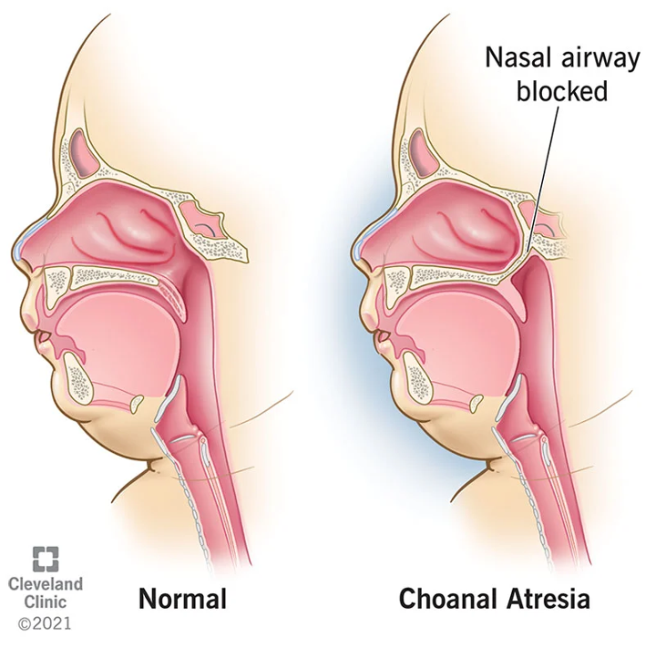
Choanal atresia:
bilateral or unilateral (50-60% uni)
Blocks posterior nasal flow
Compensate with oral breathing – have episodes of apnea cyanosis (changing colors)
This can occur with other dx like CHARGE
Infants are primarily nasal breathers (some say obligatory, debated)
Evaluate via nasopharyngoscopy
How could Choanal atresia impact feeding?
suck swallow breathe cycle
Why would SLPs maybe be the first to figure Choanal atresia out? (in the absence of an overt syndrome?)
not giving them time seeing it from the feeding and speech point of view, not just focused on medical
Choanal atresia Treatment
surgical repair during early infancy
stents- short term and post operatively
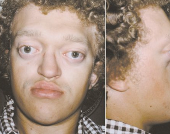
Midface hypoplasia
Associated with craniosynostosis and Crouzon and Apert Syndrome
Incomplete development of the middle of the face
the maxilla, cheekbones and eye sockets have not grown as much as the rest of the face
Large eye appearances
Surgical repair may include midface advancement
Early as 3, best to wait until post puberty
Maxilla could be pushed back into the base of skull at rest or stress of feeding accentuates the narrow space
Midface hypoplasia treatment
plastic surgery repair
Crouzon Syndrome
A disorder characterized by the early fusion of certain skull bones. This prevents average growth of the skull, which can affect the shape of the head and face.
common symptoms:
1. Bulging eyes
2. Abnormal head shape
3. Visual or hearing difficulties
4. Flatten the middle of the face
5. Dental Problems
Stridor at rest for infants or children with severe midface hypoplasia (without a cleft) and/or during feeding
Why?
What is our scope?
Not enough space for them to breath
speech and swallowing
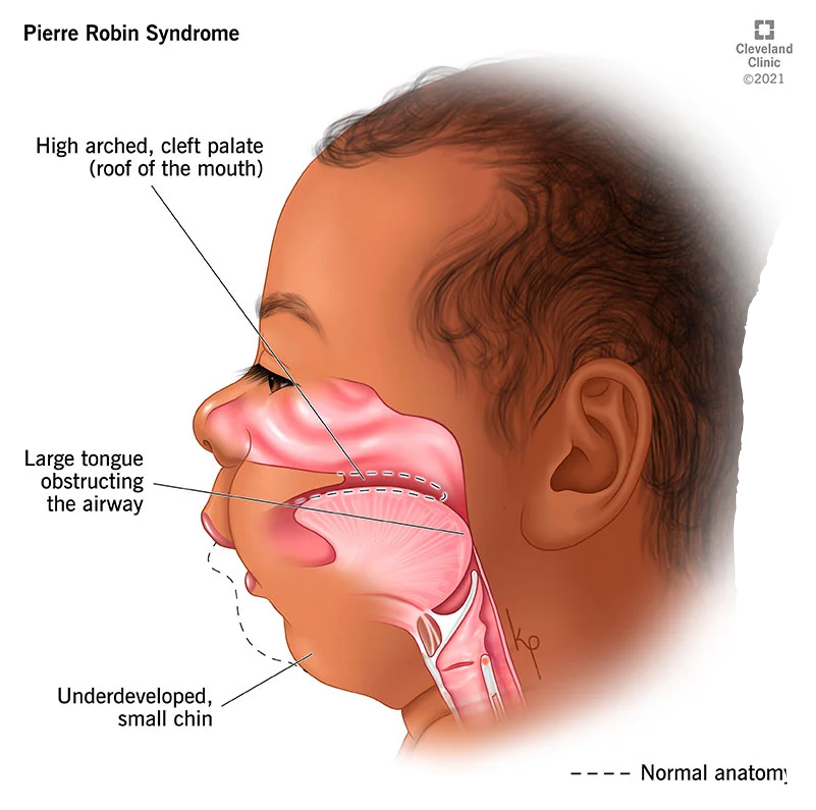
PRS: Pierre Robin Sequence/Syndrome
chin in neck folds
Triad of symptoms
Mandibular hypoplasia
Micrognathia
Glossoptosis (backward, downward, placement of tongue)
Cleft palate (can co-occur with cleft)
T or F
While PRS is equally common in males and females, there is a higher incidence among twins, Natal teeth, or teeth that are present at birth.
T
What are potential respiratory issues with PRS?
What about feeding?
Why?
Sometimes not aspiration
Every time it was extracted from the bottle, the tongue would get stuck in the airway
obstructing the airway every time they are eating
could not coordinate breathing and eating
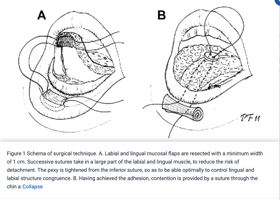
PRS interventions: based on how severe breathing and swallowing are
Tongue tip adhesions (glossopexy)
Addresses glossoptosis but doesn’t change the mandible
attaches the tip of the tongue to the bottom lip (to keep the tongue from falling back)
Mandibular distraction osteogenesis (better outcomes than TLA)
Quicker achievement of oral feeding
lengthens the jaw to help open the airway
An incision (cut) is made through the skin under the jawline, and then the jawbone is carefully separated so a distraction device can be attached to the bone. This happens on each side of the jaw.
The two small rods (called distractor arms) protrude (stick out) from the chin or from behind the jawline.
each distractor arm will be turned a full 360 degrees starting the day after surgery and is repeated every eight hours for about 10 days.
jawbone will grow to fill the space.
trach
Mandibular distraction osteogenesis
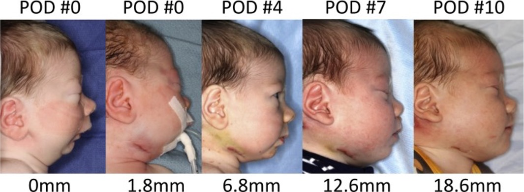
best management for prs
cleft and craniofacial team
slp role: feeding and swallowing
preop: proper feeding
postop: maintaining good feeding
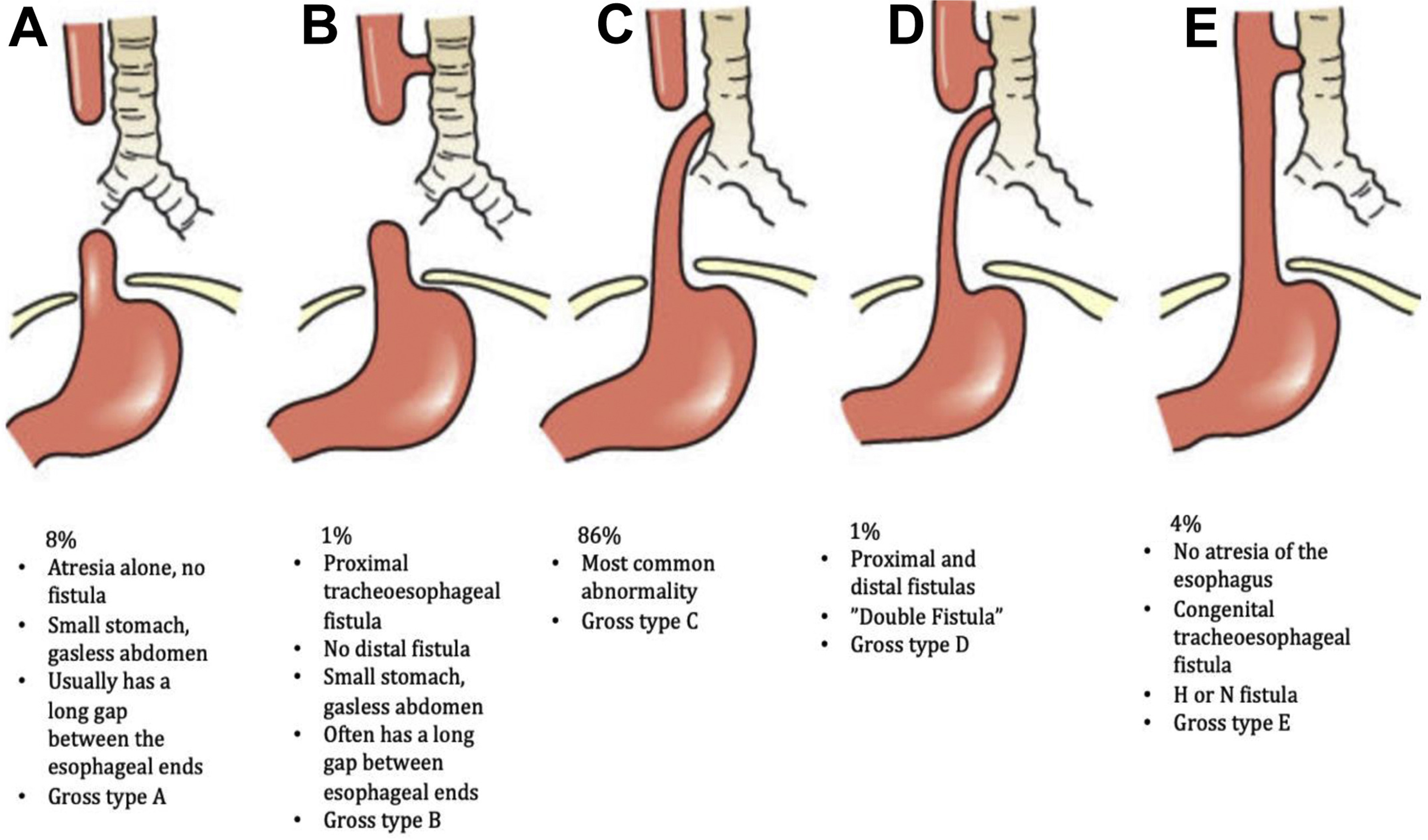
Tracheoesophageal Fistula and Esophageal Atresia
Typical are happening together
not connecting to each other
During early development, the esophagus and trachea begin as a single tube that normally divides into two adjacent passages between four and eight weeks after conception. If this separation does not occur properly, EA/TEF
abnormal connection in one or more places between the esophagus and trachea-TEF Occurs commonly with EA
The upper esophagus does not connect (atresia) to the lower esophagus and stomach. Almost 90 percent of babies born with esophageal atresia also have a tracheoesophageal fistula (TEF)
VACTERL syndrome
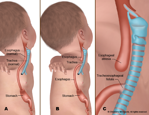
EA mother usually has polyhydramnios- why?
swallowing helps regulate the amniotic fluid
sign of there is a condition
How could TEF and EA present in a newborn initially?
spit it back up
fills- overfollows- go into lungs
possible throwing up
TEF/EA Interventions
In most cases, the Surgeon removes the abnormal connection between the trachea and esophagus and then sews together the two ends of the esophagus.
Can result in a narrowing of the esophagus/stenosis following repair
SLPs have to watch swallowing due to scar shrinkage (CAN NARROW)
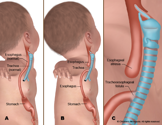
atresia
pouch
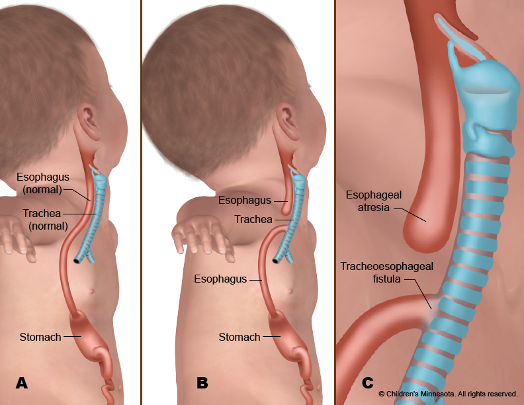
fistula
hole
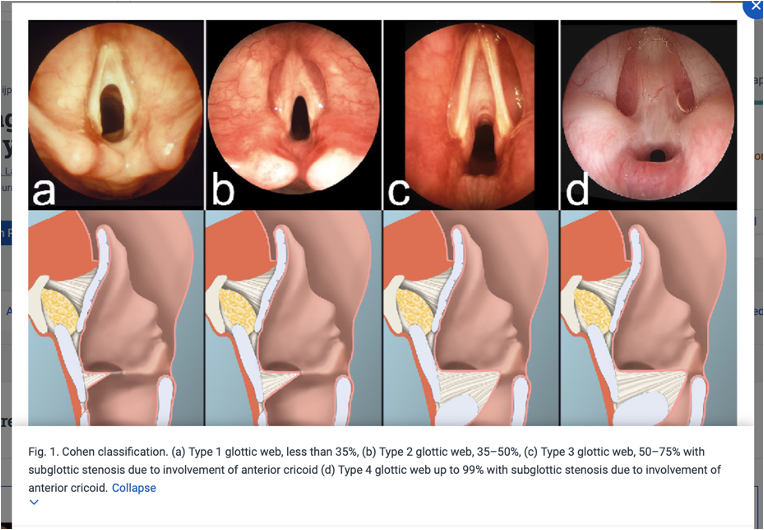
Laryngeal web: rare congenital disease
Stridor
Common in 22q
A fibrous layer of tissue of varying thickness that develops in the larynx, present at birth
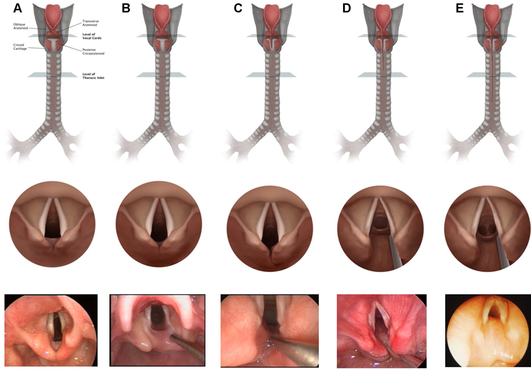
Laryngeal cleft
Dx by respiratory symptoms: stridor, dysphagia
Isolation or part of another dx (ex VACTERL)
anomalous connection between the larynx and hypopharynx
Normally, when the larynx develops, it is completely separate from the esophagus, and a Laryngeal cleft occurs when these structures do not develop normally in an embryo and there is an opening connecting the larynx and the esophagus (incomplete separation of larynx/trachea and pharynx/esophagus
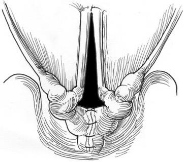
Laryngeal cleft repairs
Injection (inject gel into the space)
Surgical/suture repair (possible stitches)
SLP role Laryngeal Cleft
probably will have difficulty down the line
difficulty with textures
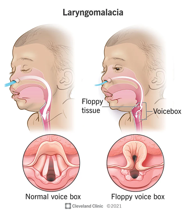
Laryngomalacia
The most prevalent cause of stridor in infants
Laryngomalacia ranks as the most prevalent cause of infant stridor.
leading to an inward collapse of the supraglottic airway with inspiration
Multiple causal theories:
Neurologic dysfunction: suggesting that altered laryngeal tone occurs by abnormal integration of the laryngeal nerves.
support from a pathologic study showing increased supraglottic nerve diameter in patients with severe laryngomalacia
Other theories, such as an imbalance of demand-supply upon infant inhalation, require further study.
Researchers have not found reflux to be a causative factor for laryngomalacia, but nearly 60% of infants have concomitant acid reflux disease.
thought to cause irritation and edema of the upper airway, potentially worsening obstruction.
Mild Laryngomalacia
Never dx formally with endoscopy by ENT, likely “managed” by PCP/Pediatrician, so dx is usually labeled suspected.
Diagnosed formally by ENT
Via Flexible fiberoptic laryngoscopy-the gold standard
ability to directly assess the dynamic collapse of the supraglottic airway during awake respiration.
Laryngomalacia is often found to have
shortened aryepiglottic folds that tether the epiglottis posteriorly, an omega-shaped epiglottis, and/or redundant arytenoid tissue that prolapses over the glottis.
Laryngomalacia Management
Medical
mild/moderate: monitor respiratory status
More severe: Supraglottoplasty
A laser or surgical instrument may be used to remove obstructive tissue in the upper larynx conservatively.
Also, keep an eye on feeding
Most common in young infants
Possible outgrowing of one
Feeding is usually the biggest issue with Laryngomalacia.. why?
Using what you know about infant feeding interventions, what might we consider?
ENT is basing their surgery on you
harder time breathing:
Suck swallow breathing
would stop breathing
aspiration
may need more dysphagia intervention after surgery
Bronchopulmonary Dysplasia
Common in premature infants born before 34 weeks GA and on a ventilator for a prolonged period
Abnormal development of lung tissue characterized by inflammation and scarring
Feeding/swallowing characteristics:
Incoordination of suck/swallow/breath
Inability to sustain suck for 2 minutes
Persistence of immature suck pattern
Difficulty initiating movements
Arhythmical
Apena
can be defined as the cessation of respiratory effort lasting more than 20 seconds or lasting a shorter duration but accompanied by bradycardia or cyanosis
Central (depressed respiratory center with no efferent output)
obstructive (obstruction to airflow causing inadequate ventilation)
mixed (both central and obstructive).
Apena causes
Craniofacial anomalies (Pierre Robin sequence, Beckwith-Wiedemann syndrome, Apert syndrome, Treacher Collins syndrome)
Chronic nasal obstruction (severe septal deviation, allergic rhinitis, nasal polyps)
Down syndrome
Metabolic abnormalities (mucopolysaccharidosis)
Infections (supraglottis, croup, bronchiolitis, pneumonia)
This can occur in premature infants due to neurological immaturity
Pediatric Tracheostomy
Performed for children with complex medical conditions to manage:
Upper airway obstruction
Syndromes (treacher Collins, PRS, Bechwith-Wiedemann)
Congenital anatomical abnormalities (web, paralysis of VF, laryngomalacia)
Infectious disease causing swallowing
tumors
Need for prolonged ventilation
Respiratory issues cause reduced gas exchange at the level of the lungs.
Neuromuscular conditions
cuffed vs. uncuffed trcah
Try to go cuffless with children due to having less risk
Neurological Dx considerations
Static vs. Progressive
Therapy expextations
Preservation vs. advancement vs monitoring for decline and compensation
Neurological progressive dx
Digresses
west disease
CP
MLD/ALD
Neurological static dx
stays the same or improves
Cerebral Palsy (CP)
2:1000 children
Dysphagia is VERY common in CP
85% of children 18-36 mo of age with CP had dysphagia (Arvedson 2019)
Increased degree of motoric impairment→ Increased likelihood of dysphagia
Those dependent on feeding have the most severe dysphagia (Arvedson 2013)
Extreme prematurity is associated with CP and dysphagia
Heterogeneous group
Oral and Pharyngeal phase concerns related to problems of trunk/shoulder and head control
tone/spasticity
CP feeding difficulties
Abnormal oropharyngeal tone, reduced strength, frequent coughing and choking
Overall, poor feeding development increases the risk of malnutrition and dehydration.
Between 30 and 99% of children with CP have feeding and swallowing disorders.
Without effective treatment for dysphagia, the risk of malnutrition, failure to thrive, respiratory compromise, or even death greatly increases.
Major 3 abnormalities of swallowing in CP
Decreased ROM/mobility of the tongue
Delayed initiation of the swallow
Reduced pharyngeal motility
All 3 above result in timing and coordination dysfunction in the pharyngeal phase
CP has a high likelihood of:
Tongue thrust
Prolonged and increased bite reflex
Atypical reflex of tonic bite may be present
Strong gag reflex
Tactile hypersensitivity in/around mouth
drooling
CP has mix of compensatory and habilitative strategies such as:
Positioning
Diet Modification
Specialized tools
Oral sensorimotor interventions such as those discussed in treatment module
Find their own individual strengths and capitalize
ex.unilateral weakness feed on the strong side
Don’t just end at compensatory, should try to promote maximal potential/skill development
NAT
non-accidental trauma (shaken baby syndrome)
Acquired Brain Injury Diagnosis
May Include any of the following:
Seizures
Strokes
TBI
NAT
At Risk for the following:
Poorly coordinated oral and pharyngeal phase
Weakness
sensory/afferent nerve involvement
Silent aspiration
Consider Dysphagia exercises based on MBS finding
Acquired Brain Injury Diagnosis common symptoms
Consider alert/awakeness, can change rapidly immediately after injury
Ability to follow direction
Attention concerns
Impulsivity
seizures/postictal stage
Positioning
Tone (baclofen etc)
Team with medical and other professionals
Atypical motor patterns
Neuromuscular Disease and Swallowing Physiology Caveats
Have to consider the nature of the disease
Acquired vs. congenital
Is it progressive?
Rehabilitative intervention vs. habilitative
Notes on MBS findings with clients with neuromuscular disease:
Frequent deep penetration on MBS will LIKELY result in aspiration due to the NATURE of their swallowing deficits
Increased likelihood of residue with thicker textures
may not be able to clear the residue → aspiration
unable to handle negative sequelae associated with aspiration
Proceed cautiously
With Significant Impairment Consider
Quality of Life
Joy
Caregiver goals
Holistic view/picture
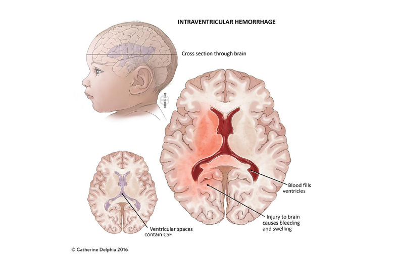
IVH: Intra ventricular Hemorrhage
can lead to CP
Intraventricular
within the ventricles
Hemorrhage
excessive bleeding
IVH has _ grades
4
IVH Grade 1
Bleeding occurs just in a small area of the ventricles.
(like a bruise to NICU)
IVH Grade 2
Bleeding also occurs inside the ventricles.
(bruise and blood)
IVH Grade 3
Ventricles are enlarged by the blood
(like a balloon) Severe
IVH Grade 4
Bleeding into the brain tissues around the ventricles.
(invading brain tissue) Severe
IVH Grades 3 and 4 are the most serious
Increased risk for long-term brain injury
Hydrocephalus
Cerebral spinal fluid in the brain may develop after severe IVH
Cerebral Palsy
Cerebral = brain
Palsy = motor weakness, motor discoordination
Damage to the brain during or directly after birth that impacts motor control
Walking
Playing
Feeding
Swallowing
Talking
Gastrointestinal disorders considerations
Hospital: GI tract is a long, continuous tube with valving
Tummy (feeding/swallowing) : Oral, Esophageal, Stomach, Duodenum, Jejunum, Small bowel, Large bowel
Brain: Mucosal receptors all the way down the “tube” can influence appetite (Irritation sensitive, Irritation can slow /at risk peristalsis)
Caution: Pressure sensitive system
NEC
Children who are born full term with severe cardiac system mimics at risk for this
Complex heart disease
Heart is having a hard time oxygenating everything
GI Developmental Considerations: younger than 34 weeks GA increased risk
GERD
Reduced or poor gastric emptying
Increased gastric residuals
Immature motility
Immature intestines = injury = Necrotizing Enterocolitis (NEC)
Feeding Intolerance (broad term, many meanings)
Feeding Intolerance in Infants = GI
Ger/GERD (Gastroesophageal Reflux (Disease)
Milk Protein Allergy
Constipation
Eosinophilic Esophagitis (EOI)
Necrotizing Enterocolitis (NEC)
Gerd
Involuntary retrograde passage of gastric contents into the esophagus
Frequency of Gerd
70-85% of infants have some degree of GERD in first 2 months of life
Resolves as infant matures
Symptoms of GERD (ask the parents)
•Pain and/or discomfort
•Facial grimacing
•Arching (pain response or learned response)
•Head turn (not have a direct path, escape feeling)
•Sandifer’s Reflex (confined to infancy18-36 months)
•Poor weight gain
•Emesis
•Crying
•Poor PO intake (oral intake)
•Coughing
•Nasal congestion
•Color change
•BRUE (Breif resulting unexplained event) (ALTE: Apparent life-threatening event)
GERD know the why
•Aerophagia
•Immature LES
•Short or narrow esophagus
•Low Vagal tone
•Formula and/or Breast Milk
•Hernia
•Overfeeding
•Positioning
Gerd can be misdiagnosed as
Aspiration
Aspiration can be misdiagnosed as
GERD
How to identify GERD
observation of clinical signs/symptoms
instrumental testing
Complications of GERD
PO refusal
Stress
negative compensatory behaviors
frequent changes
airway compromise
failute to Thrive
High risk populations for GERD
•Premature Infants
•Congenital Heart Disease
•Upper Airway Disorders
•Neurological Disorders
•Genetic Differences
•Lung Disease
Treatment options that are considered for GER/GERD
Meal size (smaller)
Meal frequency (more frequent)
Thickening formula (research shows this is good now)
Introduction of solids
Upper body strength and control
Medication (after trying everything else)
Tube placement to rule out etiology (NG Tube)
Fundoplication (controversial)
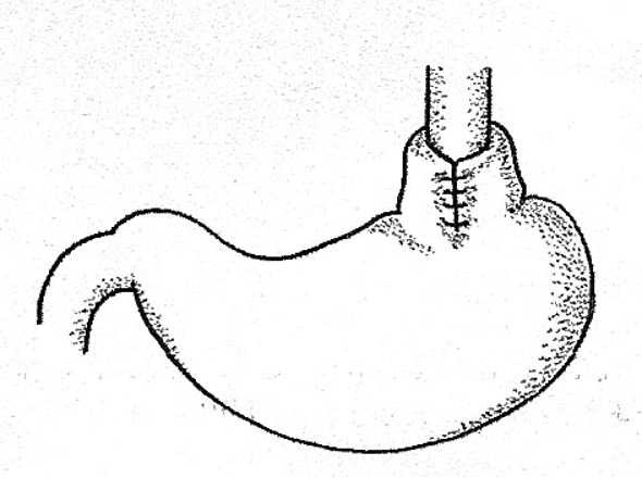
Nissen fundoplication
A procedure involves wrapping a part of the stomach around the lower part of the esophagus to treat certain gastrointestinal conditions. (continuous gaging)
if too tight, never eat enough due to the stomach's inability to expand
Delayed Gastric Emptying
Movement of gastric contents from fundus of stomach to pylorus requires muscular action
Abnormal or inconsistent muscular activity in stomach can lead to delays in emptying
Can contribute to GERD, projectile vomiting, and persistent vomiting
Esophageal Stricture
Narrowing of esophagus
May be congenital or acquired
Response to chronic acid associated with GERD
Difficulties may present with pureed or solid foods that require larger diameter for bolus passage
Bolus cannot pass may cause gagging or vomiting
Gold standard for imaging : Upper GI study
Cricopharyngeal Achalasia
•Occurs when UES fails to open despite swallow initiation and hyolaryngeal elevation.
•Presents as feeding difficulty
•Pharyngo-nasal backflow
•vomiting
•Recurrent aspiration
•Associated with Esophageal dysmotility
Failed relaxation of the LES
•Can be tied to neuromuscular disorders
•CP bar on VFSS/MBS
•Treatment includes:
Botulinim injections, dilatations, myotomy
NEC/Short Bowel Syndrome
NEC: Necrotizing Enterocolitis
Poor perfusion to part of the bowel in premature infants
Can lead to perforation and sepsis
Resection of infected bowel
Can result in Short Bowel Syndrome (other cause of SBS)
Unique Challenges
Lack absorption
May require TPN
Liver disease
growth/weight
Hydration
GI discomfort interrupts hunger/satiation cycle
Total Parenteral Nutrition (TPN)
also known as intravenous or IV nutrition feeding, is a method of getting nutrition into the body through the veins. In other words, it provides nutrients for patients who do not have a functioning GI tract or who have disorders requiring complete bowel rest.
EoE: Eosinophilic Esophagitis
Can be present in esophagus, stomach or intestine.
Autoimmune disorders precipitated by ingestion of food protein.
The body perceives certain proteins as invaders, causing eosinophils to gather, degranulate, and destroy the epithelium of the esophagus.
Environmental allergies are thought to be a complicating factor and may also play a role in the gathering of eosinophils.
You can have EE and GER
Diagnosis is complicated for EoE
15-20 eosinophils per high powered field
Biopsies needed at several sites along distal and proximal sites on the esophagus
Eosinophils are NOT naturally occurring in esophagus
EGD may be completed and note:
Furrowing
Strictures/ swelling
Rings and webs in the esophagus
T or F
Visual findings alone are insufficient to diagnose an EE disorder— there must be a confirmation biopsy!
T
EoE: Medical Treatment Considerations
Elemental diet
Neocate One +
Neocate Junior
Pediatric EO28 Splash
Elecare
PurAmino
Elimination diet (based on repeated scoping)
Remove Dairy, Soy, Wheat, Eggs, Peanuts, Tree Nuts, Fish, and Shellfish (and some subtract corn) from the diet for 6-12 weeks. Supplement with elemental formula.
Re-scope and eosinophil counts.
Repeat with each new food
Food and symptom diary is often requested
Swallowed aerosolized corticosteroids
Elemental Formulas:
These formulas contain 100% broken down proteins and are especially for those infants with suspected milk protein allergy. These infants are often fussy, seemingly cry in pain, have blood in their stools and/or poor weight gain. These formulas are not typically available to purchase on the shelves of a grocery store. They do not contain any intact proteins (only the individual amino acids or building blocks of proteins) that can cause allergy or intolerance reactions.
EoE
Allergic process
Typical allergies present in 40% of cases
Males make up 70-80% of cases
Spectrum of severity
Solid food dysphagia
food impaction
Not well managed by reflux meds alone
Reflux is “different” allergic in etiology
Management important to prevent severe digestive system damage, including impaction and need to dilatation
Can have oral sensory based feeding difficulties and extremely restricted diet
Result of eating→ pain/negative association loop with trigger foods and/or need to have very restricted diet
Why treat eosinophilic disorders?
Anatomical changes in the esophagus
Failure to thrive/poor growth
Children restrict diet due to pain and discomfort.
Feeding selectivity
Some children have extreme food selectivity due to pain and esophageal changes and food impactions.
It can result in severe damage if not controlled or can result in severe reactions
Furrowing of the esophagus etc
FPIES •Food Protein Induced Enterocolitis Syndrome
GI specific allergy
Occurs in infancy
FPIES: typical triggers
cow’s milk protein
soy
cerals