Monitoring devices
 Call Kai
 Learn
Learn Practice Test
 Spaced Repetition
 Match
Match Flashcards
 Knowt Play
1/70

There's no tags or description
Looks like no tags are added yet.
Name | Mastery | Learn | Test | Matching | Spaced | Call with Kai | Chat |
|---|
No analytics yet
Send a link to your students to track their progress
71 Terms

multiparameter machine
What is perfusion
circulation of blood thru tissues
List the order of the cardiac circulation
deoxygenated blood → body → vena cava → RA →Tricuspid valve → RV → Pulmonic valve → Pulmonary artery → Lungs (O₂ gained) → Pulmonary veins → LA → Mitral valve → LV → Aortic valve → Aorta → Body
Understand and be able to explain this formula: CO = SV X HR
CO: amount of blood heart pumps in one minute
cannot be measured directly → pulse, or BP
SV: amount of blood pumped by one ventricle per beat
HR: number of beats per minute
How can we increase CO
↑ HR and SV
but if HR too high → heart having less time to fill b/w beats → ↓ SV
What is it called when there is a fast HR
tachycardia
How do you increase stroke volume?
stronger contractions
more blood in heart
increasing heart space by giving fluids
What is the reaction of the body/CO = SV X HR when experiencing dehydration?
↓ preload (blood filling heart) → less stretch → ↓ SV = ↓ CO
body response is increasing HR
Arteries, veins, and arterioles
Arteries: elastic tissue, stretch and snap back
Veins: thin walls, huge capacity to hold blood
Arterioles: smooth muscle, relax and contract
Understand and be able to explain this formula: BP = CO x SVR
SVR: systemic vascular resistance, amount of resistance blood flow encounters
CO: amount of blood pumped by heart per minute
What can affect systemic vascular resistance SVR
Diameter of blood vessels : ↑ SVR → ↑ Resistance → Narrow vessels = vasoconstrict
length
viscosity : how think blood is → polycythemia= ↑ SVR
What changes in the short-term in SVR
diameter of blood vessels
What will the body do to shift blood to important organs?
vasoconstrict to less important organs (toes/fingers) and direct blood to lungs, liver, brain, kidneys
What is expected for the blood pressure to happen during anesthesia
expected to be lower due to anesthetic drugs
what we can do to affect CO
increase or Decrease HR or SV
↑CO
Atropine → ↑HR → CO ↑
IV fluids → ↑ SV → CO ↑
↓ CO
Bradycardia =↓ HR
Dehydration = ↓ SV → ↓ CO.
What can we do to affect BP
BP ↑
Increase CO → Increase HR or SV.
Increase SVR → Vasoconstriction.
BP ↓
Decrease CO or SVR.
Propofol, acepromazine, inhalants = Vasodilation → ↓ SVR → ↓ BP.
what can we do to affect SV
SV ↑
Give IV fluids → ↑ preload
SV ↓
Dehydration → ↓ Preload → ↓ SV
Blood loss → ↓ Preload → ↓ SV
What can we do to affect SVR
SVR ↑
Vasoconstrict → ↑ SVR. ex. Dexmedetomidine, xylazine
SVR ↓
Vasodilation → ↓ SVR. ex. Propofol, acepromazine, inhalants
what does hypotension mean and what are the causes? drugs related?
means low BP. causes from
drugs: inhalants, acepromazine, propofol, ACE inhibitors
blood loss (hypovolemia)
dehydration
What is dose dependent hypotension and how does that relate to how you would administer anesthesia?
the higher the dose of anesthetic drug the more BP to decrease
give lowest effective dose anesthesia and adjust to maintain anesthesic depth
Too much anesthesia → Vasodilation → SVR ↓ → BP ↓ (hypotension).
What is anticipated in surgery with excessive bleeding?
lower BP
pale MM (vasoconstrict)
tachycardia b/c heart making up for blood loss
Hypertension
not common under anesthesia, caused b/c of
too light, feeling pain, fear
vasoconstriction → dexmedetomidine = bradycardia, hypothermia
What would you expect to happen if an animal was too deep under anesthesia?
Central eye position
Pupil dilated
Loss of all reflexes
HR ↓ (bradycardia).
BP ↓ (hypotension).
What would you expect to happen if an animal was too light under anesthesia?
Tense jaw tone
Central eye position
Normal pupil size
HR ↑ (tachycardia).
BP ↑ (hypertension).
What color mm would you expect to see with vasoconstriction and vasodilation? what are their causes
Vasoconstriction: pale mm
Impairs peripheral perfusion
hypothermia
hyperventilation
Vasodilation: pink or red mm
improves peripheral
hypotension
hyperthermia
What are some actions you can take in response to hypotension in surgery?
We can reduce anesthetic depth to increase BP
give fluids to preload ↑ → SV ↑ → CO ↑ → BP may increase
control blood loss using suction or cautery to stop bleeding
Blood pressure monitoring and what do systolic and diastolic measure?
allows numeric assessment of diastolic, systolic, and mean arterial pressure (MAP), measured in MMhg
Systolic : pressure in blood vessels when heart beats
diastolic : pressure in blood vessels when heart rests
What are the normal systolic, diastolic and MAP ranges
Systolic normal range is 110-160 MM of mercury
Diastolic normal range 60-90 MM of mercury.
Mean arterial pressure (MAP) normal range is 85-120 MM of mercury
What is the general guidelines regarding systolic and MAP
systolic has to be greater than 180 MMHG
minimally acceptable is MAP 60
What are types of blood pressure measurement devices?
Direct arterial line
doppler → ultrasound waves by transducer detect motion of arterial wall, only gets systolic reading, cuff
oscillometric → measure oscillations within a cuff bladder and able to obtain MAP, cuff
How do you measure a BP cuff? How will the readings be affected if you choose the incorrect size?
40% of limb in dogs and 30% in cats
too large= falsely lower BP
too small = falsely higher BP
What are the three areas of a normal ECG
P wave: atrial depolarization → triggers atria contraction
QRS wave: ventricular depolarization → triggers ventricular contraction
T wave: ventricular recovery
What is the meaning of sinus, supraventricular, ventricular, premature
Sinus - comes from SA node
Supraventricular - comes from above the SA node where QRS normal
Ventricular - comes from ventricle where QRS abnormal (wide and bizzare)
Premature - comes early
What is the difference between SA node and AV node
SA node is the pacemaker of the heart starting electrical impulse
AV node stimulates contractions
Electrocardiograph ECG
measures electrical impulses of heart muscle and help detect arrhythmias
can give false signals if pt is not responding and not best info for quality of beat
which leads go where and which lead we typically use in surgery
(think snow and grass are at ground, Christmas at the end of year) pt always in R lateral recumbency
White: right forelimb
Green : right hindlimb
Black : left forlimb
Red : left hindlimb
There are 6 lead ECG but LEAD II used as main lead to read ECG
What are the two types of conductions
conductive gel (U/S gel) - does not dry out
Alcohol - cooling, need to reapply

Cardiac arrhythmias -
Normal sinus rhythm


Cardiac arrhythmias -
Sinus arrhythmia
normal in dogs not in cats
HR speeds up on inspiration

Cardiac arrhythmias -
Sinus bradycardia
slow HR but all p,t, and qrs waves normal equal

Cardiac arrhythmias -
Ventricular Premature complexes VPCS
abnormal beats coming from ventricular myocardium
not usually treated
“wide and bizarre” QRS
Cardiac arrhythmias -
1st degree AV block

conduction thru AV node delayed
PR interval prolonged

Cardiac arrhythmias -
2nd degree AV block
no QRS wave
disruption of AV nodal conduction

Cardiac arrhythmias -
3rd-degree AV block
P and QRS are independent
What conditions can cause bradycardia?
anesthetic depth → too deep
hyperkalemia
hypothermia
drugs → opioids, a 2 agonist
vagal tone
What conditions can cause tachycardia?
drugs → atropine
anesthetic depth → too light
pain
hypoxia
hypercapnia
What is the vagal tone? What are changes in vagal tone? Two reflexes?
the activity of the vagus nerve, key component of PNS
increase tone= lower HR
decrease tone= high HR
oculovagal and carotid
What is meant by vasovagal? which type are dogs prone?
there is a drop in HR and BP in response to excitement
brachycephalic and athletic dogs - high vagal tone
What is the result of hard intubation
result in a vagal response
Two types of catherters
over-the-needle catheter
through-the-needle catheter
Which type of monitoring is most critical
respiratory monitoring
Type of monitor -
Apnea monitor
located b/w ET tube and breathing circuit and it measures the lapse time in seconds since the previous breath
one sound for normal breathing and a different sound for absence of breathing
alarm sounds different if exceeding 10 secs
Type of monitor -
Pulse oximetry
measures HR and oxygen saturation, % of hemoglobin that carries oxygen
light passes thru arterial bed and measures if hemoglobin is saturated with oxygen
place oximetry in hairless minimally pigmented area like tongue, ear
>95% but desire of >98%
What does pulse oximetry need, when would you not have one
Needs a pulsatile arterial bed
during faint pulse
drug like dexmedatomdine → vasoconstriction → decre peripheral blood flow
What causes inaccurate pulse ox
vasoconstriction, occlusion of pulsatile arterial bed or dark pigmented skin
What is the relationship between SPO2 and HR
they both need to match, if HR does not match SPO2 (peripheral oxygen saturation) then sensor is incorrect
Type of monitoring -
Capnometry
measures level of CO2 (waste product) in pt’s exhaled breath or ETCO2
normal ETCO2 = 35-45 mmHg
also used to confirm ET tube placement/ CPR compressions
What stimulates breathing?
CO2 drives respiration NOT the lack of O2

What is hypoventilation, what would CO2 do?
breathing too slowly or shallowly in which CO2 not being exhaled → buildup in blood
CO2 will increase and have a ETCO2 = 60
causes: narcotic OD, CNS dysfunction, heavy sedation
What can cause hypercapnia
hypoventilation → hypercapnia (result of too much CO2), ETCO2=60
apnea - propofol and alfaxone → ↓ Respiratory drive
expiratory one-way valve (flutter valve)
CO2 absorber exhausted
dead space

What is hyperventilation and how would CO2 look like?
breathing too fast or too deeply, removes too much CO2 from body,
CO2 ↓, ETCO2 = 25
leads to hypocapnia
What causes hypocapnia? And actions to take?
hyperventilation → hypocapnia
panting
esophageal intubation
Give breaths , asses anesthetic depth , check CO2 absorber
SCENARIO
An animal is hyperventilating under anesthesia
The technicians gives breath, but instead of giving deep breaths, she gives multiple shallow breaths
The animal stops breathing
Why?
experiences hypocapnia → decrease CO2 (its main driver for respiration) → causing apea (stop breathing)
making ETCO2 low = 25
What is the difference b/w mainstream and sidestream capnometry?
MAINSTREAM
quicker results but adds dead space, measure directly, EMMA
SIDESTREAM
removes gas from circuit but problem w smaller pt, has a lag time and moisture from condensation

What is happening in this graph
the pt is rebreathing CO2
causes: flowmeter too low, malfunction flutter valve, or exhausted CO2 absorber

What is the meaning of the CO2 capnometry wave
There is presence of a leak
causes: leaky ET cuff
artificial airway too small for pt
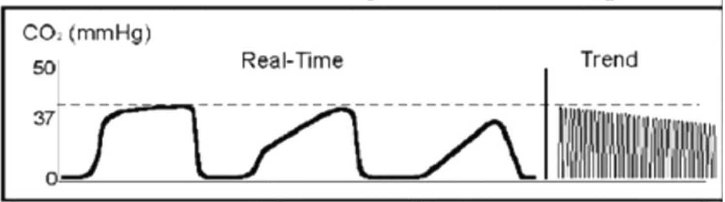
Meaning of this wave
“shark fin” obstruction in airway
causes: airway kinked, presence of foreign body, obstruction, or bronchospasm
What are the three types of deadspace and what contributes it
Anatomic - involves upper airway structures that do not participate in gas exchange → nasal passagess, larynx, trachea
Alveolar - Alveoli receive ventilation but w little or no blood flow (perfusion) → ineffective gas exchange.
Mechanical
Mainstream
What two types of make up physiologic dead space ?
anatomic and alveolar dead space
these do not particpate in CO2 or O2 exchange
Mechanical dead space, what can contribute to increasing this? how to reduce ?
it is the space in a breathing circuit where both inhalation and exhalation passes thru common path, like oxygen mask or pt monitor adaptors or Et tubes extending beyond the incisors.
makes pt possibly inhale unfiltered exhaled gas
Exhausted CO2 absorber or malfunctioning one-way valves
never 0
Reduce by minimizing # of connectors to tube, do not use face mask, pt < 6 kg Mechanical dead space refers to the volume within a breathing circuit where inhaled and exhaled gases mix, which can lead to rebreathing of exhaled gases. To reduce mechanical dead space, one can minimize the number of connectors in the tubing system, avoid using face masks, and ensure appropriate equipment for small patients.
What makes up total deadspace
anatomic, alveolar, and mechanical