Pediatric Ultrasound- FINAL Exam Review
 Call Kai
 Learn
 Practice Test
 Spaced Repetition
 Match
 Flashcards
 Knowt Play
1/24

There's no tags or description
Looks like no tags are added yet.
25 Terms

What structure is the yellow arrow pointing to?
lateral ventricle
cavum septum pellucidum
pons
cerebellum
b
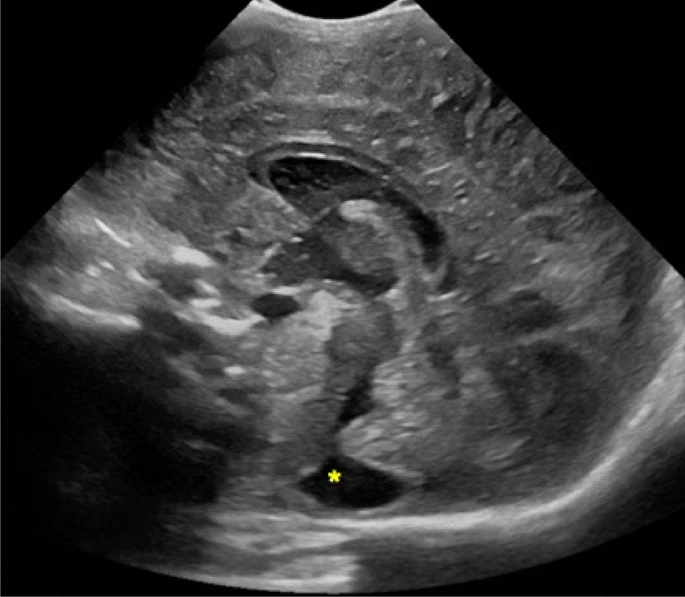
Identify the structure noted at the yellow asterisk.
frontal lobe
corpus callosum
lateral ventricle
cisterna magna
d

What sonographic plane is used in this neonatal brain image?
oblique
transverse
sagittal
coronal

What is the bright, echogenic structure in this neonatal brain image? (yellow arrows)
lateral ventricle
choroid plexus
thalamus
germinal matrix
b

In this sagittal image, what is the structure highlighted in the pink dotted line?
germinal matrix
thalamus
corpus callosum
cerebellum
c

What fontanelle is being used for this encephalogram scan?
anterior fontanelle
posterior fontanelle
mastoid fontanelle
sphenoid fontanelle
a

You are most lateral in the neonatal brain. What structure is the white arrow pointing to?
tentorium
choroid plexus
sylvian fissure
cerebellum
c

You are visualizing the choroid plexus in this coronal view.
What part of the brain are you in?
posterior
lateral
midline
anterior
a
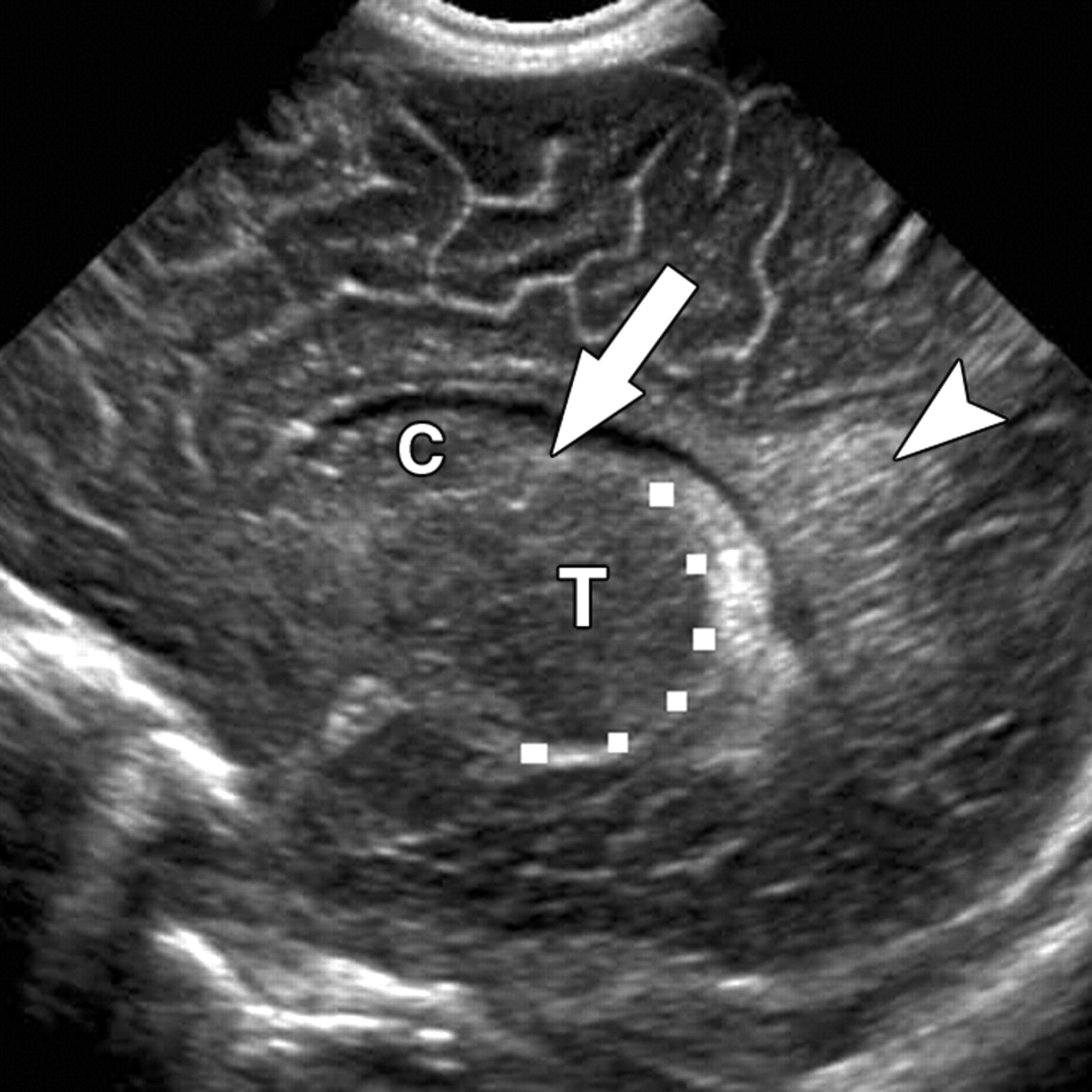
The arrow is pointing to an important area within the neonatal brain. What is another name for the caudothalamic groove?
germinal matrix
genu
choroid plexus
corpus callosum
a
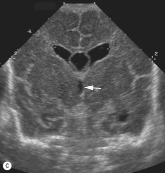
Which ventricle is the arrow pointing to?
fourth ventricle
lateral ventricle
third ventricle
anterior ventricle
c

Identify the structures at the white arrows.
third ventricles
cavum septum pellucidums
Sylvian fissures
lateral ventricles
d
Which fontanelle provides the best views for a neonatal encephalogram?
anterior
posterior
temporal
mastoidal
a
Which artery is important to doppler and assess during a neonatal encephalogram?
middle cerebral artery
anterior cerebral artery
internal carotid artery
posterior communicating artery
b
What are raised ridges or folds within the brain?
separations
sulci
gyri
ventricles
c
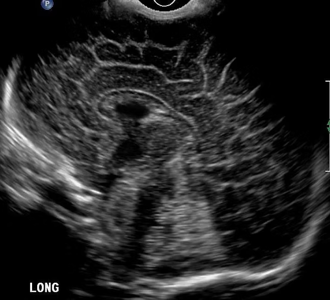
Which structure is an important landmark in this sagittal midline image of the brain that clearly identifies you're in the correct spot?
caudate nucleus
Sylvian fissure
choroid plexus
cerebellum
d
What is pyloric stenosis?
inflammation of the pylorus
thickening of the pyloric muscle causing gastric outlet obstruction
spasm of the pyloric sphincter
congenital absence of the pylorus
b
At what age does pyloric stenosis commonly present?
newborn 0-1 week
2-6 weeks of age
6-12 months
1-2 years
b
Which symptom is classically associated with pyloric stenosis?
bloody diarrhea
projectile non-bilious vomiting
bilious vomiting
constipation
b
What measurement suggests pyloric stenosis?
anterior wall <3mm
posterior wall <3mm
channel >16mm
channel <10mm
c
Pyloric stenosis is more common in females.
true
false
b
In what plane do you see a target sign with pyloric stenosis?
longitudinal
sagittal
prone
transverse
d
What is the normal wall thickness for the pylorus?
>5mm
>3mm
<3mm
=5mm
c
You see contents passing through the pyloric channel but the walls are thickened. What could this possibly represent?
thickened pyloric stenosis
intussusception
pylorospasm
appendicitis
c
What plane do you see the fluid contents passing through the pylorus channel?
transverse
caudal
longitudinal
diagonal
c
What is a true clinical sign of pyloric stenosis?
weight gain
weight loss
itchy stomach
vomiting only
b