BIOL 2101 Lab – Muscular System Study Guide
 Call Kai
 Learn
 Practice Test
 Spaced Repetition
 Match
 Flashcards
 Knowt Play
1/96

Earn XP
Description and Tags
Comprehensive vocabulary review flashcards for BIOL 2101 Lab Muscular System covering muscle histology/structure, the 75 major human muscles organized from head to toe (with locations, actions, and layers), and key muscle groups.
97 Terms
Tendon
Dense connective tissue cord attaching muscle to bone.
Fascia
Connective tissue sheet located between neighboring muscles.
Epimysium
Connective tissue wrapping the whole muscle.
Perimysium
Connective tissue wrapping each fascicle.
Endomysium
Thin connective tissue around each individual muscle fiber.
Fascicle
A bundle of muscle fibers.
Muscle fiber
A single skeletal muscle cell; long, striated, with many nuclei.
Striations
Light and dark bands seen on skeletal muscle fibers.
Nuclei (skeletal muscle)
Multiple nuclei per fiber, pushed to the edge (peripheral).
Myofibrils
Long contractile rods packed inside each muscle fiber.
Sarcolemma
Plasma membrane of the muscle fiber.
Sarcoplasmic reticulum (SR)
Network of membrane tubes that stores calcium.
Mitochondria (muscle)
Organelles that make ATP for muscle contraction.
Thick filament
Contractile filament composed of myosin.
Thin filament
Contractile filament composed of actin.
Epicranius (frontal & occipital bellies)

Orbicularis oculi
Superficial ring around the eye; closes eye (blink, squint).

Orbicularis oris
Superficial ring around the mouth; closes and purses lips.

Zygomaticus major
Superficial muscle extending from cheekbone to corner of mouth; produces smiling.

Buccinator
Deep muscle inside the cheek, deep to masseter's front edge; compresses cheek (blowing, chewing).

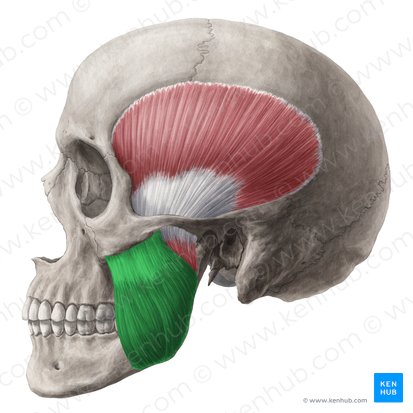
Masseter
Superficial muscle located at the angle of the jaw; closes jaw (elevates mandible).
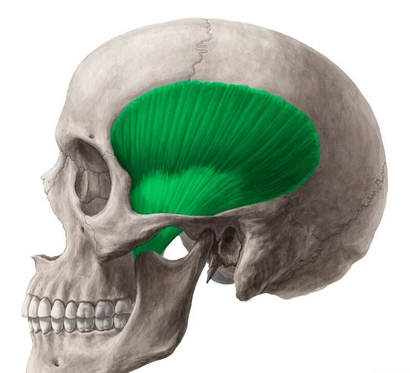
Temporalis
Superficial muscle on the side of the skull above the ear; closes jaw.
Platysma
Superficial thin sheet over the front of the neck; pulls down corners of mouth and tenses neck skin.

Sternocleidomastoid
Superficial diagonal strap from sternum and clavicle to behind the ear; flexes neck; one side turns head to the opposite side.
Splenius capitis
Deeper muscle at the back of neck, between SCM and trapezius; extends head.

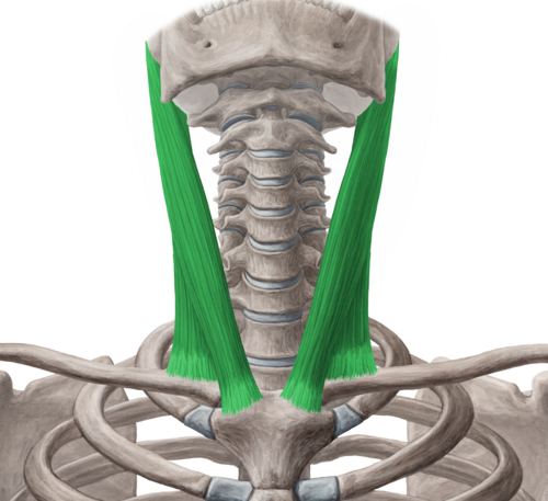
Scalenes (anterior, middle, posterior)
Superficial muscles on the side of the neck, running from cervical vertebrae to ribs 1–2; raise ribs 1–2 (breathing) and bend neck sideways.

Digastric
Superficial muscle with two bellies under the chin and jaw; opens jaw and raises hyoid.

Mylohyoid
Deeper muscle on the floor of the mouth, deep to digastric; raises hyoid and floor of mouth (swallowing).

Sternohyoid
Superficial narrow strap from sternum to hyoid, next to midline; lowers hyoid.

Omohyoid
Superficial muscle with two thin bellies extending from scapula to hyoid; lowers hyoid.

Pectoralis major
Superficial large fan across the upper chest; flexes, adducts, and medially rotates arm.

Pectoralis minor
Deeper muscle under pec major, extending from ribs to coracoid process; pulls scapula forward and down.

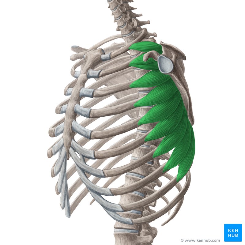
Serratus anterior
Superficial 'saw-tooth' slips on the side of the ribs; pulls scapula forward (punching).
External intercostals
Deeper muscles between ribs whose fibers run down and forward; raise ribs (inhalation).

Internal intercostals
Deeper muscles between ribs, deep to externals with fibers at right angles to them; lower ribs (forced exhalation).

Diaphragm
Deeper dome separating thoracic and abdominal cavities; main muscle of inhalation.

Rectus abdominis
Superficial vertical 'six-pack' on each side of the linea alba; flexes vertebral column (crunch).

External oblique
Superficial outermost side muscle whose fibers run down and in ('hands in pockets'); compresses abdomen, flexes and rotates trunk.

Internal oblique
Deeper muscle deep to external oblique with fibers at right angles to it; compresses abdomen, flexes and rotates trunk.

Transversus abdominis
Deepest layer of the abdominal wall with fibers running horizontally; compresses abdomen.

Trapezius
Superficial large diamond over upper back and neck; raises, retracts, and lowers scapula; extends head.
Latissimus dorsi
Superficial broad sheet over lower and mid back to the arm; extends, adducts, and medially rotates arm (pull-ups).
Levator scapulae
Deeper muscle on side and back of neck to top corner of scapula; raises scapula (shrug).
Rhomboids (major & minor)
Deeper muscles between spine and medial border of scapula, deep to trapezius; retract scapula (squeeze shoulder blades).
Erector spinae (iliocostalis, longissimus, spinalis)
Deeper group consisting of three columns running along the spine; extends vertebral column.
Quadratus lumborum
Deeper muscle in the lower back between iliac crest and rib 12; bends trunk sideways.
Deltoid
Superficial rounded cap over the shoulder; abducts arm (raise arm to the side).
Supraspinatus
Deeper muscle located above the spine of the scapula, under trapezius; starts abduction of arm.
Infraspinatus
Superficial muscle located below the spine of the scapula; laterally rotates arm.
Teres minor
Superficial small muscle just below infraspinatus; laterally rotates arm.
Subscapularis
Deeper muscle on the front surface of the scapula, facing the ribs; medially rotates arm.
Teres major
Superficial muscle below teres minor, from scapula's lower corner to humerus; extends, adducts, and medially rotates arm.
Coracobrachialis
Deeper small muscle extending from coracoid process to upper medial humerus; flexes and adducts arm.
Biceps brachii
Superficial muscle on the front of the arm with two heads; flexes elbow; supinates forearm.
Brachialis
Deeper muscle deep to biceps on the lower front of the humerus; flexes elbow (main flexor).
Triceps brachii
Superficial muscle on the entire back of the arm with three heads; extends elbow.
Brachioradialis
Superficial muscle on the lateral forearm, extending from humerus to radius; flexes elbow.
Pronator teres
Superficial short diagonal muscle just below the elbow, anterior; pronates forearm (palm down).
Flexor carpi radialis
Superficial anterior forearm muscle toward the thumb side; flexes and abducts wrist.
Palmaris longus
Superficial anterior forearm muscle in the middle with a thin tendon to palm (absent in some people); flexes wrist.
Flexor carpi ulnaris
Superficial anterior forearm muscle on the pinky side; flexes and adducts wrist.
Flexor digitorum superficialis
Deeper middle layer muscle of the anterior forearm with tendons to fingers 2–5; flexes fingers.
Pronator quadratus
Deeper small square muscle near the wrist in the deepest layer; pronates forearm.
Supinator
Deeper muscle wrapping around upper radius; supinates forearm (palm up).
Extensor carpi radialis longus
Superficial posterior forearm muscle on thumb side, next to brachioradialis; extends and abducts wrist.
Extensor carpi ulnaris
Superficial posterior forearm muscle on the pinky side; extends and adducts wrist.
Extensor digitorum
Superficial middle posterior forearm muscle with tendons to fingers 2–5; extends fingers.
Gluteus maximus
Superficial largest muscle of the buttock; extends thigh (climbing stairs, standing up).
Gluteus medius
Superficial upper outer buttock muscle, above and partly under glut max; abducts thigh and keeps pelvis level when walking.
Gluteus minimus
Deeper muscle deep to gluteus medius; abducts thigh.
Piriformis
Deeper muscle from sacrum to top of femur, below glut minimus; laterally rotates thigh.
Iliopsoas (psoas major + iliacus)
Deeper muscle extending from lumbar spine and inside of the ilium to the femur; flexes thigh (strongest hip flexor).
Tensor fasciae latae
Superficial muscle on upper lateral thigh, front of the iliotibial tract; abducts thigh and tenses IT tract.
Sartorius
Superficial long strap crossing the thigh diagonally from hip to inner knee; flexes, abducts, and laterally rotates thigh (sitting cross-legged).
Rectus femoris
Superficial muscle straight down the middle of the anterior thigh; extends knee; flexes thigh.
Vastus lateralis
Superficial muscle on the lateral (outer) anterior thigh; extends knee.
Vastus medialis
Superficial muscle on the medial (inner) thigh forming a 'teardrop' above the knee; extends knee.
Vastus intermedius
Deeper muscle lying deep to rectus femoris; extends knee.
Adductor longus
Superficial muscle on the upper inner thigh; adducts thigh.
Adductor magnus
Deeper largest adductor on the inner thigh, seen best from behind; adducts thigh.
Gracilis
Superficial thin strap along the most medial thigh; adducts thigh; flexes knee.
Biceps femoris
Superficial muscle on the lateral posterior thigh with two heads; flexes knee; extends thigh.
Semitendinosus
Superficial muscle on the medial posterior thigh with a long cord-like tendon; flexes knee; extends thigh.
Semimembranosus
Deeper muscle deep to semitendinosus on the medial posterior thigh; flexes knee; extends thigh.
Tibialis anterior
Superficial muscle on front of the shin, just lateral to the tibia; dorsiflexes and inverts foot.
Extensor digitorum longus
Superficial anterior leg muscle lateral to tibialis anterior with tendons to toes 2–5; extends toes; dorsiflexes foot.
Fibularis longus
Superficial muscle on upper lateral leg; everts and plantar flexes foot.
Fibularis brevis
Superficial muscle on lower lateral leg, under fibularis longus; everts and plantar flexes foot.
Gastrocnemius
Superficial calf muscle with two heads (medial & lateral); plantar flexes foot (tiptoe); flexes knee.
Soleus
Superficial flat muscle deep to gastrocnemius, visible at its sides; plantar flexes foot.
Rotator cuff ('SITS')
Muscle group comprising Supraspinatus, Infraspinatus, Teres minor, and Subscapularis.
Quadriceps femoris
Anterior thigh group comprising Rectus femoris, vastus lateralis, vastus medialis, and vastus intermedius.
Hamstrings
Posterior thigh muscle group comprising Biceps femoris, semitendinosus, and semimembranosus.
Triceps surae (calf)
Calf muscle group comprising Gastrocnemius and soleus (which share the calcaneal tendon).
Hip adductors
Group of muscles that adduct the thigh, consisting of Adductor longus, adductor magnus, and gracilis.
Suprahyoid muscles
Muscles located above the hyoid that raise the hyoid, comprising Digastric and mylohyoid.
Infrahyoid muscles
Muscles located below the hyoid that lower the hyoid, comprising Sternohyoid and omohyoid.