Maya - Dental Pulp Biology and Odontogenesis: Development, Anatomy, Innervation, and Pathology
 Call Kai
 Learn
Learn Practice Test
 Spaced Repetition
 Match
Match Flashcards
 Knowt Play
1/53

Earn XP
Description and Tags
Operative 2 Exam
Name | Mastery | Learn | Test | Matching | Spaced | Call with Kai | Chat |
|---|
No analytics yet
Send a link to your students to track their progress
54 Terms
What embryonic cells are the origin of the oral cavity and teeth?
Neural crest cells, which give rise to the dental lamina
List the stages of odontogenesis in order. (6)
.
Which forms first, dentin or enamel?
Bud →
Cap →
Bell →
Dentinogenesis →
Amelogenesis →
Appositional dentin and enamel →
.
Dentin forms first (dentinogenesis occurs before amelogenesis)
Morpho-differentiation vs. histo-differentiation?
Morpho: development of the shape; crown develops first (tooth forms crown-down)
.
Histo: differentiation at the cellular level
"What structures are labeled in the histo-differentiation (bell stage) photomicrograph?

When does root formation occur, and what are the 3 primordial root terms?
"After dentinogenesis and amelogenesis
1. Cervical loop
2. Hertwig's epithelial root sheath
3. Epithelial root diaphragm
How do the cell rests of Malassez form, and what is their clinical significance?
"Epithelial root sheath breaks up → dental sac CT contacts dentin → forms cementoblasts and PDL
.
Epithelial cells left in the PDL = cell rests of Malassez
Can form lateral periodontal cysts later in life
Lateral vs. accessory canals: location, function, contents, and endo management?
Lateral: coronal 2/3; accessory: apical 1/3 (increased here)
.
Direct contact between pulp and PDL
.
Do NOT contain pulp tissue
Sealed during obturation
What are the 2 ways lateral/accessory canals form?
1. Break in the root sheath before dentin forms
2. Blood vessel traveling between dental sac and dental papilla
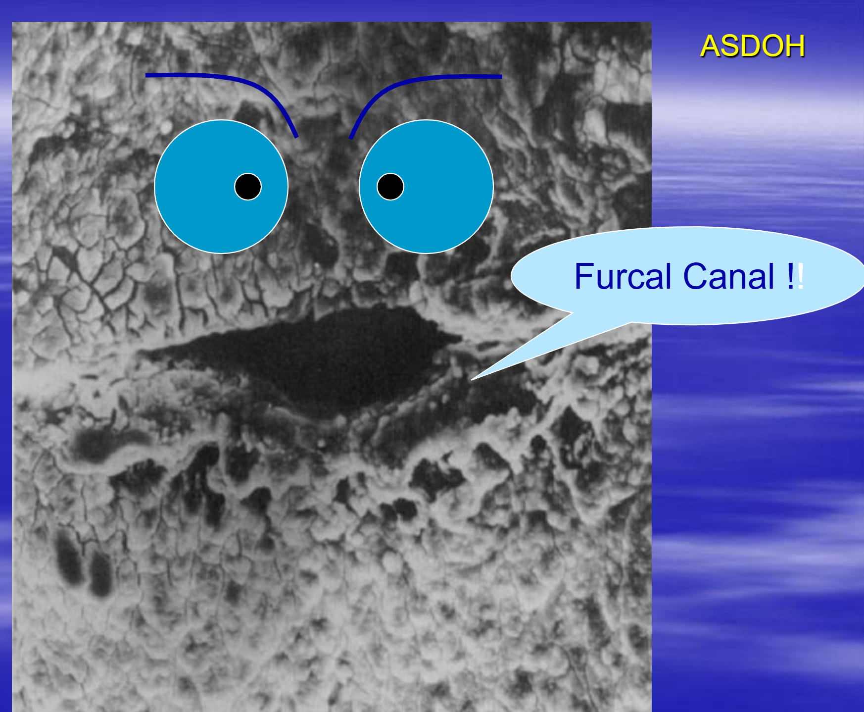
What is the accessory canal opening into the furcation of a multirooted tooth called?
Furcal canal
What is cementum?
Hard tissue that covers the root; where the PDL attaches
What is the apical foramen, how far is it offset from the apex, and what does it contain?
Opening at the end of the root where pulp contacts PDL
Offset 0.5-1 mm
Contains the neurovascular bundle
What is the apical foramen's size in a mature tooth, and how does it change with age?
0.3-0.6 mm
.
Changes location and gets smaller due to cementum deposition
What is an apical delta?
An apical foramen with several accessory canals
What are the 3 histologic components of the pulp?
Cells
Fibers
Ground substance
List the layers of the pulp-dentin complex from dentin inward (6)
Dentin →
Predentin →
Odontoblastic layer →
Cell-free (cell-poor) zone →
Cell-rich zone →
Central pulp (pulp proper)
What is in each zone of the pulp?
Odontoblastic layer: odontoblasts lining predentin
.
Cell-free: no cells
.
Cell-rich: undifferentiated mesenchymal cells, fibroblasts, blood vessels, defense cells
.
Pulp proper: central core
List the 6 cells of the healthy pulp.
Fibroblasts
Mesenchymal cells
Odontoblasts
Histiocytes
Dendritic cells
Lymphocytes
Pulpal fibroblasts: prevalence, location, and products?
Most common pulp cell
.
Mostly in the cell-rich layer
.
Produce collagen (I and III) and ground substance
.
Per lecture notes: differentiate into odontoblasts that make dentin
Odontoblasts: uniqueness, product, parts, and connections?
Unique to pulp; produce dentin
.
Cell body + Tomes' (odontoblastic) process, which extends into dentin to the DEJ
.
Joined by desmosome-like junctions
How do odontoblast shape and organization vary by region?
Coronal: columnar, most organized
.
Middle 1/3 (mid-root): cuboidal, well organized (just above cell-rich zone)
.
Apical: squamous, least organized
What are pulpal defense cells, and what irritants trigger them?
Cells of the inflammatory response in the pulp due to an irritant
.
Irritants: caries, trauma, operative procedures
What is pulpal ground substance like, and what is its function?
Similar to connective tissue
a glycoprotein medium
Transports nutrients and oxygen
Pulpal fibers: collagen types, sources, elastic fibers, and distribution?
Dentin: type I (odontoblast)
.
Pulp: types I and III (fibroblast)
.
No elastic fibers (still being investigated)
.
Apical region has the most collagen
What are the components of the pulpal extracellular matrix, and what do tenascin and fibronectin do? (3)
Water
Glycosaminoglycans,
Collagen (I, III),
non-collagenous proteins (tenascin, fibronectin): - - Tenascin/fibronectin: substrate adhesion glycoproteins for cell attachment, spreading, and migration
Describe the pulpal nerve supply (fiber types, locations, efferent function).
Afferent sensory: A-delta (peripheral pulp), C fibers (central pulp)
.
Efferent motor: sympathetic (parasympathetic questionable) → smooth muscle contraction
Characteristics and associated diagnosis of A-delta fibers (slide titled ""A(alpha)"")?
Low threshold, myelinated, high conduction velocity
.
At the pulp-dentin border (peripheral)
Sharp, lightning, non-lingering pain → Reversible pulpitis
Characteristics and associated diagnosis of C fibers?
High threshold, non-myelinated, low conduction velocity
.
Through the central pulp
Throbbing, dull, lingering, poorly localized pain
→ (Symptomatic) irreversible pulpitis; needs endodontic therapy
If pulpal nerves extend only 100-200 µm into dentin, why does drilling just past the DEJ hurt?
Sensory nerves can extend into the dentinal tubule alongside Tomes' process
What are the 3 theories of dentin sensitivity, and which is most common?
Direct innervation: nerve fibers in the tubule
Odontoblastic receptor: nerves and odontoblasts are close
Hydrodynamic (most common): fluid movement in tubules triggers nerves; basis of thermal testing
Pulpal arterial supply: importance, origin, and branches?
Determines vitality
.
Origin: internal maxillary artery
.
Branches: posterior, middle, and anterior superior alveolar; inferior alveolar
How do arterioles enter and travel through the pulp, and where are vessels most numerous?
Enter through the apical foramen and lateral canals, course toward the crown
.
Most numerous in the subodontoblastic zone
Pulpal venous return: drainage, vein features, exit, and where flow is greatest?
Drains to pterygoid plexus and internal maxillary vein
.
Veins: larger, not innervated, thin walled; A-V shunts present
.
Exit through apical foramen and lateral canals
.
Blood flow greatest in coronal pulp
What are the functions of pulpal A-V shunts? (2)
1. Bypass the capillary bed during inflammation → ↓ intrapulpal BP
.
2. Help control normal pulpal vasculature
In the pulpal microcirculation, what bypasses the true capillaries, and what controls entry into them?
A-V shunt connects arteriole to venule
.
Precapillary sphincters (open/closed) control capillary entry
What are the 5 functions of the pulp, and which is most important?
1) Induction: produces predentin; stimulates initial ameloblast formation
.
2) Formation: continuously forms secondary dentin (most important)
.
3) Nutrition: dentinal fluid nourishes dentin
.
4) Defense: forms reparative (tertiary) dentin in response to irritant
.
5) Innervation: innervates dentin
What are the age changes of the pulp?
Calcification of chamber and canals → smaller canals
Blood vessels and nerves decrease and mineralize
Collagen and reticular fibers form bundles
Diffuse and localized calcification
Is pulpal calcification (e.g., pulp stones) correlated with symptoms?
No; it is not an indication for RCT
What are the 2 types of pulpal calcification and where does each occur?
Pulp stones (denticles): chamber
.
Diffuse/linear: radicular (canals), along nerves, vessels, or collagen bundles
How are pulp stones classified? (5)
1) True: smooth;
2) False: concentric layers
3) Free: in chamber;
4) Attached: to chamber wall;
5) Embedded: in chamber wall
What is the immune response to small, large, and prolonged carious exposures?
Small: APCs, random migration of T cells
Large: PMNs, B-cell activation
Prolonged: specific T cells in effector phase eliminate bacteria
Why is the pulp called a non-compliant environment?
Tooth is hard and non-expandable, so it can't swell → intrapulpal pressure increases
What immune cells are in the inflamed pulp?
PMNs; T, B, and NK cells
Plasma cells making IgG and IgA against microbes in the carious lesion
What is the key message about carious pulp exposures?
By the time there is a carious exposure, it's too late (necrosis) → endo tx or extraction
.
Do NOT direct pulp cap a carious exposure
What were the outcomes of direct pulp caps on carious exposures in Barthel 2000?
5 yr: 45% failure, 18% questionable, 37% success
.
10 yr: 80% failure, 7% questionable, 13% success
How does the pulp respond to a deep cavity prep, and what is the usual outcome?
Normal pulp (a)
.
Odontoblasts destroyed, acute inflammation, cell division in the cell-rich zone (b)
.,
Pulp recovers in the vast majority (c), with reparative dentin
How is any dental procedure (e.g., crown prep) viewed pulpally?
As iatrogenic trauma that can cause reversible pulpitis
What factors in full-coverage restoration can lead to pulp involvement?
Prep: frictional heat, vibration, desiccation
.
After prep: cleansing/sterilization, acid etching, impressions, restorative materials, insertion and cementation, marginal leakage → Pulpal inflammation → necrosis
What are the 2 categories of pulpal reactions?
Vascular and neural
Due to the pulp's low compliance, what sequence leads to necrosis? (5)
↑ Blood flow →
↑ tissue pressure →
↓ blood flow (stasis) →
toxic metabolites →
localized necrosis (→ RCT or extraction)
What causes the rise in intrapulpal pressure, and why does it lead to necrosis?
Vascular permeability, cellular infiltrate, vascular stasis
.
Pulp in rigid walls → stasis and microabscess → necrosis
What are the 4 defense mechanisms of the pulp?
1. AV shunting
2. Secondary dentinal mechanisms
3. Reactionary and reparative dentin formation
4. Immune reactions
How does pulpal blood flow respond to an insult such as grinding?
↑ Flow through AV shunts and redistribution of flow
Secondary vs. reactionary/reparative dentin: when does each form?
Secondary: physiological, throughout life
Reactionary/reparative: in response to an irritant
Where does reparative dentin form, and how does it affect pulp space?
Under large restorations; pulp horns, canal, and chamber shrink