mycology exam 3
 Call Kai
 Learn
 Practice Test
 Spaced Repetition
 Match
 Flashcards
 Knowt Play
1/90

There's no tags or description
Looks like no tags are added yet.
91 Terms
Ameba
Single-celled parasite that moves by means of cytoplasm extensions called
pseudopods (false feet)
Trophozoite
The motile feeding stage of amebas that multiply by binary fission
Cyst
The non-motile, non-feeding stage of amebas that are capable of survival
outside their hosts and of initiating human infection
Karyosome
Central mass of chromatin inside select amebic
nuclei
Peripheral chromatin
Ring of chromatin that surrounds the nuclei of
select amebas
Chromatoid bar
Oblong structures containing condensed RNA
material in the cytoplasm of select amebic cysts
Glycogen mass
A cytoplasmic area without defined borders that is
believed to represent stored food and is usually visible
in young cysts
Excystation
The transformation of amebic cysts into amebic
trophozoites
ileocecal area of the intestine
Encystation
The transformation of amebic trophozoites into amebic
cysts
Specimen of choice for recovering intestinal amebas
stool
Specimen of choice for recovering extraintestinal amebas – varies by
species but includes:
Mouth scrapings
• Cerebrospinal fluid (CSF)/brain tissue
• Corneal scrapings
Entamoeba histolytica/ Entamoeba
dispar TROPH
Ingests RBCs
avg size: 12-25 um
one nucleus, pseudopodia

Entamoeba histolytica /
Entamoeba dispar
CYST
1-4 nuclei, mature cysts have 4
avg size 12-28 um

Entamoeba histolytica /
Entamoeba dispar other info
toxic megacolon
Entamoeba hartmanni -
Trophozoite
one nucleus
ingested bacteria may be present
avg size 8-12 um

Entamoeba hartmanni - Cyst
avg size 7-9 um
1-4 nuclei
smiley face
rounded chromatoid bars

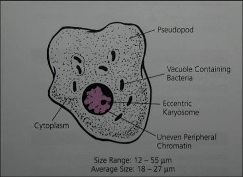
Entamoeba coli - Trophozoite
avg size 18-27 um
pseudopods
one nucleus
large, irregular karyosome
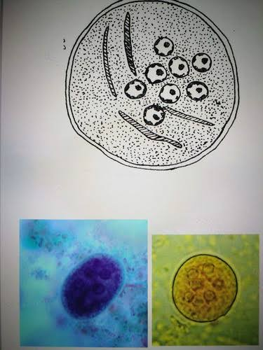
Entamoeba coli - Cyst
avg size 12-25um
1-8 nuclei
large, irregular karyosome
Endolimax nana - Trophozoite
avg size 7-10 um
one nucleus
pseudopods

Endolimax nana - Cyst
avg size 7-10 um
NO peripheral chromatin
1-4 nuclei, 4 most common

Iodamoeba bütschlii -
Trophozoite
avg size 12-18 um
one nucleus
NO peripheral chromatin
bacteria, yeast, other debris may be found in cytoplasm

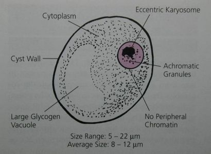
Iodamoeba bütschlii - Cyst
avg size 8-12 um
one nnucleus
well defined glycogen mass
naegleria fowleri- trophozoite
sluglike, blunt psedopods
one nucleus
one central karyosome
ameboid: pseudopods, large karyosome, 8-22 um
AMEBOFLAGELLATE: flagella, 7-15um

Naegleria fowleri - Cyst
9-12um, cyst wall
central karyosome, peripheral chromatin

Naegleria fowleri other info
CSF giemsa stain for dx
Three known morphologic forms:
• Ameboid trophozoite
• Flagellate form
• Cyst
• Ameboid trophs are only form known in humans.
Warm bodies of water, including lakes, streams, ponds, swimming pools
• Contaminated dust
• Highest prevalence – summer months
• It grows best at higher temperatures up to 115°F (46°C)
Primary amebic meningoencephalitis (PAM)
Acanthamoeba Species -
Trophozoite
avg size 25 um
sluggish, spinelike pseudopods
one nucleus

Acanthamoeba Species - Cyst
DOUBLE CELL WALL (outside has ragged edges)
8-25um

acanthomoeba other info
DOUBLE CELL WALL
CSF – standard methods
• Other samples:
• Brain tissue
• Corneal scrapings
EYE INFECTION-contacts
Granulomatous amebic encephalitis (GAE)
• Slowly developing symptoms include:
• Headaches
• Seizures
• Stiff neck
• Nausea and vomiting
Sporozoa
A group of protozoa that have no obvious means
of locomotion
Macrogametocyte
Female sex cell of Plasmodium spp
Microgametocyte
Male sex cell of Plasmodium spp.
Merozoite
Asexual sporozoa trophozoite
Ring form
Ringlike structure that appears initially after
invasion of RBC by Plasmodium spp
Schizogony
Asexual multiplication that occurs in humans prior
to invasion of the red blood cells
Schizont
Morphologic form responsible for the development
and maturing of merozoites critical to parasite
survival
Sporogony
Sexual multiplication that occurs in mosquitoes
Sporozoite
Infective stage transferred by vector
Immature schizonts
Growing chromatin and
visible cytoplasmic
material surrounds the
growing chromatin.

mature schizont
Fully developed
merozoites emerge
(asexual sporozoa
trophozoites)
Cytoplasmic material is
presumed to be absent
except with Plasmodium
vivax.

plasmodium life cycle
Vector is the mosquito genus Anopheles.
Vector transfers the infective *sporozoites
with blood meal.
Sporozoites enter liver to multiply.
Schizogony (asexual multiplication)
Also known as **exoerythrocytic cycle
Liver cells rupture to release merozoites.
Merozoites infect red blood cells.
*Erythrocytic cycle
Merozoites can go on to:
Infect new RBCs
Develop into microgametocytes and
macrogametocytes
Be destroyed by the immune system
Timing of blood collection
in between paroxysms
p. vivax
paroxysms Q 48hrs
p.ovale
paroxysms Q 48 hrs
p.malariae
paroxysms Q 72hrs
p.falciparum
q 36-48 hrs
Plasmodium vivax
AKA: Benign tertian malaria, vivax malaria
infects young RBCs causing distorted shape
ring form-single chromatin dot with ring surrounding vacuole
Most widely distributed of the Plasmodium
species and found in tropical, subtropical, and
temperate regions
Paroxysms occur every 48 hours.
avg 16 merozoites

plasmodium ovale
AKA Benign tertian
malaria, Ovale malaria
Differences from P.
vivax:
Larger ring
Thicker & more
amoeboid ring
Multiple rings can
infect 1 RBC
avg 8 merozoites per infected RBC
infects only young and immature cells, causing deformed, ragged edge cells
ROSETTE APPEARANCE of merozoite
Found primarily in tropical Africa as well as Asia
and South America
Q 48hr

Plasmodium malariae
BAND FORM, BIRDS EYE
6-12 merozoites
only infects mature RBCs, no distortion
Rings seen less often.
Developing trophozoites and immature and
mature schizonts are more common.
Found in subtropic and temperate regions
There are no known relapses.

Plasmodium falciparum
Synonyms:
Black water fever
Malignant tertian malaria
Aestivoautumnal malaria
Subtertian malaria
Falicparum malaria
avg 24 merozoites
Ring form: HEADPHONE configuration
CRESCENT SHAPE
may infect all age RBCs, no distortion
Infections – in late summer and early fall
Epidemiology
Limited to tropical and subtropical regions
36 to 48 paroxysms
Deadliest form of malaria
• Large amounts of toxic cellular debris
• May enter kidneys, brain, and liver
• May result in coma and death

Plasmodium knowlesi
can form bands

Babesia spp
Develops within RBC, from the trophozoite, to
form a Maltese cross appearance
Produces sporozoites, which are taken up by
the tick vector
Vector is a tick of the genus Ixodes
Humans considered accidental host
Merozoites undergo binary fission to produce
more sporozoites that ultimately infect new
RBCs.
Specimen of choice – Giemsa-stained
peripheral blood films
Thick blood smear for screening
Thin blood smear for differentiating Babesia
species from Plasmodium species
Timing of blood collection not critical
Babesia microti and
Babesia divergens
MALTESE CROSS
Trophozoite
• Resembles the ring form of Plasmodium species
• Absence of malarial pigment and dots
Merozoite
• Four trophozoites mature to
form a Maltese cross.
• Sexual stage occurs in the
tick (vector).
• Asexual stage occurs in
animals and humans.
Specimen of choice – Giemsa-stained
peripheral blood film
• Thick and thin blood films
B. divergens tends to be more severe and fatal if
untreated

B. microti
Southeast New
England, Midwest
and Mexico
• Most common tick
vector – Ixodes
scapularis
• Primary reservoir –
white-footed mouse
B. divergens
European countries
• Has been found in the
Nantucket area
• Tick vector – Ixodes ricinus
• Reservoir hosts – cattle and
rabbits
Axostyle
Rod-like support structure in select flagellates
Axoneme
Intracellular portion of a flagellum
Cytostome
Rudimentary mouth present in some flagellates
Median bodies
Comma-shaped structures inside Giardia
intestinalis
Structure associated with energy, metabolism, or
support
Undulating membrane
Fin-like structure
Costa
Rod-like structure located at the base of the
undulating membrane
Flagellate life cycle
troph, cyst form
Ingestion of infected cysts when present =
initiation of human intestinal infection
Trophozoites in intestinal tract
Actively multiply by asexual binary fission
Changes in intestinal environment –
excystation, when cysts are present
Giardia intestinalis troph
troph: BILATERAL SYMMETRY
avg size: 10-15um
falling leaf motility
2 ovoid nuclei, each w a large karyosome, 2 median bodies, 2 axonemes, sucking disk
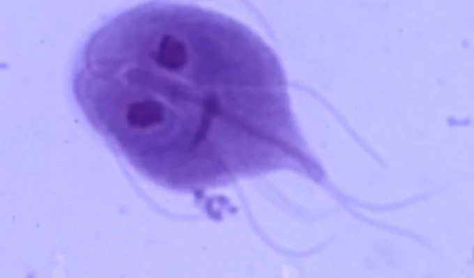
Giardia intestinalis cyst
10-12um
immature cyst has 2 nuclei, mature has 4
median bodies

Chilomastix mesnili
Trophozoite
avg length: 8-15um
PEAR SHAPED
four flagella
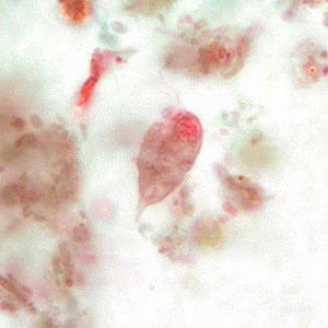
Chilomastix mesnili - Cyst
7-10um x3-7um
clear hyaline knob
LEMON SHAPED

Dientamoeba fragilis
Trophozoite
contains ingested bacteria
8-12um
2 nuclei
Symptoms vary and include:
Diarrhea and abdominal pain
– Diarrhea alternating with constipation
Bloody or mucoid stools
Possible theory: D. fragilis may be transmitted
via select helminth eggs (Chapter 8):
• Enterobius vermicularis (pinworm)
• Ascaris lumbricoides (large intestinal roundworm)
Only known morphologic form – trophozoites

Trichomonas vaginalis
Trophozoite
costa, axostyle, undulating membrane ½ body length, 4 flagella
8-15um

Hemoflagellate
Single-celled parasite that moves by means of tail-
like extensions of cytoplasm known as a flagellum
(pl. flagella) that resides in blood and tissue
Blepharoplast
Basal body structure located in the cytoplasm of
the hemoflagellates
Kinetoplast
An umbrella term that refers to the blepharoplast
and small parabasal body located in the cytoplasm
of the hemoflagellates
Chaga’s Disease
A common name for the disease caused by
Trypanosoma cruzi
Amastigote
Oval, non-flagellated morphologic form found in some of the
hemoflagellate life cycles

Promastigote
Long, slender hemoflagellate morphologic form containing a free
flagellum

Epimastigote
Long, spindle-shaped, hemoflagellate morphologic form equipped with
a free flagellum and an undulating membrane that extends over ½ the
body length.

Trypomastigote
Long and slender to study hemoflagellate morphologic form of
Trypanosoma spp. that typically looks like the letter C or U on
permanent stain. It has a full body length undulating membrane.

espundia
leishmaniasis braziliensis
Leishmania braziliensis Complex
Biopsy of infected ulcer = specimen of choice
• Microscopy:
Specimen of choice for recovery of amastigotes
Arthropod vector
• Sand fly species: Lutzomyia and
Psychodopygus
Infective stage for human
• Promastigotes

Leishmania donovani Complex
Screening test: Montenegro skin test
Darkening of the skin (kala-azar – black fever)

Trypanosoma brucei gambiense
West African sleeping sickness
Tsetse fly: Glossina palpalis and
Glossina tachinoides
Diagnostic stage for humans
• Trypomastigotes
Infective stage for tsetse fly
• Trypomastigotes – tsetse fly picks them up during
blood meal.
West African (Gambian) sleeping sickness
• Painful chancre surrounded by white halo at bite site
lymphadenopathy
Winterbottom’s sign

Trypanosoma brucei rhodesiense
Synonyms:
East African
sleeping sickness
Rhodesian
trypanosomaisis
Giemsa-stained blood preparations
• Trypomastigotes*
Microscopic examination of CSF
• Trypomastigotes
Tsetse fly species: Glossina morsitans and Glossina
pallidipes
Reservoir hosts:
• Cattle
• Sheep
• Wild game animals

Trypanosoma cruzi
Chaga’s disease
American trypanosomiasis
Arthropod vector
• Kissing (reduviid) bug
Mode of transmission
• Kissing bug defecates near blood meal
site.
• Kissing bug subsequently rubs it into the
skin via the scratch in preparation for its
blood meal.
Central and South America
• Highest prevalence – Brazil
Erythematous nodule
(chagoma)
Myocarditis
• Enlargement of the colon (megacolon) or esophagus
(megaesophagus)

Balantidium coli - Trophozoite
CILIA!!!!
35-50um
2 nuclei
cystosome mouth

Balantidium coli
Cyst
52-55um
2 nuclei
DOUBLE CYST WALL

Isospora belli (AKA Cystoisospora)
Oocyst
double cell wall
2 mature sporocysts each contain 4 sporozoites
30×12um

Cryptosporidium parvum
Oocyst
4-6um
ROSETTE SHAPE
4 sporozoites
waterparks, community swimming pools, day care
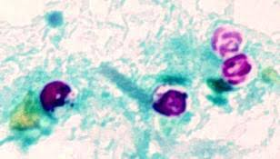
Blastocystis hominis
Vacuolated Form
Whole vacuole pushes nuclei to edge of cell
5-32um

Cyclospora cayetanensis
Oocyst
Oocysts to autofluoresce under ultraviolet
light microscopy
7-10um
2 sporocysts each w 2 sporozoites

Microsporidia Spore
TUBULES
1-5um
Most cases occur in AIDS patients.
• Haiti
• Zambia
• Uganda
• The United Kingdom
• The U.S.
• The Netherlands

Toxoplasma gondii
Tachyzoites and Bradyzoites
crescent shape
Serologic methods performed on blood
samples
Definitive host – cat
Intermediate hosts –
rats or mice
nfected child may experience:
– Hydrocephaly, microcephaly

Pneumocystis jiroveci
Trophozoite
2-4um
ameboid shape
4-8 nuclei

Pneumocystis jiroveci
Cyst
4-8 nuclei
4-12 um
AIDS patients often develop Kaposi’s sarcoma
(malignant skin disease).