LAB 2. ERYTHROCYTE INDICES & RETICS COUNT
 Call Kai
 Learn
 Practice Test
 Spaced Repetition
 Match
 Flashcards
 Knowt Play
1/73

There's no tags or description
Looks like no tags are added yet.
74 Terms
Mean Cell Volume (MCV)
Mean Cell Hemoglobin (MCH)
Mean Cell Hemoglobin Concentration (MCHC)
ENUMERATE
Erythrocyte Indices
blood film/smear examination
RBC indices must always be counter checked with a __
RBC count
Hemoglobin
Hematocrit
These values are used in calculating the different erythrocyte indices
MEAN CELL VOLUME
average volume of an erythrocyte
reflects the RBC diameter/size of the RBC
femtoliters (fL)
10-15 L
MCV Unit of measurement:

MCV Formula
80-100 fL
MCV Reference range
NORMAL MCV (80-100 fL) = NORMOCYTIC
INCREASED MCV (>100 fL) = MACROCYTIC
DECREASED MCV (<80 fL) = MICROCYTIC
Interpretation of the value of MCV
NORMAL MCV (80-100 fL) = ___
INCREASED MCV (>100 fL) = ___
DECREASED MCV (<80 fL) = ___
MEAN CELL HEMOGLOBIN
average weight (content) of hemoglobin in a red blood cell
Expresses the mass of hemoglobin per cell and it parallels the MCHC value.
picograms (pg)
10-12 g
MCH Unit of measurement

MCH Calculation
26-32 pg
MCH Reference range
MEAN CELL HEMOGLOBIN CONCENTRATION
average concentration of hemoglobin in each individual red blood cell
Reflecting RBC staining intensity or color and the amount of central pallor.
grams per deciliter (g/dL)
MCHC Unit of measuremen
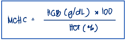
MCHC Calculation
32-36 g/dL
MCHC Reference range
NORMAL MCHC (32-36 g/dL) = NORMOCHROMIC
DECREASED MCHC (<32 g/dL) = HYPOCHROMIC
INCREASED MCHC (>36 g/dL) = SPHEROCYTIC
MCHC Interpretation:
NORMAL MCHC (32-36 g/dL) = ___
DECREASED MCHC (<32 g/dL) = ___
INCREASED MCHC (>36 g/dL) = ___
normal MCV
SOURCES OF ERROR:
Dimorphic population of RBCs → ___
↑ MCV
SOURCES OF ERROR:
↑ reticulocytes → ____
spherocytes
SOURCES OF ERROR:
MCHC = 36-38 g/dL → check for ___
hemoglobin value
SOURCES OF ERROR:
MCHC >38 g/dL → analytical problem → check ___
cold agglutinins
SOURCES OF ERROR:
Markedly ↑ MCH → presence of ___
↓ RBC Count
↑ MCV, MCH, MCHC
(Inversely proportional)
Relationship of RBC count with the RBC indices
incubate specimen at 37 C for 15 minutes prior to analysis
Corrective action for suspected presence of cold agglutinins
MCH
rarely used RBC index when classifying and differentiating anemias
Microcytic; hypochromic
Iron deficiency anemia,
anemia of inflammation,
thalassemia,
Hb E disease and trait,
sideroblastic anemia
Interpret the results and the clinical diagnosis:
MCV: <80 fL
MCHC: <32 g/dL
Normocytic; normochromic
Hemolytic anemia,
myelophthisic anemia,
bone marrow failure,
chronic renal disease
Interpret the results and the clinical diagnosis:
MCV: 80-100 fL
MCHC: 32-36 g/dL
Macrocytic; normochromic
Megaloblastic anemia,
chronic liver disease,
bone marrow failure,
myelodysplastic syndrome
Interpret the results and the clinical diagnosis:
MCV: >100 fL
MCHC: 32-36 g/dL
RETICULOCYTE COUNT
test that assesses erythropoietic activity of the bone marrow.
RETICULOCYTE
Is the last immature RBC stage
It lost its nucleus, but retains a residual or small amount of RNA within its ribosomes in the cytoplasm
2-3 days
1 day
Retics spend __ in the bone marrow, and _ in the PB
New Methylene Blue
Brilliant Cresyl Blue
Supravital stains used for Retics count
Wright’s stain
In what stain are retics not detected?
Supravital staining
___ is a process wherein the cells are being stained in their living state.
reticulum
Retics → Incubated in NMB, BCB → dye-ribonucleoprotein complex → dark filamentous strands called ___
reticulocyte
any nonnucleated RBC containing 2 or more particles of blue-stained, granulofilamentous material
Whole blood w/ EDTA
Capillary blood
Specimen/s used for reticulocyte count
room temperature
10 minutes
Incubate blood spx with the supravital stain at ___ for 3 to ___.
1000 RBCs
oil immersion
RETICS COUNT:
Count ___ under the ___ objective lens

Reticulocyte count Formula
Miller Ocular Disc
disk insrted in the eyepiece of the microscope
have 2 squares (one large square [Square A] & 1 small square [Square B])
permits a rapid survey of erythrocytes; provides a more precise reticulocyte count
1/9
Miller Ocular Disc: area of smaller square is ___ the area of the larger square
112 cells
Miller Ocular Disc: minimum of ___ should be counted in the small square

Retics count using the Miller Ocular Disc Formula
0.5 % – 2.5 %
Ref range for Retics count
↑ amount of blood sample added to the staining solution
if not, it will be very pale → falsely decreased
Correction factor/action in Retics count for Anemic patients
↓ amount of blood sample added to the staining solution
if not, it will be deeply stained and masks the retics → falsely decreased
Correction factor/action in Retics count for Polycythemic patients
falsely decreased
Improperly mixed solution prior to smear preparation will lead to __ retics count
F
RNA remnants are NOT refractile.
T or F
RNA remnants are refractile.
Adjust fine focus
disappears when out of focus = retics
stays when out of focus = refractive artifacts
How to differentiate RNA residues in retics from refractive artifacts
staining
falsely decreased
↑ glucose level or the used of heparin → inhibits ___ → pale cells → ___ reticulocyte count
Falsely increased
Presence of RBC inclusions: Heinz, Howell-Jolly, and Pappenheimer bodies → __ retics count
Aplastic anemia
Aplastic crises of hemolytic anemia
Chemotherapeutic or radiation-induced hypoproliferation
Pernicious anemia
Decreased erythropoiesis
ENUMRATE
Clinical applications of Decreased Reticulocytes
Blood loss
Crisis associated with hemolytic anemia
Subsequent to treatment of pernicious anemia, folic acid deficiency, or iron deficiency
ENUMRATE
Clinical applications of Increased Reticulocytes
Absolute Reticulocyte Count (ACR)
It is the actual number of reticulocytes in 1 liter (L) or 1 microliter (μL) of blood.
ACR Formula
NOTE: The calculated result has to be converted from 10^12/L to 10^9/L (move 3 decimal points to the right)

20 – 115 × 109/L
ACR Ref range
T
T or F
The value of ARC is not commonly used in clinical laboratories & the percent reticulocyte is mostly used.
anemia
Reticulocyte percentage should be corrected incase of __.
CRC
hematocrit
In anemia, the retics % is falsely elevated because the total number of circulating RBCs is decreased. The ___ corrects this by adjusting the reticulocyte percentage according to the patient's ___.

CRC Formula
CRC of approximately 1%
Ref interval for normal hematocrit using the CRC
CRC of 2% to 3%
Ref interval for Hematocrit value of 35 %
CRC of 3% to 5%
Ref interval for Hematocrit value of ≤35 %
RETICULOCYTE PRODUCTION INDEX (RPI) / SHIFT CORRECTION
a general indicator of the rate of erythropoiesis increase above normal in anemias
considered clinically more useful than CRC.
Inversely proportional
The shorter time they spend in BM they have longer time spending in the peripheral blood.
Relationship between the time spent of Retics in the BM and the PB
Inversely proportional
↑ Hct ↓ Correction factor/ Time in PB
Relationship between the Hct value and the (correction factor) maturation time in PB

RPI Formula
RPI > 3 (adequate bone marrow response)
RPI < 2 (inadequate bone marrow response)
Ref Interval for RPI
Chronic hemolysis
recent hemorrhage
and response to therapy
Clinical application of Elevated RPI
Bone marrow failure (e.g., aplastic anemia)
ineffective erythropoiesis (e.g.,vitamin B12 or folate deficiency) → Megaloblastic anemia
Clinical application of Decreased RPI