Cardio Vid 4

0.0(0)
Studied by 0 people
call kaiCall Kai
learnLearn
examPractice Test
spaced repetitionSpaced Repetition
heart puzzleMatch
flashcardsFlashcards
GameKnowt Play
Card Sorting

1/26

flashcard set

Earn XP

Description and Tags

Cardiology Exam 2

27 Terms

1
New cards

Capillary Network (Blood Comes & Goes from; Function)

- Blood is supplied to each capillary network by arterioles.

  • Blood leaves the capillary network by draining into venules.

  • Major function: Exchange of nutrients and wastes with surrounding tissues.


<p>- Blood is supplied to <span style="color: rgb(255, 0, 0);"><strong>each capillary network by arterioles</strong></span>.</p><ul><li><p>Blood leaves the capillary network by <span style="color: rgb(140, 142, 255);">draining into venules.</span></p></li><li><p>Major function: <span style="color: rgb(1, 255, 19);"><strong>Exchange of nutrients and wastes with surrounding tissues</strong></span>.</p></li></ul><p></p>
2
New cards

Entire Network → Blood and Lymphatic Capillaries
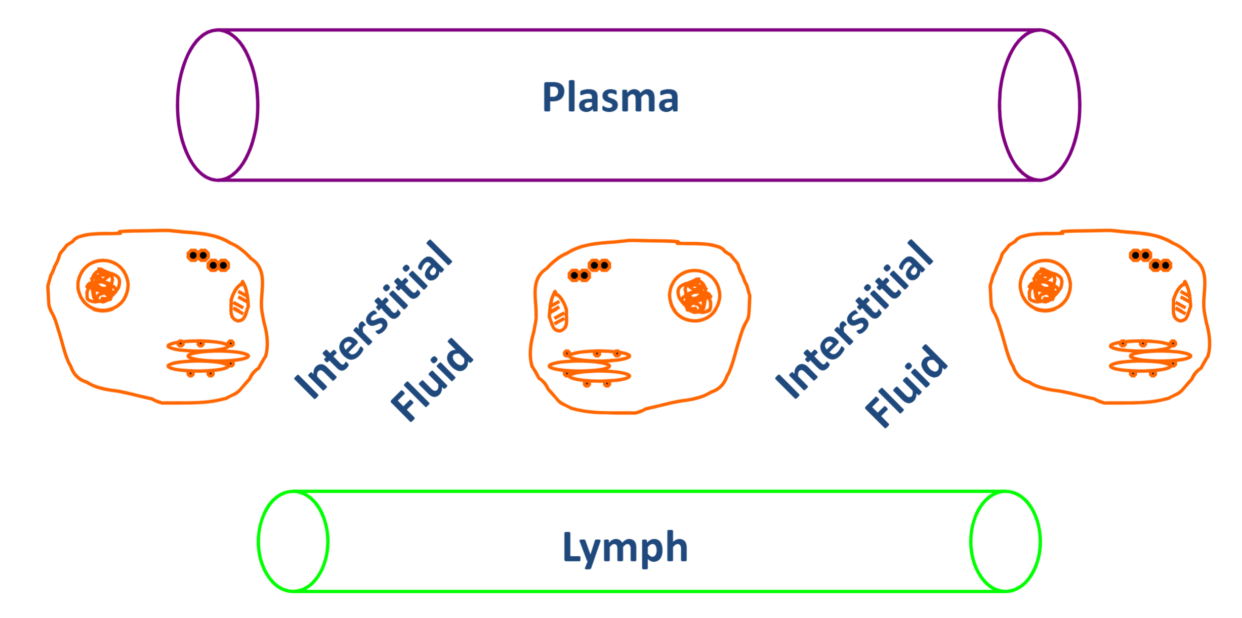
knowt flashcard image
3
New cards

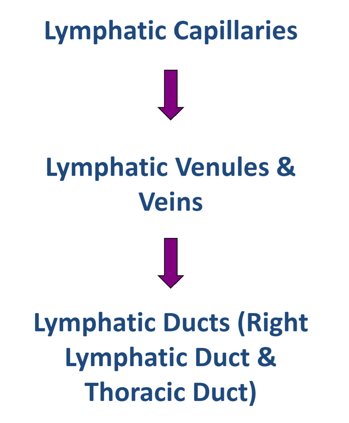
Lymphatic Network

Lymph is drained away from the tissues by the lymphatic network.

4
New cards

Lymphatic Network (Steps)

knowt flashcard image
5
New cards

Lymph is a fluid that is:

  • Created in the interstitial spaces of tissues.

  • Returned to the blood circulation through the lymphatic system.


6
New cards

Lymphatic Vessels → Veins & Arteries

knowt flashcard image
7
New cards

How do we NORMALLY exchange things between the vasculature & interstitium?(3)

  1. Diffusion across endothelial cell membranes.

  2. Movement through fenestrations.

  3. Transport via small vesicles (pinocytosis).


<ol><li><p><span style="color: rgb(37, 255, 64);"><strong>Diffusion across endothelial cell membranes.</strong></span></p></li><li><p>Movement through <span style="color: rgb(255, 35, 35);"><strong>fenestrations</strong></span>.</p></li><li><p>Transport via <span style="color: rgb(223, 7, 255);"><strong>small vesicles (pinocytosis</strong></span>).</p></li></ol><p></p>
8
New cards

Three Types of Capillaries

  1. Continuous Capillaries

  2. Fenestrated Capillaries

  3. Sinusoidal Capillaries

.

The further on the list the larger the capillaries.


<ol><li><p><span style="color: rgb(78, 255, 0);"><strong>Continuous </strong></span>Capillaries</p></li><li><p>Fenestrated Capillaries</p></li><li><p><span style="color: rgb(255, 0, 0);"><strong>Sinusoidal </strong></span>Capillaries</p></li></ol><p>.</p><p>The further on the list the larger the capillaries.</p><p></p>
9
New cards

Fluid movement across clefts and fenestrations is driven by Starling forces (or pressures) across the capillary wall: (4 Factors)

knowt flashcard image
10
New cards
term image

Capillary hydrostatic pressure

11
New cards
term image

Interstitial hydrostatic pressure

12
New cards
term image

Capillary oncotic pressure

13
New cards
term image

Interstitial oncotic pressure

14
New cards

If you can explain this picture, you will understand A LOT

about capillary exchange

knowt flashcard image
15
New cards

Arterial End (Capillary exchange) (4)

- On the arterial end, Pc > πc → Net filtration (fluid leaves the capillary).

<p>- On the arterial end, <span style="color: rgb(0, 255, 25);"><strong>Pc &gt; πc</strong></span> → Net filtration (fluid leaves the capillary).</p>
16
New cards

Venous End (Capillary exchange) (4)

  • Why did Pc decrease? Fluid leaves the capillary at the arterial end, causing capillary hydrostatic pressure (Pc) to decrease as blood travels toward the venous end.

  • Why did πc increase? Proteins remain inside the capillary while fluid leaves, increasing the protein concentration and therefore capillary oncotic pressure (πc).
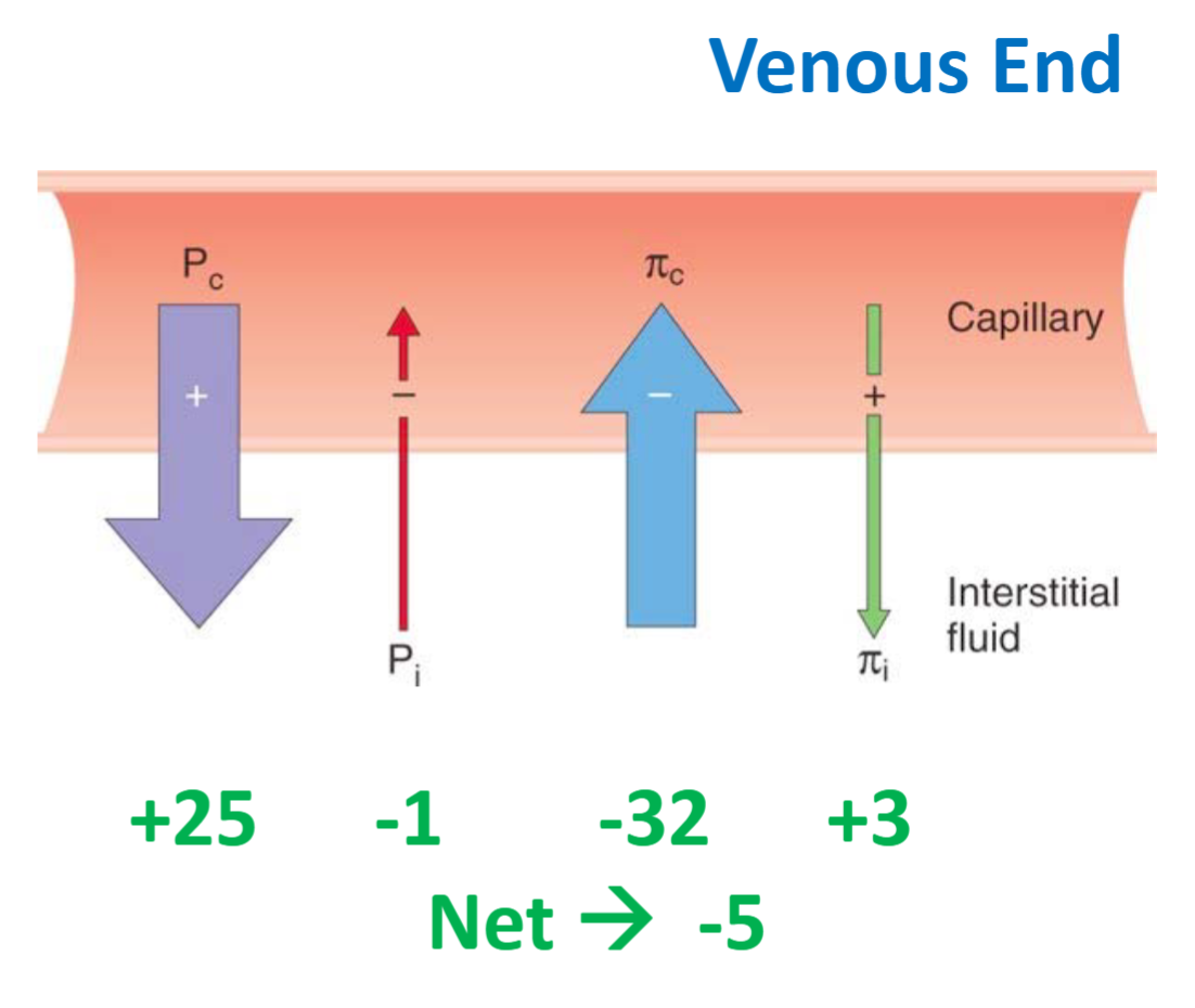

<ul><li><p>Why did Pc decrease? <span style="color: rgb(0, 255, 25);"><strong>Fluid leaves the capillary at the arterial end, causing capillary hydrostatic pressure (Pc) to decrease</strong></span> as blood travels toward the venous end.</p></li><li><p>Why did πc increase? Proteins remain inside the capillary while fluid leaves, <span style="color: rgb(255, 76, 76);"><strong>increasing the protein concentration and therefore capillary oncotic pressure (πc).</strong></span></p></li></ul><p></p>
17
New cards

Is the amount of fluid that leaves the capillary on the arterial end equal to the amount of fluid that enters the capillary on the venous end?

No, more fluid leaves the capillary on the arterial end than enters on the venous end

18
New cards

Lymphatic Vessels (Three Steps)

knowt flashcard image
19
New cards

Thoracic duct

  • The thoracic duct collects lymph from most body regions.

  • It ascends into the root of the neck on the left side.

  • It empties into the left subclavian vein near the junction with the left internal jugular vein.
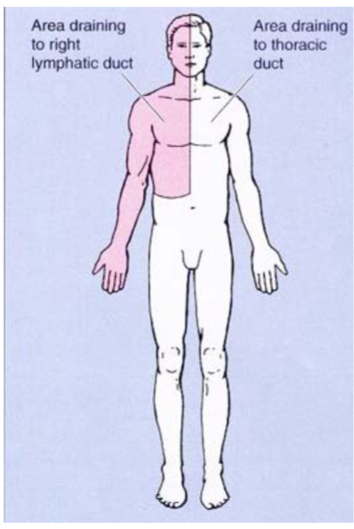

<ul><li><p>The thoracic duct collects lymph from most body regions.</p></li><li><p>It ascends into the root of the neck on the left side.</p></li><li><p><span style="color: rgb(0, 255, 5);"><strong>It empties into the left subclavian vein near the junction with the left internal jugular vein.</strong></span></p></li></ul><p></p>
20
New cards

Right Lymphatic duct

  • The right lymphatic duct receives lymph from the right side of the head, neck, and trunk.

  • It also receives lymph from the right upper extremity.

  • It empties into the right subclavian vein.
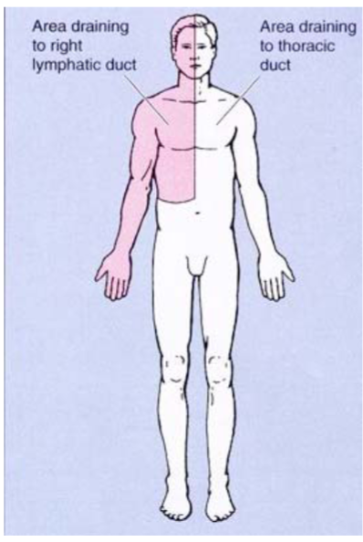

<ul><li><p>The right lymphatic duct receives lymph from the right side of the head, neck, and trunk.</p></li><li><p>It also receives lymph from the right upper extremity.</p></li><li><p>It empties into the right subclavian vein.</p></li></ul><p></p>
21
New cards

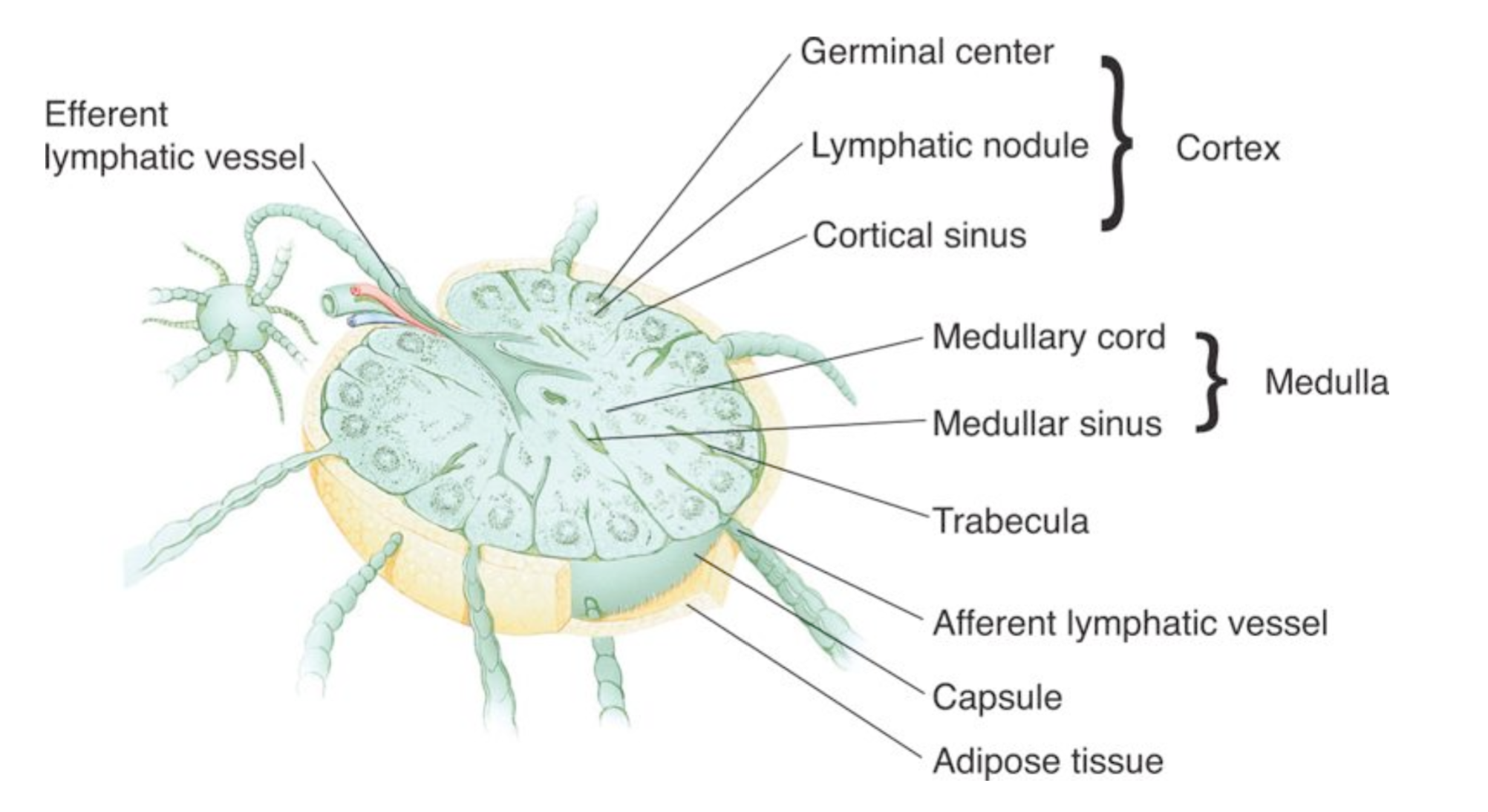
Lymph Nodes

- Lymph nodes are “beads” or “swellings” along certain lymphatic vessels.

- They act as filters.

22
New cards

Lymph Node Anatomy

knowt flashcard image
23
New cards

Lymphatic Drainage of the Breast (6 Most prominent spots for lymph nodes)

Lymph nodes are most extensive in the

1) axillary (armpit)

2) breast

3) neck

4) abdomen

5) thorax

6) pelvis, and groin regions.

<p>Lymph nodes are most extensive in the </p><p>1) axillary (armpit)</p><p>2) breast</p><p>3) neck</p><p>4) abdomen</p><p>5) thorax</p><p>6) pelvis, and groin regions.</p>
24
New cards

Edema (2 Types)

Palpable swelling due to expansion of extracellular fluid

<p>Palpable <span style="color: rgb(0, 255, 46);"><strong>swelling due to expansion of extracellular fluid</strong></span></p>
25
New cards

Pitting Edema

- Pressing a finger into the skin over accumulated fluid creates a temporary depression.

- Normal skin and subcutaneous tissues quickly rebound when the pressure is released.
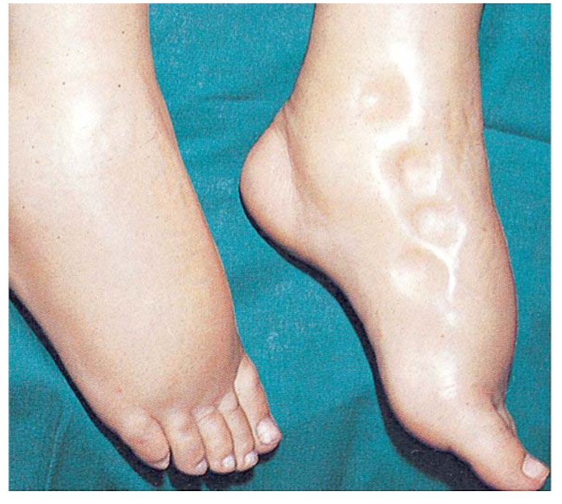
<p>- Pressing a finger into the skin over accumulated fluid creates a temporary depression.</p><p>- Normal skin and subcutaneou<span style="color: rgb(2, 255, 53);"><strong>s tissues quickly rebound when the pressure is released.</strong></span></p>
26
New cards

Non-Pitting Edema

  • Excess fluid in the interstitium becomes infiltrated with proteins or other materials.

  • This prevents the fluid from moving freely within the tissue spaces.


<ul><li><p>Excess fluid in the interstitium<span style="color: rgb(128, 255, 57);"><strong> becomes infiltrated with proteins or other materials.</strong></span></p></li><li><p>This prevents the fluid from moving freely within the tissue spaces.</p></li></ul><p></p>
27
New cards

Edema can be caused by? (4)

  • Edema occurs when interstitial fluid volume exceeds the lymphatics' capacity to return fluid to circulation.

  • It can be caused by:

    • Excess filtration (↑ Pc).

    • Decreased absorption (↓ πc).

    • Increased capillary permeability.

    • Blocked lymphatics.