Blood Protozoans
 Call Kai
 Learn
 Practice Test
 Spaced Repetition
 Match
 Flashcards
 Knowt Play
1/47

There's no tags or description
Looks like no tags are added yet.
48 Terms
Promastigote
stage of Leishmania spp., found within the insect vector

Epimastigote
stage typical of Trypanosoma spp., in the insect vector

Trypomastigote
stage typical of the bloodstream form of Trypanosoma spp.
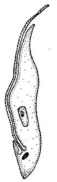
Amastigote
intracellular stage of T. cruzi and Leishmania spp.

Trypanosoma brucei - Epidemiology
T. brucei (T.b.) gambiense (tropical West Africa and Central Africa)
human and animal reservoir (human most important)
T. brucei (T.b.) rhodesiense (East Africa)
human and animal reservoir (animal most important)
Life Cycle of African Trypanosomiasis
infection occurs through bite of the tsetse fly which injects the trypomastigote (infective stage)
trypomastigote enters the lymphatics and passes to bloodstream where it is carried throughout body (diagnostic stage)
fly takes a blood meal and becomes infected with trypomastigotes
parasite multiples in fly midgut, leaves the midgut and transforms into epimastigote
epimastigote reaches the salivary glands where it transforms into trypomastigotes

Infective stage of African Trypanosomiasis
trypomastigote
Diagnostic Stage of African Trypanosomiasis
trypomastigote enters the lymphatics and passes to bloodstream
Trypanosoma brucei - Clinical Syndrome
African sleeping sickness (T.b. rhodesiense) - more acute
painful at bite site, can become a red sore, also called a chancre
haemolymphatic stage
meningoencephalitic stage
African sleeping sickness (T.b. gambiense) - more chronic
occasionally, within 1 to 3 weeks, the infective bite develops into a chancre
similar symptoms as that seen by T.b. rhodesinse
Haemolymphatic stage
fever, severe headaches, irritability, extreme fatigue, swollen lymph nodes, and aching muscles and joints
Meningoencephalitic stage
progressive confusion, personality changes, and other neurologic problems occur after infection has invaded the central nervous system. if left untreated, infection becomes worse, and death will occur within months
Trypanosoma brucei - Diagnosis
microscopic staining of body fluid (including blood or lymph node aspirate) for trypomastigote
small kinetoplast located at the posterior end, a centrally located nucleus
Trypanosoma brucei - Treatment and Prevention
pentamidine/suramin
used to treat the first stage of disease
nifurtimox-eflornithine
used to treat the second stage of the disease
education and control the breeding of the tsetse fly
wear long pants/sleeves that are neutral colors (attracted to bright colors)
use insect repellent
Trypanosoma cruzi - Epidemiology
distribution: central/south america (rarely in southern parts of US)
estimated 6-8 million people in Mexico, Central American and South America are infected
Human and animal reservoirs (mostly wild animals; but dogs may be a significant reservoir)
Vector for Trypanosoma cruzi
reduviid bug (triatomine; kissing bug)
associated with animal nesting areas; the bugs are found in houses made from materials such as mud, adobe, straw, and palm thatch; bugs emerge at night
bugs defecate after they ingest blood and parasite in feces enter body through mucous membranes or breaks in skin
Trypanosoma cruzi - Transmission
congenital transmission
blood transfusion
organ transplantation
consumption of uncooked food that is contaminated with triatomine bug feces
Life Cycle of T. Cruzi
infection occurs when the vector takes a blood meal and releases trypomastigote in its feces near the site of the bite (infective)
trypomastigotes invade cells near site of inoculation and differentiate into intracellular amastigotes which will multiply and become trypomastigotes which are release into the bloodstream (diagnostic stage)
trypomastigotes infect cells from a variety of tissues and transform into intracellular amastigotes (alternative diagnostic stage)
vector takes a blood meal and becomes infected with trypomastigotes which transform into epimastigotes
epimastigotes multiply and differentiate in the midgut and become trypomastigotes in the hindgut

infective and diagnostic stage of T. cruzi
trypomastigotes
Alternative diagnostic stage of T. cruzi
intracellular amastigotes
Trypanosoma cruzi - Clinical Syndrome
Chaga’s disease (American Trypanosomiasis)
Acute phase: mild to asymptomatic
fever and local swelling where the parasite entered the body
Chronic phase: no organisms in the bloodstream; organism enters the heart, liver, spleen, brain and lymph nodes
asymptomatic for life OR symptomatic (20%)
Trypanosoma cruzi - Diagnosis
microscopic staining of blood or CSF for trypomastigote (only acute stage)
large, subterminal or terminal kinetoplast, a centrally located located nucleus
antibody detection (chronic stage)
microscopic staining of biopsied tissue for amastigotes
molecular analysis (only acute stage)
Trypanosoma cruzi - Treatment and Prevention
benzidazole and nifurtimox
education and vector control (improve housing to eliminate bugs)
screen blood and tissue donors
Leishmania - Epidemiology
distribution: found in parts of the tropics, subtropics, and southern Europe (varies with the species)
number of new cases of cutaneous leishmaniasis each year in the world is about 1 million; numbers of new cases of visceral leishmaniasis is abt 300,000
human and animal reservoir: dogs and rodents
Leishmania - Vector
sand flies
Leishmania - Infections
cutaneous leishmaniasis (skin sores)
visceral leishmaniasis (kala-azar) (affect internal organs of the body; spleen, liver and bone marrow)
mucocutaneous leishmanisasis (partial or total destruction of facial muscous membranes)
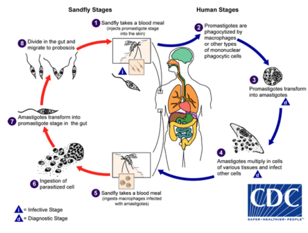
Life Cycle of Leishmaniasis
infection occurs when the vector takes a blood meal and injects the promastigotes (infective stage)
promastigotes get phagocyted by macrophages and transform into amastigotes
amastigotes multiply in cells of various tissues (diagnostic stage) and proceed to infect other phagocytic cells
vector takes a blood meal and becomes infected with amastigotes in macrophages which transform into epimastigotes in the gut
epimastigotes multiply and migrate to proboscis
Leishmania - Cutaneous
within ~2 weeks, a red papule at bite site; lesion becomes irritated with intense itching and begins to enlarged and ulcerate - lesion may heal without treatment within months but usually leaves a disfiguring scare
Leishmania - Visceral (Kala-azar)
within a few weeks (or even years), a gradual onset of fever, diarrhea and anemia - chills that resemble malaria are common; organisms invade liver, spleen, kidney
if untreated can become a lethal disease in a few weeks or may persist as a chronic disease and lead to death in 1 or 2 years
Leishmania - Diagnosis
cutaneous:
microscopic staining of tissue for amastigotes
molecular analysis
visceral:
microscopic staining of tissue or bones marrow for amastigotes
antibody detection
molecular analysis
Leishmania - Treatment and Prevention
cutaneous: skin lesions may heal on their own (take months/years and leave an ugly scar)
miltefosine
visceral:
amphotericin B by IV infusion
education and protection from vector
wear long-sleeves/pants
insect repellent
use of mosquito net (need smaller hole sizes)
Plasmodium (Malaria) - Epidemiology
distribution: worldwide in tropics/subtropics
in 2022 an estimated 249 million cases occurred worldwide with an estimated 608,000 death
Africa accounts for 95% of malaria cases and 96% of malaria deaths (children under 5 years of age accounted for ~80% of the deaths)
~2000 cases and 5 deaths each year in the US
human reservoir
Plasmodium - Vector and Species
vector: anopheles mosquito
major species: Plasmodium vivax, P. falciparum, P. malariae, P. ovale
P. falciparum is most severe and life-threatening; most prevalent in Africa
P vivax is dominant in most countries outside of Africa
Plasmodium - Transmission
blood transfusions (only 1 case every 2 years in US)
congenital transmission (rare)
“airport” malaria
infected mosquitos that are transported rapidly by aircraft from a malaria-endemic country to a non-endemic country
bite a local resident
Life Cycle of Plasmodium
infection occurs when the vector takes a blood meals and injects the sporozoite (infective stage)
sporozoites infect liver cells and mature into schizonts which rupture and release merozoites
merozoites infect red blood cells and continue to mature, resulting in a variety of developmental stages of the parasite (diagnostic stage)
the late-stage gametocytes are ingested by the vectors multiply (sporogonic cycle) with later stages of the parasite developing into oocysts which grow, rupture and release sporozoites
sporozoites go salivary glands of the vector

Plasmodium - Infection
incubation: 7 to 30 days
erythrocytes (RBCs) infected:
P. vivax and P. ovale infect younger reticulocytes (density of infected cells rarely exceeds 3%)
individuals who lack the duffy antigen on the surface of their RBCs are protective against P. vivax
P. malariae infects older RBCs
P. falciparum infects ALL erythrocytes (can produce a high density of infected RBCs)
individuals with sickle cell trait are predominately protective from infection
Malaria - Clinical Syndromes
fever, chills, sweats, headaches, nausea and vomiting, body aches, and general malaise
benign tertian fever (every 48 hrs): P. vivax and P. ovale
benign quartan fever (every 72 hrs): P. malariae
malignant tertian fever: P. falciparum (fever is usually continuous or briefly remittent)
3 stages of malaria paroxysm
“cold": shaking chills
“hot”: temperature to 40 C or high, systemic symptoms
“sweating”: diaphoresis (sweating), fever resolves, fatigue
Severe malaria manifestations
cerebral malaria
severe anemia
hemoglobinuria
pulmonary edema
abnormalities in blood coagulation and thrombocytopenia
cardiovascular
Plasmodium - Diagnosis
microscopic staining of blood
thick (detect parasite) and thin (species-level identification) smear preparations
must determine parasitemia
antibody detection
antigen detection (rapid diagnostic tests)
molecular assays
Plasmodium - Treatment
all patients should be treated
resistance to commonly used chloroquine is occurring
utilize alternative treatment for resistant strains
atovaquone-proguanil or artemether-lumefantrine
Plasmodium - Prevention
education
malaria prophylaxis
can delay the appearance of malaria symptoms by weeks or months
P. vivax and P. ovale, dormant liver stage parasites, the liver stages may reactivate and cause disease months after the infective mosquito bite; relapse for as long as 3 to 4 years
control mosquito population
use mosquito nets, insect repellent, wear long sleeves/pants
vaccine
for children living in regions with moderate to high P. falciparum transmission
Babesia spp. - Epidemiology
distribution:
mostly found in North America followed by China
highest prevalence in the US is along the northeastern seaboard
human and animal reservoir (mostly mouse)
tick vector (Ixodid)
most common species in US: B. microti
Life Cycle of Babesia
infection occurs the vector takes a blood meal and injects the sporozoite (infective stage)
sporozoites enter erythrocytes and reproduce (diagnostic stage)
humans are considered “dead-end hosts”
ticks acquire sporozoite by taking a bite of the mouse and the parasite multiplies in the tick (sporogonic cycle)
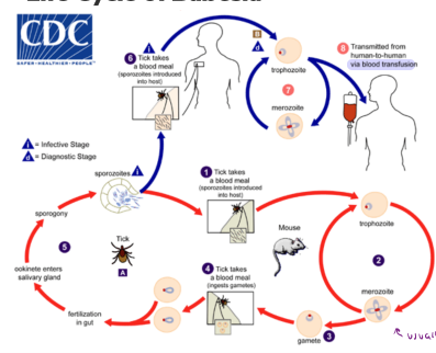
Infective and diagnostic stage of Babesia
sporozoite
Babesia spp. - Clinical Syndromes
most infections are probably asymptomatic
incubation: 1 to 4 weeks
general malaise, fever, headache, chills, sweating, fatigue, and weakness
as infection progresses get destruction of erythrocytes, thus anemia develops (can see renal failure)
more severe disease in immunocompromised, individuals with no spleen and the elderly
Babesia spp. - Diagnosis
microscopic staining of blood smear
must distinguish from Plasmodium
antibody detection
Babesia spp. - Treatment and Prevention
asymptomatic persons typically do not receive treatment
high risk individuals and patients that received parasite by blood transfusion should be treated
education
wear long sleeves/pants and use insect repellent
remove ticks within a few hours