Thorax + Osteology

0.0(0)
Studied by 0 people
call kaiCall Kai
learnLearn
examPractice Test
spaced repetitionSpaced Repetition
heart puzzleMatch
flashcardsFlashcards
GameKnowt Play
Card Sorting

1/41

encourage image

There's no tags or description

Looks like no tags are added yet.

42 Terms

1
New cards

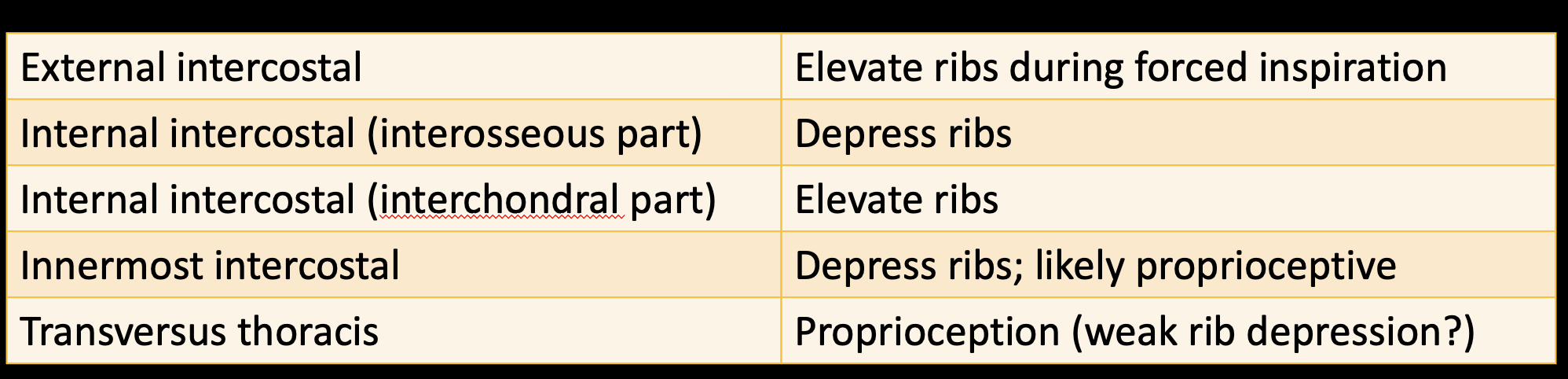
muscle fiber direction external vs internal intercostals

-external: medial and inferiorly (“hands in your pockets”; insert on inferior ribs); more superficial; elevate ribs

-internal: lateral and superiorly (“hands on your heart”; insert on superior rib); more deep; depress ribs

  • Interosseous part: most, between bones

  • Interchondral part: farthest anterior, between cartilage of ribs and sternum


<p><strong><mark data-color="blue" style="background-color: blue; color: inherit;">-external: </mark></strong><mark data-color="blue" style="background-color: blue; color: inherit;">medial and inferiorly</mark> (“hands in your pockets”; insert on inferior ribs); more superficial; elevate ribs</p><p><strong><mark data-color="blue" style="background-color: blue; color: inherit;">-internal: </mark></strong><mark data-color="blue" style="background-color: blue; color: inherit;">lateral and superiorly</mark> (“hands on your heart”; insert on superior rib); more deep; depress ribs</p><ul><li><p><strong>Interosseous part:</strong> most, <mark data-color="red" style="background-color: red; color: inherit;">between bones</mark></p></li><li><p><strong>Interchondral part</strong>: <mark data-color="red" style="background-color: red; color: inherit;">farthest anterior, between cartilage of ribs and sternum</mark></p></li></ul><p></p>
2
New cards
<p>actions of intercostals</p>

actions of intercostals

*interchondral part help external intercostals elevate ribs
<p>*interchondral part help external intercostals elevate ribs</p>
3
New cards

divisions of the thoracic cavity

-The thoracic cavity is divided into 2 pulmonary cavities and the mediastinum

*bounded by ribs and things ribs attach to

<p>-<span style="font-family: &quot;Calibri Light&quot;;">The thoracic cavity is divided into <strong>2 pulmonary cavities and the mediastinum</strong></span></p><p><span style="font-family: &quot;Calibri Light&quot;;">*bounded by ribs and things ribs attach to</span></p>
4
New cards

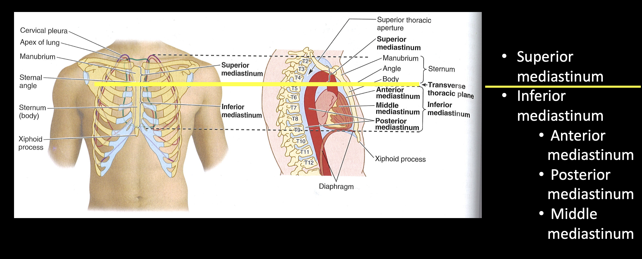
divisions of the mediastinum

-Superior mediastinum

***Transverse Thoracic Plane*** (imaginary line @sternal angle/T4)

-Inferior mediastinum

•  Anterior mediastinum

•  Posterior mediastinum

•  Middle mediastinum
<p><strong>-Superior mediastinum</strong></p><p>***<strong>Transverse Thoracic Plane</strong>*** (imaginary line @sternal angle/<mark data-color="green" style="background-color: green; color: inherit;">T4</mark>)</p><p><strong>-Inferior mediastinum</strong></p><p>•&nbsp; Anterior mediastinum</p><p>•&nbsp; Posterior mediastinum</p><p>•&nbsp; Middle mediastinum</p>
5
New cards

What vertebral level indicates the transition between superior versus inferior mediastinum?

T4

-Transverse Thoracic Plane
6
New cards
<p>contents of the mediastinum</p>

contents of the mediastinum

*sup: + trachea

<p>*sup: + trachea</p>
7
New cards

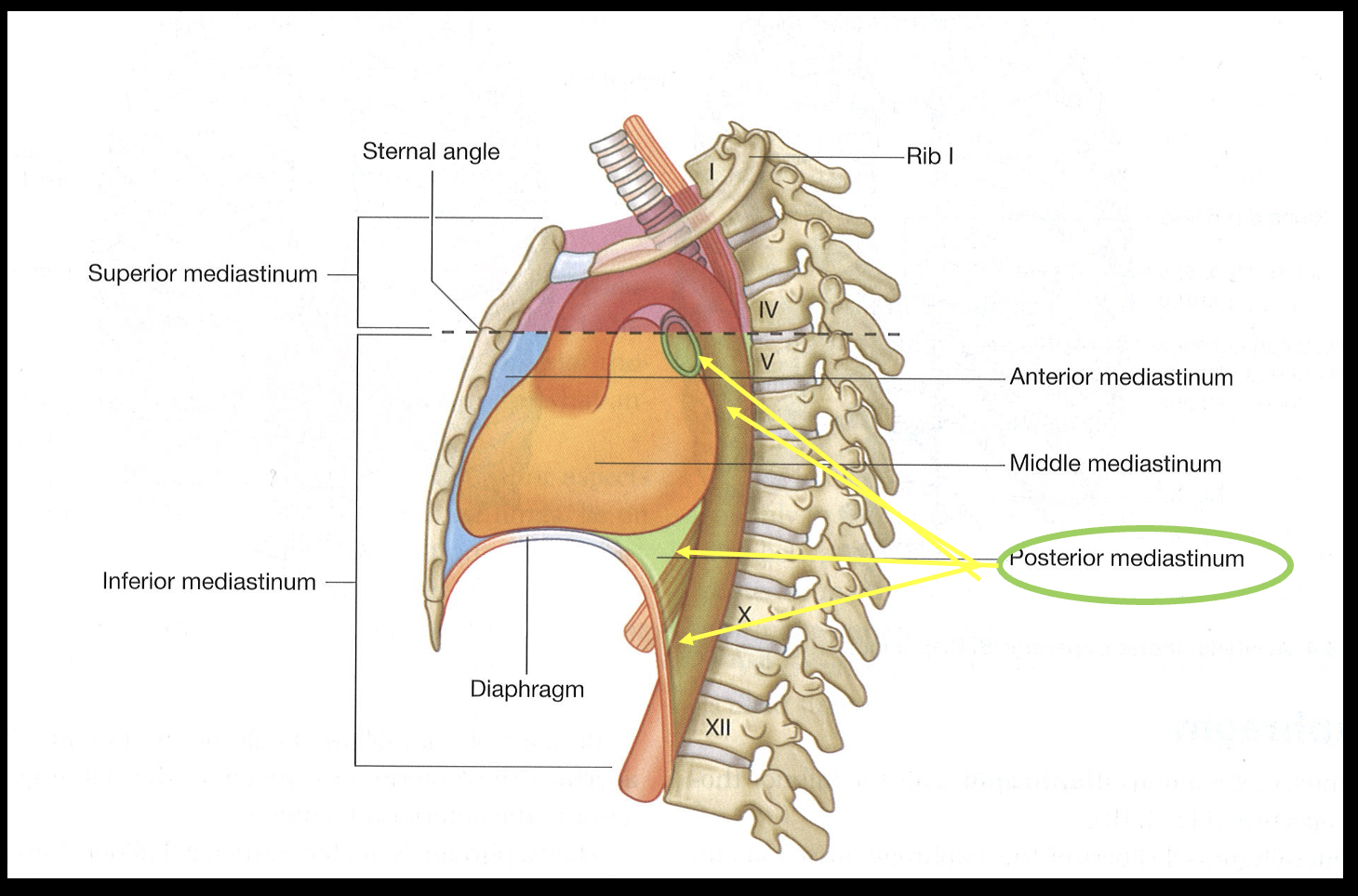
contents of posterior mediastinum

-Thoracic aorta

-Esophagus

-Thoracic duct

-Azygos veins

-Sympathetic trunks

*inf line and post heart
<p><em>-Thoracic</em> aorta</p><p>-Esophagus</p><p>-Thoracic duct</p><p>-Azygos veins</p><p>-Sympathetic trunks</p><p>*inf line and post heart</p>
8
New cards
<p>identify in posterior mediastinum</p>

identify in posterior mediastinum

*associated w/ posterior abdominal wall

  1. SNS chain: ANS to organs of thorax and abdomen

  2. Greater Splachnic nerve: sympatehtic fibers in abdomen; from SNS chain

  3. Esophageal plexus: formed by vagus n, warps around esophagus

  4. Esophagus

  5. Thoracic aorta


<p>*associated w/ posterior abdominal wall</p><ol><li><p><strong>SNS chain</strong>: ANS to organs of thorax and abdomen</p></li><li><p><strong><mark data-color="red" style="background-color: red; color: inherit;">Greater Splachnic nerve: </mark></strong><em><mark data-color="red" style="background-color: red; color: inherit;">sympatehtic</mark></em><mark data-color="red" style="background-color: red; color: inherit;"> fibers in abdomen; from SNS chain</mark></p></li><li><p><strong>Esophageal plexus:</strong> formed by vagus n, warps around esophagus</p></li><li><p><strong>Esophagus</strong></p></li><li><p><strong>Thoracic aorta</strong></p></li></ol><p></p>
9
New cards

thoracic duct

Posterior Mediastinum

-largest lymph in body

-between esophagus and aorta

-Empties into junction of these 2 veins (In the neck: LEFT Internal jugular v.  and subclavian v.)

<p>Posterior Mediastinum</p><p>-largest lymph in body</p><p><mark data-color="red" style="background-color: red; color: inherit;">-between esophagus and aorta</mark></p><p>-Empties into junction of these 2 veins <mark data-color="red" style="background-color: red; color: inherit;">(In the neck: </mark><strong><mark data-color="red" style="background-color: red; color: inherit;">LEFT </mark></strong><mark data-color="red" style="background-color: red; color: inherit;">Internal jugular v.&nbsp; and subclavian v.)</mark></p>
10
New cards

azygos system of veins

Posterior Mediastinum

-if veins have hard time getting to vena cava

-intercostal v → azygous vein → superior vena cava

-accessory hemi-azygos v (upper left side) + hemi-azygos v (lower left side)=variable present!

<p>Posterior Mediastinum</p><p>-if veins have hard time getting to vena cava</p><p><strong>-intercostal v → azygous vein → <mark data-color="red" style="background-color: red; color: inherit;">superior</mark> vena cava</strong></p><p><strong>-accessory hemi-azygos v</strong> (upper left side) + <strong>hemi-azygos v</strong> (lower left side)=<mark data-color="purple" style="background-color: purple; color: inherit;">variable present!</mark></p>
11
New cards

contents of the superior mediastinum

-Thymus (remnants)

-Great vessels

-Trachea

-Esophagus; Thoracic duct (*most in posterior)

-Important nerves

*inbetween lungs, sup to plane

<p>-Thymus (remnants)</p><p>-Great vessels</p><p><mark data-color="red" style="background-color: red; color: inherit;">-Trachea</mark></p><p>-Esophagus; <mark data-color="red" style="background-color: red; color: inherit;">Thoracic duct</mark> (*most in posterior)</p><p>-Important nerves</p><p>*inbetween lungs, sup to plane</p>
12
New cards

thymus

Superior Mediastinum

-immune system

-atrophy w/ age; largest in peds

<p>Superior Mediastinum</p><p>-immune system</p><p>-atrophy w/ age; largest in peds</p>
13
New cards

great vessels

Superior Mediastinum

-Pulmonary trunk: blood heart to pulmonary circut/lungs

-Arch of the aorta: blood heart to body

-R and L Brachiocepahlic v: venous return head and upper limb to sup vena cava

-Superior vena cava: deoxygenated blood to heart

<p>Superior Mediastinum</p><p><strong>-Pulmonary trunk:</strong> blood heart to pulmonary circut/lungs</p><p><strong>-Arch of the aorta:</strong> blood heart to body</p><p><strong>-R and L Brachiocepahlic v: </strong>venous return head and upper limb <mark data-color="blue" style="background-color: blue; color: inherit;">to sup vena cava</mark></p><p><strong>-Superior vena cava:</strong> deoxygenated blood to heart</p>
14
New cards

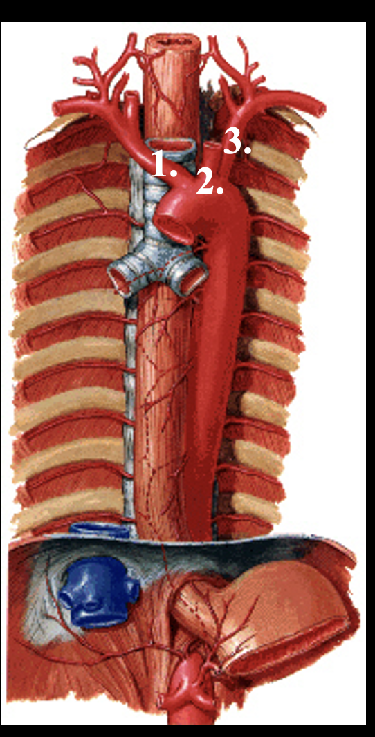
branches from arch of the aorta

1.Brachiocephalic trunk (Will branch into R. common carotid a., R. subclavian a.)

2.Left common carotid

3.Left subclavian
<p><span>1.Brachiocephalic trunk (Will branch into R. common carotid a., R. subclavian a.)</span></p><p><span>2.Left common carotid</span></p><p><span>3.Left subclavian</span></p>
15
New cards
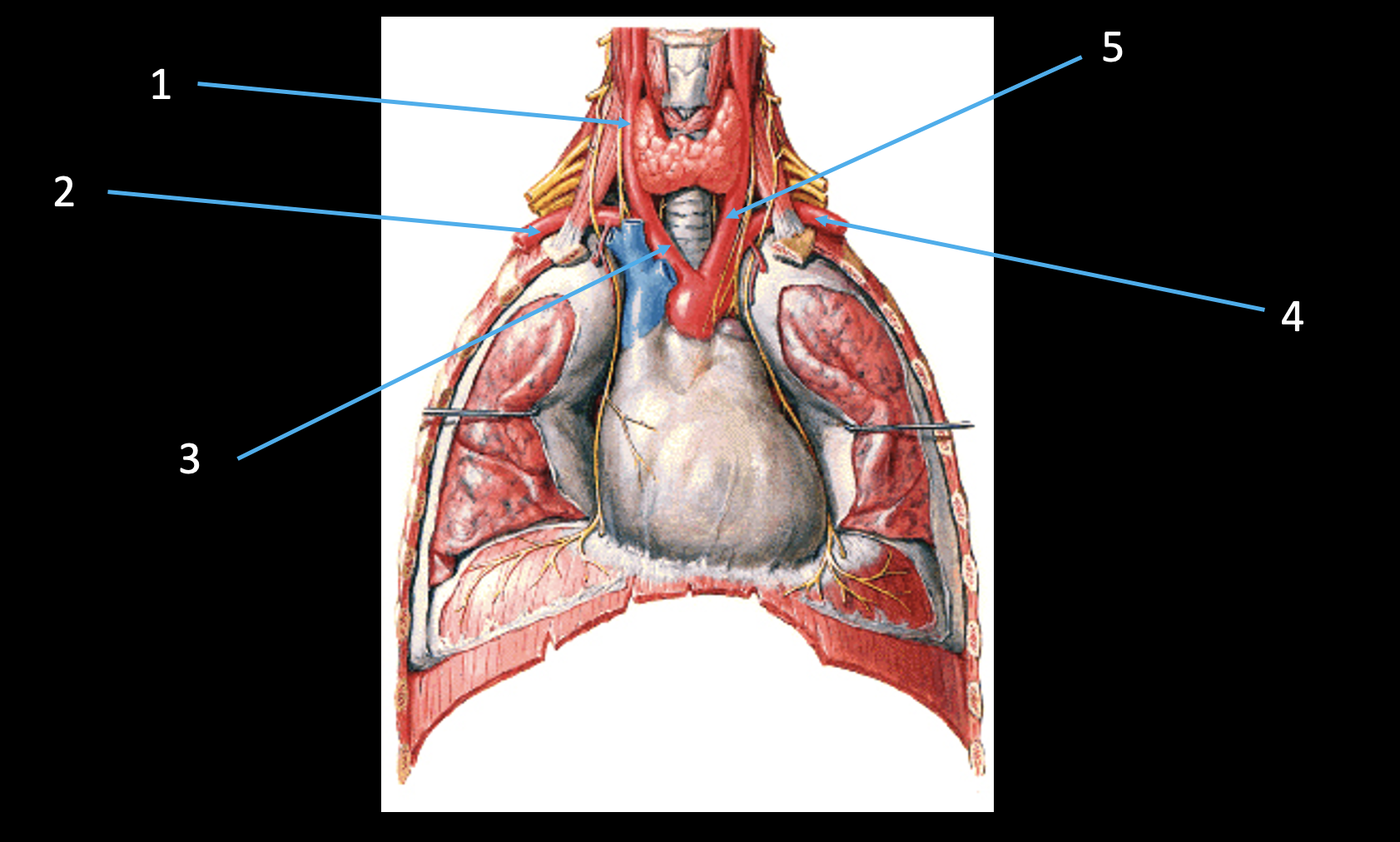
<p>identify branches of the aorta</p>

identify branches of the aorta

1.Right common carotid a.

2.Right subclavian a.

3.Brachiocephalic trunk

4.Left subclavian a.

5.Left common carotid a.

16
New cards

trachea

(only in) Superior Mediastinum

-terminates as R and L primary brachii

  • R bigger than left!

-airway

<p><mark data-color="red" style="background-color: red; color: inherit;">(only in) Superior Mediastinum</mark></p><p>-terminates as R and L primary brachii</p><ul><li><p>R bigger than left!</p></li></ul><p>-airway</p>
17
New cards

esophagus

Superior Mediastinum (some, mostly in post)

-posterior to trachea

-GI contents oral → stomach

-Vagus n forms the esophageal plexus (n to abdominal cavity)

<p>Superior Mediastinum (some, mostly in post)</p><p>-posterior to trachea</p><p>-GI contents oral → stomach</p><p><strong>-Vagus n forms the esophageal plexus</strong> (n to abdominal cavity)</p>
18
New cards

thoracic duct

Superior Mediastinum

-lymph that drains into venous system= left internal jugular vein and left subclavian vein

<p>Superior Mediastinum</p><p>-lymph that drains into venous system= <strong>left internal jugular vein and left subclavian vein</strong></p>
19
New cards

important nerves in the superior mediastinum

-Phrenic n (C3-5): to diagram; anterior to root of lung, on pericardium surface; R and L

-Vagus n (CN X): wraps around esohpagus to form esohpageal plexus; PSNS thorax and abdominal organs, R and L

  • Recurrent laryngeal n: innervates larynx; branch of vagus n; wraps around aortic arch + ligamentum arteriosum (R and L but R doesn’t wrap around aorta, around subclavian a)

  • Vagal trunk: anterior and posterior; continuation of vagus n after pass through esophageal plexus

    • Posterior/R passes posterior to esophagus; anterior/L passes anterior to esophagus


<p><strong>-Phrenic n (C3-5)</strong>: to diagram; <em>anterior to root of lung, on pericardium surface</em>; R and L</p><p>-<strong>Vagus n (CN X):</strong> wraps around esohpagus to form esohpageal plexus; PSNS thorax and abdominal organs, R and L</p><ul><li><p><strong>Recurrent laryngeal n:</strong> innervates larynx; branch of vagus n; <mark data-color="blue" style="background-color: blue; color: inherit;">wraps around aortic arch + ligamentum arteriosum</mark> <mark data-color="red" style="background-color: red; color: inherit;">(R and L but R doesn’t wrap around aorta, around subclavian a)</mark></p></li><li><p><strong>Vagal trunk:</strong> anterior and posterior; continuation of vagus n after pass through esophageal plexus</p><ul><li><p>Posterior/R passes posterior to esophagus; anterior/L passes anterior to esophagus</p></li></ul></li></ul><p></p>
20
New cards

roots of the phrenic nerve?

C3-5

*“C3-5 keep the diaghram alive”; part of cervical plexus

21
New cards

Questions

-The phrenic nerve provides motor innervation to the

-What is the function of the vagus nerve?

-respiratory diaphragm

-parasympathetic innervation to thoracic organs

22
New cards

bony thorax

-bounded by sternum, ribs, vertebre

-Superior Thoracic aperture: bounded by 1st ribs

-Inferior Thoracic aperture: 12th rib and margin costal cartiledge

<p>-bounded by sternum, ribs, vertebre</p><p><strong>-Superior Thoracic aperture: </strong>bounded by 1st ribs</p><p><strong>-Inferior Thoracic aperture:</strong> 12th rib and margin costal cartiledge</p>
23
New cards

how does the thoracic cavity change size?

-Respiratory diaphragm!: separates thoracic and abdominal cavity; dome shaped w/ large SA

  • central tendon

  • bony thorax does NOT entirely correspond to thoracic cavity

-When diaphragm contracts: becomes flat and increases thoracic cavity (and decreases abdominal cavity)

-Innervated by the phrenic nerve: neck → TC → diagrahm

<p>-Respiratory diaphragm!: separates thoracic and abdominal cavity; dome shaped w/ large SA</p><ul><li><p>central tendon</p></li><li><p>bony thorax does NOT entirely correspond to thoracic cavity</p></li></ul><p><strong>-When diaphragm contracts: becomes flat and increases thoracic cavity</strong> (and decreases abdominal cavity)</p><p>-Innervated by the phrenic nerve: neck → TC → diagrahm</p>
24
New cards

“Thoracic outlet” syndrome

-if muscles too tight etc.=compress neuromuscular structures at neck

  • including subclavian a and brachial plexus


<p>-if muscles too tight etc.=compress neuromuscular structures at neck</p><ul><li><p><mark data-color="red" style="background-color: red; color: inherit;">including subclavian a and brachial plexus</mark></p></li></ul><p></p>
25
New cards

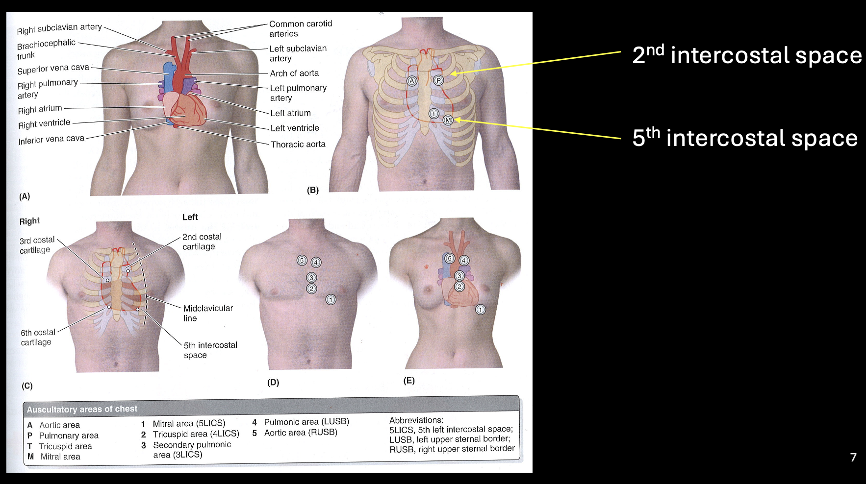
heart sounds and intercostal spaces

-2nd and 5th intercostal space
<p>-2nd and 5th intercostal space</p>
26
New cards
<p>identify the intercostal muscle layers</p>

identify the intercostal muscle layers

-innermost → internal → external intercostal

<p>-innermost → internal → external intercostal</p>
27
New cards

transversus thoracis

-on posterior thoracic wall

-proprioceptive function

<p>-on posterior thoracic wall</p><p>-proprioceptive function</p>
28
New cards

innermost intercostal mm

-vertical direction fibers

-proprioceptive function

<p>-vertical direction fibers</p><p>-proprioceptive function</p>
29
New cards

diaphragm during inspiration

-breathing in: flat, lungs taller (lungs don’t contract on own, rely on diaphragm)

-breathing out: dome, decrease thoracic cavity

<p>-breathing in: flat, lungs taller (lungs don’t contract on own, rely on diaphragm)</p><p>-breathing out: dome, decrease thoracic cavity</p>
30
New cards
<p>pt asked to hold breathe and then take x-ray. Based on this picture, which phrenic nerve is damaged?</p>

pt asked to hold breathe and then take x-ray. Based on this picture, which phrenic nerve is damaged?

Right phrenic nerve!

-B not contracted, A contracted

31
New cards
<p>identify intercostal neurovascular sturcutres</p>

identify intercostal neurovascular sturcutres

1.Intercostal nerve

  • pass inbetween intermost IC and internal IC; nerves ventral rami of spinal n

2.Thoracic aorta

3.Posterior intercostal artery

4.Internal thoracic artery (branch of?=subclavian a)

*Thoracic aorta → intercostal a

*subclavian a → internal thoracic a

32
New cards
<p>identify posterior thoracic wall</p>

identify posterior thoracic wall

1.Intercostal vein, artery, nerve

*intercostal v → azygous v → superior vena cava

2.Azygous vein

3.Sympathetic chain (*ANS/SNS throughout body)

4.Innermost intercostal mm.

33
New cards

3D Relationship Questions

1.Which intercostal neurovascular structure is the most superior?  Most inferior?

2.The intercostal neurovascular structures are found between which muscles?

3.The intercostal arteries branch from which vessel?

4.The intercostal veins drain to which vessel?

5.The intercostal nerves branch from?

1.Intercostal vein is most superior, then the artery, then the nerve is most inferior

  • v → a → n

2.Internal and innermost intercostal mm.

3.Thoracic aorta

4.Azygous vein (azygous system of veins)

5.Intercostal nerves are the ventral rami of thoracic spinal nerves

34
New cards

internal thoracic artery + terminal branches

-supply ant thoracic wall and diaphgram; commonly used for bypass surgery

-subclavian a → internal thoracic a →

  • superior epigastric a: ab wall/rectus abdominis

  • Musculophrenic a: ab wall and diapgram


<p>-supply ant thoracic wall and diaphgram; commonly used for bypass surgery</p><p><mark data-color="blue" style="background-color: blue; color: inherit;">-subclavian a → internal thoracic a →</mark></p><ul><li><p><strong>superior epigastric a:</strong> ab wall/<mark data-color="purple" style="background-color: purple; color: inherit;">rectus abdominis</mark></p></li><li><p><strong>Musculophrenic a:</strong> ab wall and diapgram</p></li></ul><p></p>
35
New cards
<p>identify</p>

identify

1.Rib (1st)

2.Manubrium of sternum

3.Body of sternum

4.Xiphoid process (of sternum)

5.Also a rib (7th)

6.Clavicle

7.Scapula

8.Thoracic vertebra

36
New cards

types of ribs: typical vs atypical

-Typical Ribs: 3-10

-Atypical Ribs: 1-2, 11-12
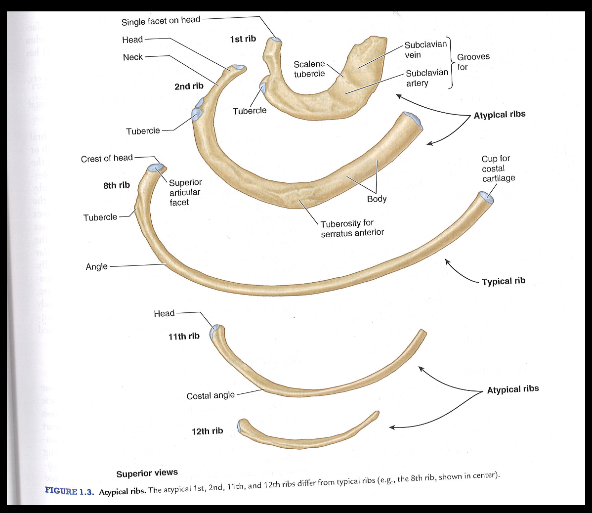
<p><span>-Typical Ribs: 3-10</span></p><p style="text-align: left;"><span>-Atypical Ribs: 1-2, 11-12</span></p>
37
New cards

types of rbis: true vs false vs floating

-Vertebrosternal (true): 1-7

-Vertebrochondral (false): 8-10

-Vetebral (floating): 11-12

<p><span>-Vertebrosternal (true): 1-7</span></p><p style="text-align: left;"><span>-Vertebrochondral (false): 8-10</span></p><p style="text-align: left;"><span>-Vetebral (floating): 11-12</span></p>
38
New cards
<p>identify rib articulations</p>

identify rib articulations

knowt flashcard image
39
New cards
<p>identify clavicle</p>

identify clavicle

knowt flashcard image
40
New cards
<p>identify sternum</p>

identify sternum

knowt flashcard image
41
New cards
<p>identify thoracic vertebra</p>

identify thoracic vertebra

knowt flashcard image
42
New cards

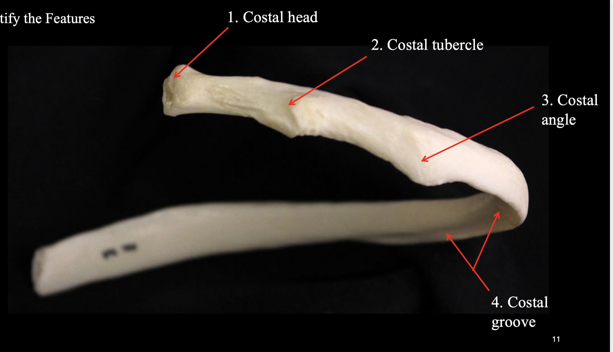
how do ribs articulate with thoracic vertebrae?

-Body of vertebrae + head of rib

-TP vertebrae + tubercle of rib

<p>-Body of vertebrae + head of rib</p><p>-TP vertebrae + tubercle of rib</p>