L6: NC Reflexes
 Call Kai
 Learn
Learn Practice Test
 Spaced Repetition
 Match
Match Flashcards
 Knowt Play
1/38

There's no tags or description
Looks like no tags are added yet.
Name | Mastery | Learn | Test | Matching | Spaced | Call with Kai | Chat |
|---|
No analytics yet
Send a link to your students to track their progress
39 Terms
Sensory receptors
What do they respond to?
What are the two parent categories?
Respond to different stimuli.
Interreceptor: senses information from within the body such as: temperature, pain, and pressure
Exteroceptors: Near the body surface, responds to external stimuli.
What are proprioceptors?
Synonymous with sensory receptors.
Respond to stimuli that informs the degree the body segment is changing, direction, and rate
What are the various muscle receptors?
Muscle spindles and Golgi Tendon Organ (GTO)
What is a muscle spindle
Location
Senses
MS is a tiny capsule filled with fluid and specialized fibers.
They are located predominantly in the muscle belly, situated in parallel with fibers direction.
Senses when the muscle is stretched.
Where are a great amount of muscle spindles located?
Are located in the muscles that control fine motor skills.
Muscle spindle anatomy
Fiber types
Nerve endings
Extrafusal fibers: normal muscle fibers
Intrafusal fibers: muscle fibers within the spindle
non-contractile center portion
the ENDS contract
Type I ending: Primary/Annulospiral ending: coiled around the middle noncontractile section
Type II ending: Flower-spray endings: coiled around the ends of noncontractile midsection
Type I ending: Primary/Annulospiral ending
sensitive to
Phasic info.
Sensitive to the velocity of change/ rate of change in length (fast or slow)
Speed in which the muscle is changing length.
Type II ending: Flower-spray endings
Sensitive to
Sensitive to static (tonic) muscle length
change of length of the muscle NOT the speed at which it changed.
How does the CNS differentiate between annulospiral n. endings and flower-spray n. endings?
The difference in the frequency of the impulse between phasic and tonic information.
What type of motor neuron interfuses the fibers of muscle spindles?
Extrafusal fibers are innervated by alpha motor neurons
Intrafusal fibers are innervated by gamma motor neurons
How is the Golgi Tendon Organ (GTO)/ Tendon Organ situated?
what does it monitor? explain
Purpose?
Is embedded “in series” in the tendon at the MTJ (weakest length)
It monitors tension. As tension increases and becomes too much for the GTO → the GTO sends information via a sensory n to CNS → causes an inhibitory response → motor n to muscle, inhibiting and relaxing it.
Protective mechanism

What are the joint and skin proprioceptors?
Pacinian Corpuscles, Ruffini endings, and cutaneous receptors.
Pacinian Corpuscles
Location
Detect
Transmits
Purpose
L: In regions around the joint capsules, ligaments, skin, and tendon sheaths
D: Rapid joint angle changes (rapid) and pressure
T: impulses to CNS for rapid changes in joint angles and pressures for brief periods of time (not continuous)
P: Predicts where body joint segment will end in space at current rate.
and appropriate readjustment in position can be anticipated and effected
Ruffini Endings
Location
Detect
L: in deep layers of skin and joint capsule
D: Continuous states of pressure in joints and skin
senses joint position and changes in joint angle, activated by mechanical deformation
Cutaneous receptors
what are the two types
Meissner corpuscles: sensation of touch
Free nerve endings: sensation of pain
Serve as proprioceptors when they show sensitivity to texture, hardness, softness, and shape, and participate in reflexes.
What are the labyrinthine and neck proprioceptors?
semicircular canals and neck proprioceptors
Semicircular canals
Located
Filled with
How is motion detected
Responsible for
L: located at perfect right angles to each other.
It is filled with endolymph and hair cells.
Hair cells detect the motion of fluid as the head moves. Information is then sent to the CNS → gives a sense of head movement and overall balance.
Responsible only for the orientation and movement of the head.
Neck proprioceptors
what are they?
Sensitive to?
Which muscles have the highest density?
Are a collection of all proprioceptors.
Sensitive to change of angle between the body and head through the neck.
Deep neck muscles have the highest density of spindles compared to other muscles
important in the maintaining balance.
How does the labyrinthine system and neck proprioceptors work independently and together.
LS detects when head movement and sends signals for the head to straighten out and align with the neck/body.
NP can override the signaling of the LS to the CNS to prevent reflexes when in control of movement (balanced).
What is a reflex movement?
→ How does it travel?
Is a specific pattern of movement without violation (voluntary intervention) from the cerebrum
Stimulus → Receptor → sensory neuron → spinal cord→ motor neuron → muscle → response
What are the two categories of neural pathways?
Monosynaptic: sensory neuron synapses to motor neuron directly
Polysynaptic: interneurons are involved
What are the proprioceptive reflexes?
Phasic reflexes
knee-jerk reflex
Tonic reflexes
Golgi tendon reflex
Asymmetrical TNR
Symmetrical TNR
Labyrinthine reflex symmetrical
Labyrinthine righting reflex
Phasic stretch reflex
Associated with..
Generally happens when…
Gives a…
Associated with annulospiral ending of the muscle spindle
Generally happens when a muscle is eccentrically lengthened and quickly (no pause) concentrically contracts.
“Neuromuscular bump” in which the muscle spindle sends an impulse for the muscle to contract in addition to a cognitive (voluntary) contraction.

Knee-jerk reflex
what type of reflex
explain both synapses
Phasic Stretch Reflex
Monosynaptic
Stimulus→ patellar tendon is hit
Receptor→ muscle spindle
Sensory neuron→ sends info to spinal cord
Spinal cord→ sensory neuron synapses and sends an excitatory stimulus motor neuron to quadriceps
Motor neuron→ delivers motor input to quadriceps
Muscles→ quadriceps contract, extending the knee
Polysynaptic
Stimulus→ patellar tendon is hit
Receptor→ muscle spindle
Sensory neuron→ sends info to spinal cord
Spinal cord→ sensory neuron synapses with interneuron which synapses to motor neuron that innervates the hamstrings sending an inhibitory stimulus
Motor neuron→ inhibitory stimulus increases threshold
Muscles → hamstrings relax

What is a tonic stretch reflex?
Associated with…
What is the neurological response…
It gives us a sense of…
Associated with the flower spray ending
The static (maintained stretched length) or slow stretching of muscle is met with a neurological response to contract, producing a longer lasting tonic contraction.
Muscles tries to contract when being stretched out and continues even after released.
TSR gives us a sense of muscle tone
What is muscle tone?
Is the state of contraction the muscle maintains/the resting length of the muscle spindles.
muscle spindles adjust to a certain level of tone (resting length) and try to maintain it → response by the muscle to want to contract back to its tone during and after the contraction is released.
Asymmetrical Tonic Neck Reflex
what type of reflex?
what is detected?
explain…
Reflex mainly occurs
reason for reflex?
Tonic stretch reflex
Right or left rotation of the head/neck is detected by joint proprioceptors of the neck.
When the head is turned to the right → joint proprioceptors detect a change in joint angle → information is sent via sensory neurons → CNS→ motor neuron of the right arm is stimulated, and contracts abductors and extensors/motor neuron of left arm is stimulated and contracts adductors and flexors.
and vice-versa
Reflex mainly occurs in babies, not adults
Believed to be a protective reflex for them to not roll face down before proper motor control.

Symmetrical Tonic Neck Reflex
What is it?
What happens → Response
What is it believed to facilitate?
Flexion or extension of the head/neck and how the arms respond accordingly.
When a baby does flexion/extension, proprioceptors of the neck send information to the spinal cord
Neck extension → extension of arms
Neck flexion→ flexion of arms
Believed to facilitate locomotion (crawling) in babies

What is the labyrinthine reflex symmetrical?
Correlated to?
Stimulus, receptors, response
Mainly seen
During TNR, neck proprioceptors also stimulate the lower extremities
Stimulus: is head/neck flexion or extension
Receptors: joint receptors in the neck
Response: lower extremity appropriate response to stimulus
TNR Extension → lower extremities extend
TNR Flexion → lower extremities flex

Labyrinthine Righting Reflex
What is the LS attempting to maintain?
What is the receptor?
Difference between babies and adults
controlled vs. uncontrolled
The LS tries to maintain the head in an upright position with gravity.
The receptor is the utricle
functions to sense when the head is not in line and stimulates the CNS to bring the head back up and in line with the neck.
In babies, their LS does not allow them to fall with their body. In every position, the head remains upright.
In adults, the LRR can be overridden, and the person’s head can follow their body when in control.
However, if a person is falling, there is a neurological response for the upper and lower extremities to contract and get themselves upright with gravity.

LRR when spinning
What causes dizziness? What is the body’s response?
Fluid in the semicircular canals is in motion as a person spins. When the person stops spinning, the fluid continues to move, causing dizziness.
Body/extremities try to restore balance by thrusting about arms, because the LS is signalling to the brain that the person is still out of balance.
What are the exteroceptive reflexes?
Flexor reflex, extensor thrust reflex, and flexor and crossed extensor reflex.
Flexor reflex
Stimulus
Receptor
Response
S: Pain
Receptor: free nerve endings
→ information is sent to the CNS→ faciliatory response to flexors close to the source of pain
Response: quick withdraw from source of pain

Extensor thrust reflex
Receptor
Explain how it works when walking/running
R: Pacinian corpuscle (at balls of feet)
When weight is shifted during walking/running on the balls of our feet, there is an increase in pressure detected. Information is sent to the CNS (spine) via a sensory neuron → synapses occur to the extensors of the hip, knee, and ankle (plantarflexors) and they contract → person thrusts themselves forward and upwards

Flexor and Crosses Extensor Reflex
Receptor
What happens to each leg
This is a reflex that mainly occurs in the lower extremities.
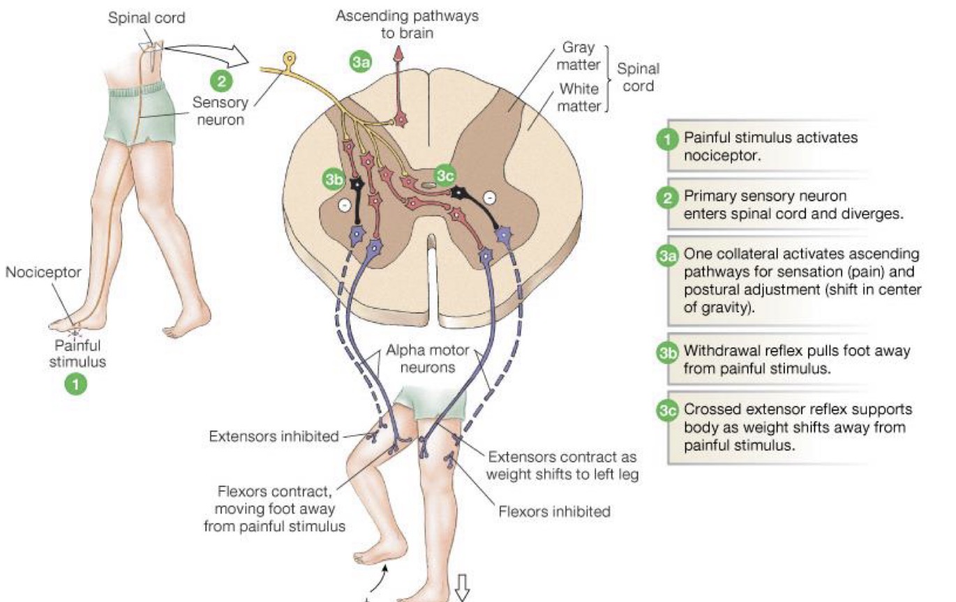
1) Free nerve ending detects painful stimulus
2) Sensory neuron sends info to spinal cord
3b) The leg that received the pain stimulus
Motor neuron to flexors gets facilitatory activation
Dorsiflexors, knee flexors, and hip flexors contract
Motor neurons to extensors are inhibited
Result: Leg is withdrawn from source of pain
However, to readjust weight, the flexor reflex occurs simultaneously with the extensor reflex
3c) Sensory neurons via interneurons synapse to motor neurons of opposite leg
Motor neurons of the extensors are stimulated and muscles contract
Flexors are inhibited
Result: Body weight is shifted and we stay upright with gravity
What is kinesthetic awareness?
When would we not have controlled movements?
Kinesthetic proprioception is like? Serves as a foundation to?
The conscious awareness of body position (or joint position) and the amount & rate of
without rapid transmission of sensory→ integrative→ and motor commands we would not have controlled movements.
Kinesthetic proprioception is like muscle memory, which serves as the foundation of voluntary motor skill learning and movement overall.
What is reciprocal inhibition?
Is it automatic? When is it not?
Agonists are stimulated by motor neurons to contract, and the antagonists are simultaneously and reciprocally inhibited.
This is automatic in reflexes and familiar movements. However, when learning a new skill or when a skill is complex, it is consciously controlled to achieve more skilled performance.
What is coactivation/reciprocal activation?
When does it happen (2)? What is happening?
When the opposite muscle group is stimulated to contract.
May happen when an unfamiliar motor task is performed, so more muscle than necessary contracts (requiring more energy).
May also happen to maintain joint stiffness, either for a movement or injury.
When does coactivation decrease?
Coactivation decreases as person learns to do reciprocal inhibition. The persons movements get more efficient (requiring less energy).