Bones and Joints
 Call Kai
 Learn
 Practice Test
 Spaced Repetition
 Match
 Flashcards
 Knowt Play
1/141

There's no tags or description
Looks like no tags are added yet.
142 Terms
Long bones
Greater in length that width
ex: bones of arm, forearm, metacarpal bones, phalanges
Short bones
length nearly equal to width
ex: bones of wrist
Bones are
the primary organ of the skeletal system
Skeletal system consists of
Bones, cartilage, and ligament
Flat bones
flat, thin surfaces, may be slightly curves
ex: some cranial bones and scapula
Irregular bones
complex shapes
ex: vertebrae
Axial skeleton
Bones along central body axis, provides support and protection
Appendicular skeleton
Bones of upper and lower limbs, facilitates movement and mobility
Most complex structure of the skeleton and how many bones it’s made of
Skull, make of 22 bones
Neurocranium bones
8 bones of this type
form the rounded cranium and surround the brain
Frontal bone, 3 parts
squamous part (flat)= forehead
orbital part= roof of orbits (where eyes are)
nasal parts= articulation for nasal bones and maxilla (arrow shaped)
Parietal bone
quadrilateral in shape on the top and sides of skull

Frontal bone

Parietal bone
What key structure does the occipital bone contain
Foramen magnum

Occipital bone
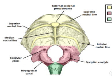
What are the 3 sections in the occipital bone?
Squamous, condylar, basilar (yellow, red, green resp)
Describe the locations of the occipital squamous, condylar, and basilar parts
squamous= posterior to magnum foramen
condylar= lateral to magnum foramen
basilar= anterior to magnum foramen

Ethmoid
Ethmoid is
Roof and nasal septum of nasal cavity
Describe the cribriform and perpendicular plates
Cribriform plate= horizontal part, roof of nasal cavity
Perpendicular plate= vertical part, superior 2/3 of nasal septum

Name the red, green, and gray side structures
Red= cribriform plate
Green= perpendicular plate
Gray side structures= ethmoidal labyrinth
What is the spenoid
Base and side of skull
Describe the 2 greater and lesser wings, and the 2 pterygoid processes
2 greater wings= large, flat projection that extend laterally from the body
2 lesser wings= smaller, upward projection from the body
2 pterygoid processes= downward projections from the junction of body and greater wings
What is the temporal bone
Sides of skull

Name the red part
Squamous (mandibular fossa)

Name the blue part
Zygomatic process (articulates with zygomatic bone)

Name the green part
Tympanic part (surrounds the external opening of the external auditory meatus)

Name the purple part
Petromastoid

Name the yellow part
Styloid process
Describe the petrous part of temporal bone
can only be viewed internally
pyramid shapes
internal auditory meatus
Viscerocranium, how many bones and function
made of 14 bones
Forms the face, oral cavity (supports teeth), and parts of nasal cavities
attachment sites for muscles of facial expression and chewing

Zygomatic: prominence of cheeks and lateral wayys of orbits
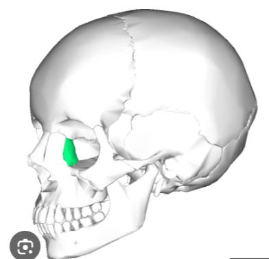
Lacrimal bone: medial wall of orbits (smallest bone in face)
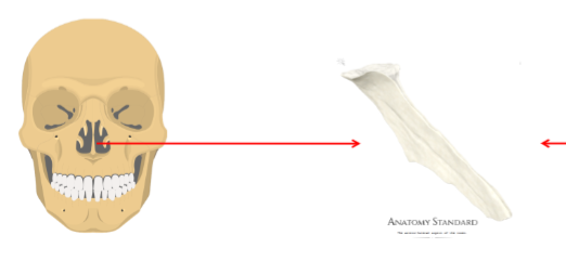
Vomer: forms the hinder and lower part of the nasal septum

Palatine: between left and right maxilla, forms lateral wall and floor of nasal cavity, and roof of oral cavity

Nasal bones

Inferior nasal conchae (inferolateral wall of nasal cavity)
4 processes of maxilla
Frontal, alveolar, palatine (anterior part of hard palate), zygomatic process

Upper= frontal process
Lower= alveolar process
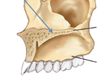
(black lines)
Upper= Palatine process
Lower= Alveolar process
Body of mandible
Body=horizontal portion
Left and right ramus
2 processes of mandible
Coronoid process and condylar process (articulates with mandibular fossa of temporal bone)

Condylar and coronoid process (resp, L-R)
Hyoid function
Attachment site for anterior neck muscles

Green= greater horn
Yellow= lesser horn
Red= body
Which articulation processes form the zygomatic arch
Temporal process of zygomatic bone and zygomatic process of temporal bone

Zygomatic bone

Foramen magnum
Cervical vertebrae
bones of neck
C1-C7
C2 articulates with occipital condyles
Thoracic vertebrae
Forms upper back region
Each articulates laterally with 1-2 pairs of ribs
T1-T12
Lumbar vertebrae
Form lower back region
L1-L5
Sacrum vertebrae region
5 fused vertebrae, S1-S5
Fused by late 20s
laterally articulates 2 hip bone
Coccyx (tailbone)
formed by 4 coccygeal vertebrae, Co1-Co4
start uniting during puberty, does not bear upper body weight
provides attachment points for muscles
4 spinal curvatures
Cervical= forward convex
Thoracic= backward convex
Lumbar= forward convex
Sacral= backward convex
Spinal curvature function
gives column some flexibility
supports weight of body and acts as natural shock absorber
Atlas (C1) functions and characteristics
supports the head, allows for nodding motion
lacks body and spinous process
anterior and posterior arches connect lateral masses
Articular facet for dens
On inner side of C1 anterior arch

Articular facet for dens
Axis (C2) functions and characteristics
produces dens
acts as atlas pivot, allows shaking head (‘no‘)

Dens
Vertebra prominens (C7) features
transition from cervical to thoracic region
longer spinous process (easily palpated)
Key features of cervical vertebrae
have transverse foramen
have bifurcated spinous processes (except for C7)
Thoracic vertebrae key features
Heart shaped bodies
Costal facet on lateral side of body and on sides of transverse processes (except T11, T12)
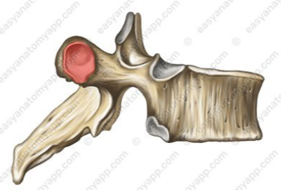
Costal facet

Thoracis spinous process (longer, angled downwards)
Lumbar vertebrae key characeristics
largest vertebrae
thicker, oval body
shorter spinous body that is perpendicular to body
bears most of the upper body weight

Spinous process of lumbar vertebra (perpendicular to body)
What is the sacrum
Anteriorly curved, triangular bone
forms posterior wall of pelvic cavity

Ala (wing of sacrum)

Superior articular process

Base of sacrum

Apex of sacrum

Auricular surface of sacrum (ear shaped on upper-lateral part of sacrum)

Anterior and posterior sacral foramina (8 total)

Transverse ridges of sacrum

Medial crest of sacrum

Sacral canal:
Whose ending is bound by sacral hiatus
Has bony projections called sacral cornua

Sacral cornu= left (landmark for epidural injection)
Sacral hiatus= upper
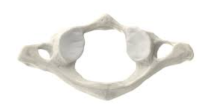
C1
Manubrium
Widest and most superior portion of sternum
has 2 clavicular notches


Jugular notch (shallow superior depression between clavicular notches)

Costal notches (articulation points for ribs)
Sternum body characteristics
longest portion of sternum
Costal notches for ribs 2-7

Sternal angle
Where manubrium and body meet
where cartilage of rib 2 attaches
key landmark in palpitation and counting ribs

Xiphoid process (projection at sternum tip)
True ribs
ribs 1-7
connect individually to costal cartilages
False ribs
ribs 8-12
not directly attached to sternum
Costal cartilage of ribs 8-10
fused to costal cartilage of rib 7
indirectly attached to sternum
Floating ribs
Ribs 11-12
without connection
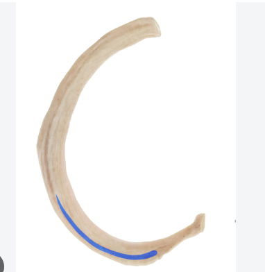
Costal groove of rib
marks path of nerves and blood vessels
Angle of rib
Where shaft begins to curve

Tubercle of rib (small knot-like projection below neck)

Neck of rib

Head of rib
Pectoral girdle consists of, and functions to
clavicles and scapulae
supports upper limbs
Clavicle functions and structure
Stabilizes shoulder joint
sternal end: roughly pyramidal
Acromial end: broad and flattened


Spine of scapula
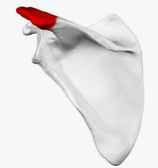
Acromion of scapula

Supraspinous fossa of scapula