CHAPTER 13: DYNAMICS OF CHANGE
 Call Kai
 Learn
Learn Practice Test
 Spaced Repetition
 Match
Match Flashcards
 Knowt Play
1/77

Earn XP
Description and Tags
sas + ppt
78 Terms
ectoderm origin
formative tissue: dental organ (epithelial)
final structure: enamel
mesoderm origin
formative tissue 1 → dental papilla
final structure → dentin & pulp
formative tissue 2 → dental sac
final structure: cementum and PDL
3 phases of gestation (lasts for 9mo)
period of ovum
period of embryo
period of fetus
period of ovum
0-14 days
fertilization to implantation
period of embryo
2-8 weeks, all major organs appear
most important because of the cell differentiation that occurs during this time
period of fetus
8-40 weeks
maturation of the newly formed organs
body changes
no year is more dramatic in terms of growth than the first year of life
most children undergo 50% increase in length, 200% increase in weight
toward the end of the first year of life → growth rate slows
age 2 → good motor skills to run, climb, walk up, down steps
birth-3y.o → the nervous system of the child grows dramatically
5 mesenchymal elevations constitute the initial feature of the face:
frontonasal process
2 maxillary process
2 mandibular process
[happens during 4th week in utero}
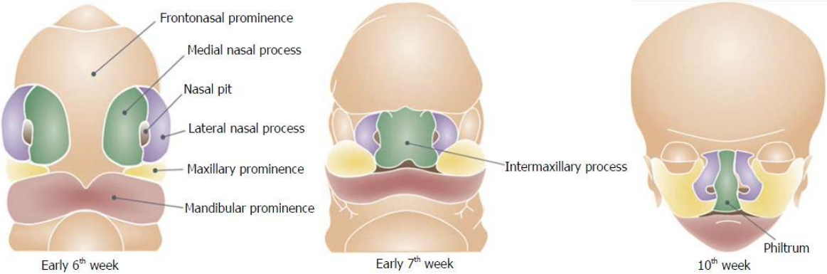
frontnasal + lateral nasal process
the fusion forms upper lip
failure to fuse may cause cleft
medial nasal process
failure to fuse may cause medial nasal cleft (hair lip)
maxillary process + mandibular process
the fusion forms the cheek
failure to fuse may cause oblique facial cleft
maxillary process + lateral nasal process
failure to fuse may cause transverse facial cleft
embryonic stage of leapfrog pattern
mandible is considerably larger than the maxilla
early fetal stage of leapfrog pattern
maxilla accelerates, outgrowing the mandible
11 weeks of leapfrog pattern
mandible surges, equaling the maxilla in size
13-20 weeks of leapfrog pattern
mandible lags again
at birth of leapfrog pattern
the infant is born with a mandible that is retrognathic (receded) relative to the maxilla
cranial vault
closer to adult size than any other part of the head
the 6 membrane-filled fontanel gaps allow compression during birth and close by ossification within 2 years
remodelling
driven by cellular mechanics:
osteoblast (bone forming) build up tissue while osteoclast (bone destroying) resorb it, expanding the jaw to accommodate teeth and sinuses
fontanels
close by ossification within 2 years after birth
refer to the gaps that separate the bones that compose the cranium bc these are not fused
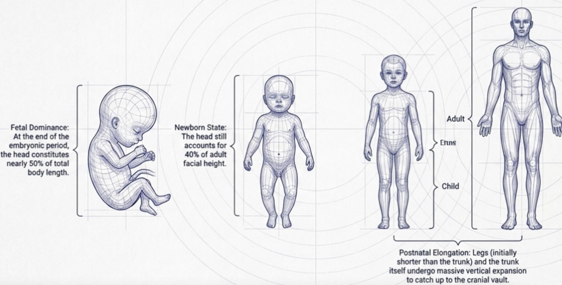
face of the newborn
tiny mouths
face appears broad and flat
underdeveloped and receded lower jaw
total face height is only 40% of adult face height
dental changes
teeth are formed by tissues originating from both ectoderm and mesoderm
ectoderm responsible for the future enamel
mesoderm responsible for the future pulp-dentin
3 formative tissues:
dental organ → forms the enamel
dental papilla → forms the dentin and pulp
dental sac → forms the periodontium (cementum, alveolar bone, PDL)
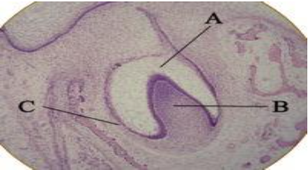
initiation
aka: bud stage
first noticed in the 6-week old fetus
expansion of the basal layer above the basement membrane

proliferation
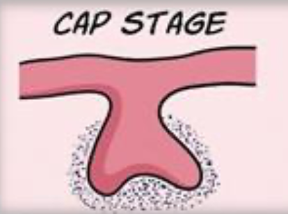
aka: cap stage
9-10 weeks in utero
the multiplication of the cells of the initiation stage and an expansion of the tooth bud → tooth germ.
this stage is characterized as the tooth germs continues to proliferate it produces as caplike appearance
histodifferentiation
11-12 weeks in utero, cells begin to specialize
this occurs for primary dentition between the 11th-12th week of prenatal development
it is characterized by the continuation of the process of proliferation, differentiation, morphogenesis
the cap continues to grow and begins to look like a bell (hence the name bell stage)

6 odontogenic steps
elongation of IEE → reciprocal induction
differentiation of odontoblasts → reciprocal induction
deposition of dentin → bulk of tooth
deposition of enamel
root formation
cementum, pdl formation
4 types of cells within the enamel/dental organ produced by differentiation
inner enamel epithelium
outer enamel epithelium
stellate reticulum
stratum intermedium

inner enamel epithelium
concave part
defines the shape of the crown
becomes ameloblasts (enamel producing) → bell stage

outer enamel epithelium
convex part
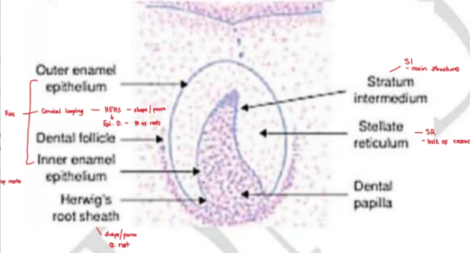
stellate reticulum
bulk of dental organ
network of star-shaped central core
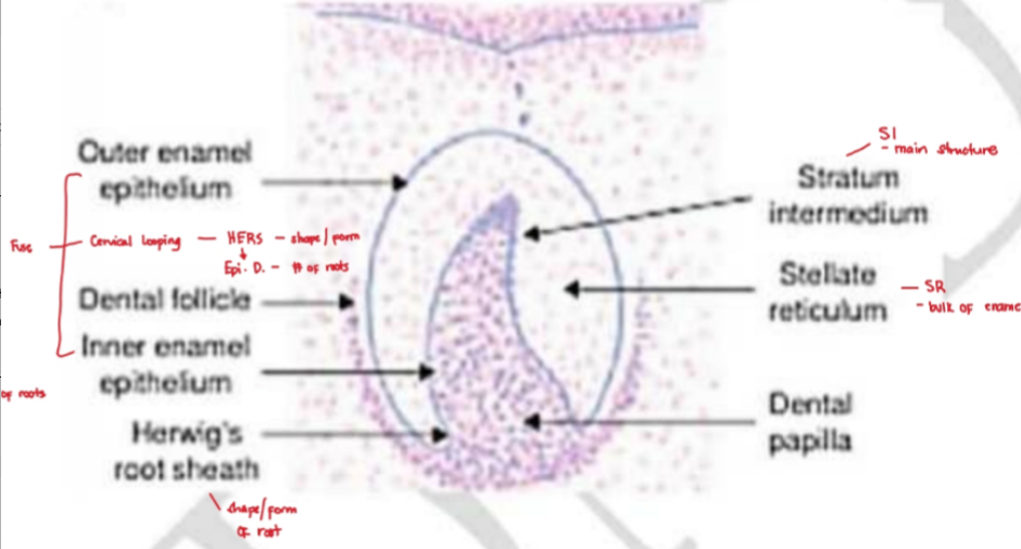
stratum intermedium
nourishes IEE
main structure for enamel formation

fuse of OEE-IEE
root development
collapse → reduced enamel epithelium
cervical loop of HERS → defines the shape/form of root
most apical segment of HERS (epithelial diaphragm) → defines the # of roots
1 → mn-mx anterior ; mn 1PM ; mn-mx 2PM
2 → mx 1PM ; mn molars
3 → mx molars
clinical anomalies in morphodifferentiation
morphological failure → peg teeth, microdontia
amelogenesis imperfecta → failure of ameloblasts to differentiate properly
dentinogenesis imperfecta → failure of odontoblasts to differentiate properly
morphodifferentiation
aka: advanced bell stage
stage at which the cells find an arrangement that ultimately dictates the final size and shape of the tooth
disturbances in this stage will result in abnormal forms & sizes of teeth (ex: peg teeth, microdontia & macrodontia)
korff’s fibers
irregular spiraling fibers from deep in the pulp that entangle with the reticular fibrils from the mesenchyme of the pulp
these long spiraling fibers assist in the structural support of the developing dentin.
apposition
occurs when the network or the tissue matrix of the tooth is formed
the organized special tissues now deposit incremental layers of enamel and dentin matrix
calcification
4% organic material and water
96% inorganic( calcium, phosphorus)
influx of mineral salts within the previously developed tissue matrix
eruption
the process of elimination of primary teeth is caused by the eruptive pressure of the permanent successor at the apex of the primary tooth and its surroundings
HERS → responsible for the size and shape of the root and eruption of tooth
tooth root is approximately 1⁄2-2/3 of its final length at the time of gingival emergence
mn incisors → are the first primary tooth to erupt
2 ½ - 3 y.o → 20 primary teeth are completed
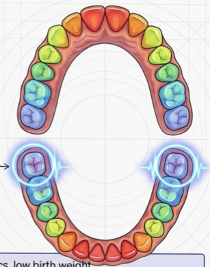
permanent 1st molars → the only teeth that show a trace of hard tissue formation at birth
3 phases of eruption:
preeruptive phase
eruptive phase (prefunctional)
eruptive phase (functional)
engine of eruption
HERS
root formation
bone remodelling
cellular proliferation
HERS
dictates the size/shape of root and drives eruption
root formation
pushes against the base of the crypt (root is only ½ - 2/3 final length at emergence)
eruption heatmap of primary dentition
6-month start → eruption sequence beings with the primary mn central incisor
age 2.5-3yrs finish → all 20 primary teeth have fully erupted into the oral cavity
6-7 y.o → 1st permanent molars (the only permanent teeth that show a trace of hard tissue formation at the exact moment of birth)
sequence highlights:
6-7 y.o: mn central incisor & first molars
9-12 y.o: canines & premolars
11-13 y.o: 2nd molars
delays in this sequence can be tied to:
genetics, low birth weight, nutritional deficiencies
specific syndromes (down syndromes, regional odontoplasia)
cellular proliferation
connective tissue of the dental papillae expands
causes of tooth eruption
root formation
growth of the jaw
proliferation of HERS
pressure from muscular action
apposition and resorption of bone
pressure at the apical part of the tooth
proliferation of the connective tissue of the dental papillae
attrition
wearing of teeth during function
normal physiologic process when teeth occlude
exfoliation
eruptive pressure stimulates the osteoclasts (bone destroying cells)
caused by eruptive pressure of the permanent successor at the apex of the primary tooth and its surroundings
nolla’s stages of tooth calcification
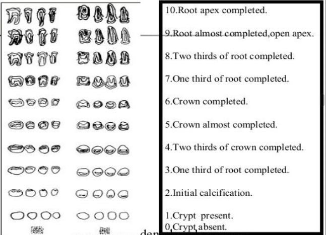
nolla’s 6 stage
crown completed
eruptive movement begins
nolla’s 7 stage
1/3 root completed
alveolar crest pierced
nolla’s 8 stage
2/3 root completed
gingival margin pierced
primary dentition development up to age 3
age 3 1⁄2 to 4 months in utero → 1st permanent molars (the only permanent teeth that show a trace of hard tissue formation)
6 weeks in utero → primary teeth begin to form
at birth → 1st molar (first tooth to show tooth germ formation)
6 mos. → primary mn incisors erupts (first primary tooth to erupt)
1 y.o → the enamel of all the primary teeth is usually completed
age 2 → all primary teeth erupted
eruption age of primary dentition

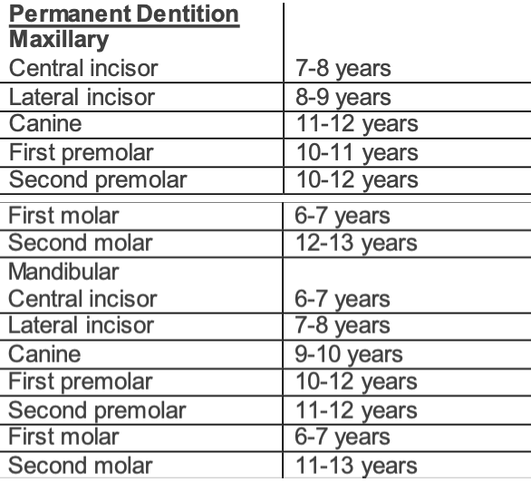
eruption age of permanent dentition
4 areas of cognitive development by Mussen&coworkers
area of perception → young infants only perceive movements, facial relationships, color
recognition of information → infants have the ability to recognize the similarity of new objects compared with old ones
ability to categorize → ability to group things together by shape, color and use as early as 1 year of age
enhancement of memory → even young infants have been shown to have some memory and recall past experiences
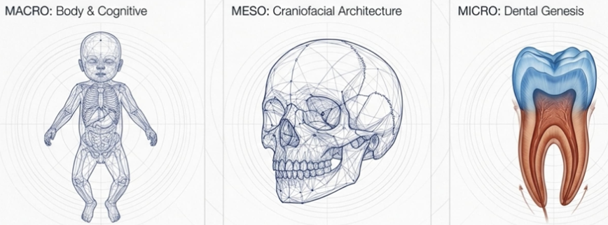
the developmental triad
the first 3 years of human life → most dramatic biological remodelling an individual will ever undergo
this atlas examines that transformation by zooming inward; from staggering mathematics of whole-body growth into the intricate assembly of the face and finally down to the precise cellular choreography of tooth formation
note:
macro → body & cognitive
meso → craniofacial architectures
micro → dental genesis
by the end of year 2:
75% adult brain capacity
brain achieves three-quarters of its adult weight
motor skills of a 6.0 mo
sits alone for 30+ seconds
motor skills of a 8.9 mo
neat pincer grasp
motor skills of a 11.7 mo
walks alone
motor skills of a 24 mo
runs, climbs stairs with both feet, kicks a ball
cognitive development of a 0-4 mo
tracks objects
primary circular reactions
cognitive development of a 9-12 mo
first real words
object permanence
finds hidden objects
cognitive development of a 12-18 mo
tertiary circular reactions
categorizes objects by shape/color
cognitive development of a 24 mo
telegraphic two-word sentences, symbolic play
cephalocaudal growth gradient
frontal-parietal fontanelles
anterior / bregma
close at 12-24 mos / 18 mos
last to close, diamond-shape
intersect of coronal & sagittal suture
frontal-parietal-sphenoidal-temporal fontanelles
close at 1-2 mo
sphenoidal / pterion
thiniest part of the skull
affects MMA → causes hematoma
parietal-occipital fontanelles
close at 2 mo
triangular-shaped
posterior / lambda
parietal-occipital-temporal fontanelles
close at 1-2 mo
mastoidal / asterion
intersect of sagittal and lambdoid suture
2 frontal suture
metopic
frontal-parietal suture
coronal
2 parietal suture
sagittal
parietal-temporal suture
squamosal
parietal-occipital suture
lambdoid
korff’s fibers
long spiraling fibers from deep pulp mesenchyme
entangle to support the developing dentin
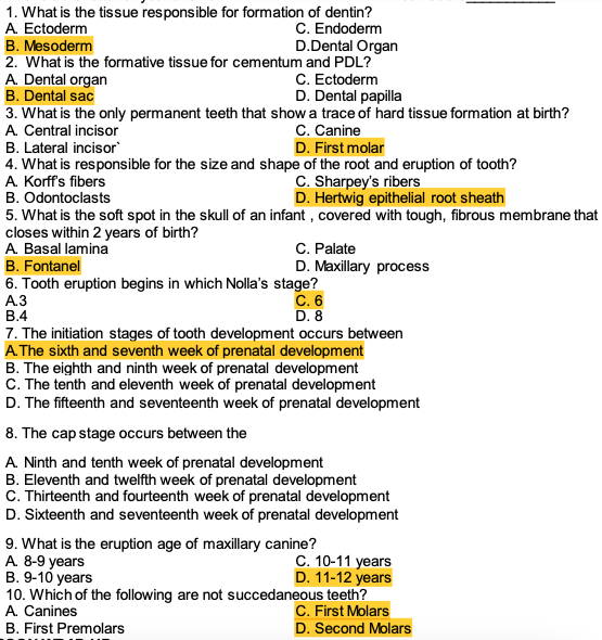
practice question in sas