Blood Flow and Abdominal Muscles - Anatomy Practical 1
 Call Kai
 Learn
 Practice Test
 Spaced Repetition
 Match
 Flashcards
 Knowt Play
1/41

Earn XP
Description and Tags
NAU OTD
42 Terms

Superior Vena Cava
large vein that carries deoxygenated blood from the upper body to the heart, specifically into the right atrium.

Inferior Vena Cava
large vein that carries deoxygenated blood from the lower body to the heart, specifically into the right atrium.

Aorta
the largest artery in the body, responsible for carrying oxygenated blood from the heart to the rest of the body.
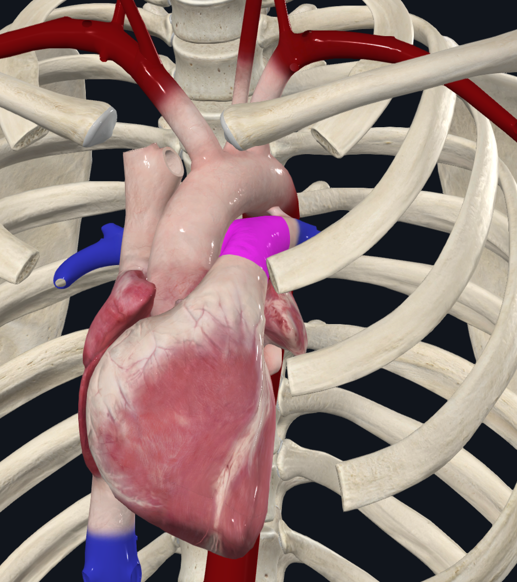
Pulmonary Trunk
Carries deoxygenated blood from the heart to the lung
Right Lung
3 lobes, oblique fissure, horizontal fissure
Left Lung
2 lobes, Oblique fissure, cardiac notch for the heart

Trachea
Connects larynx to brachial tree to allow for airflow inbetween
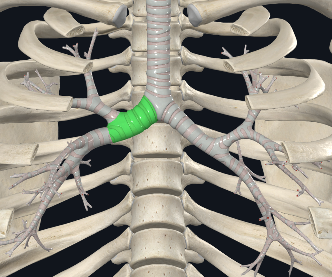
Right Main Bronchus
Shorter, wider, more vertical

Left Main Bronchus
Narrower, twice as long as R, travels more horizontal

Secondary (Lobar) Bronchi
They branch directly from the left and right main (primary) bronchi and carry air into the specific lobes of the lungs

Tertiary (Segmental) Bronchi
They branch off from the secondary bronchi and supply air to the individual bronchopulmonary segments inside each lung lobe

Alveoli
Maximum gas exchange happens between the blood and the respiratory system
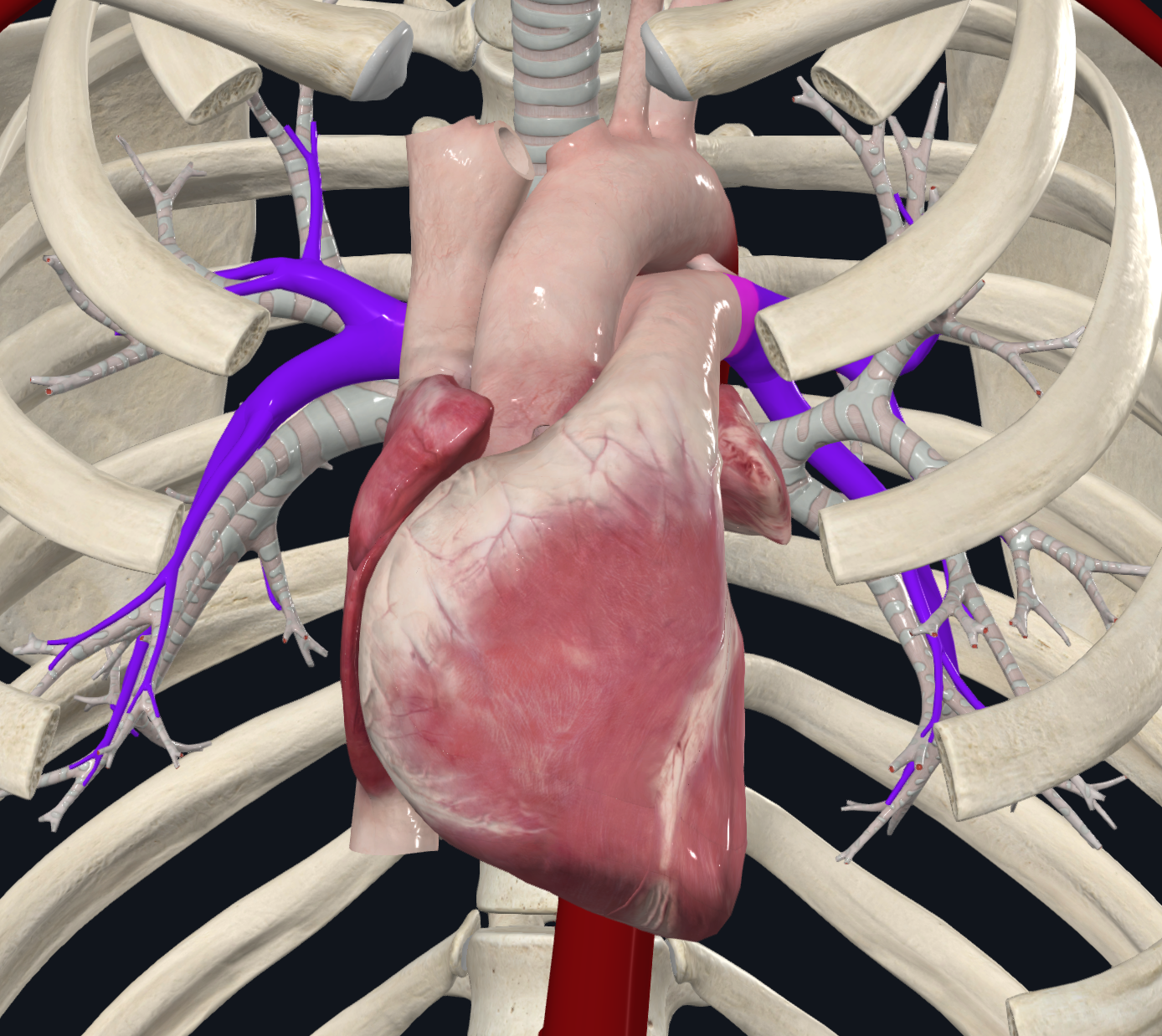
Pulmonary Arteries
Carry deoxygenated blood from the heart to the lungs for oxygenation
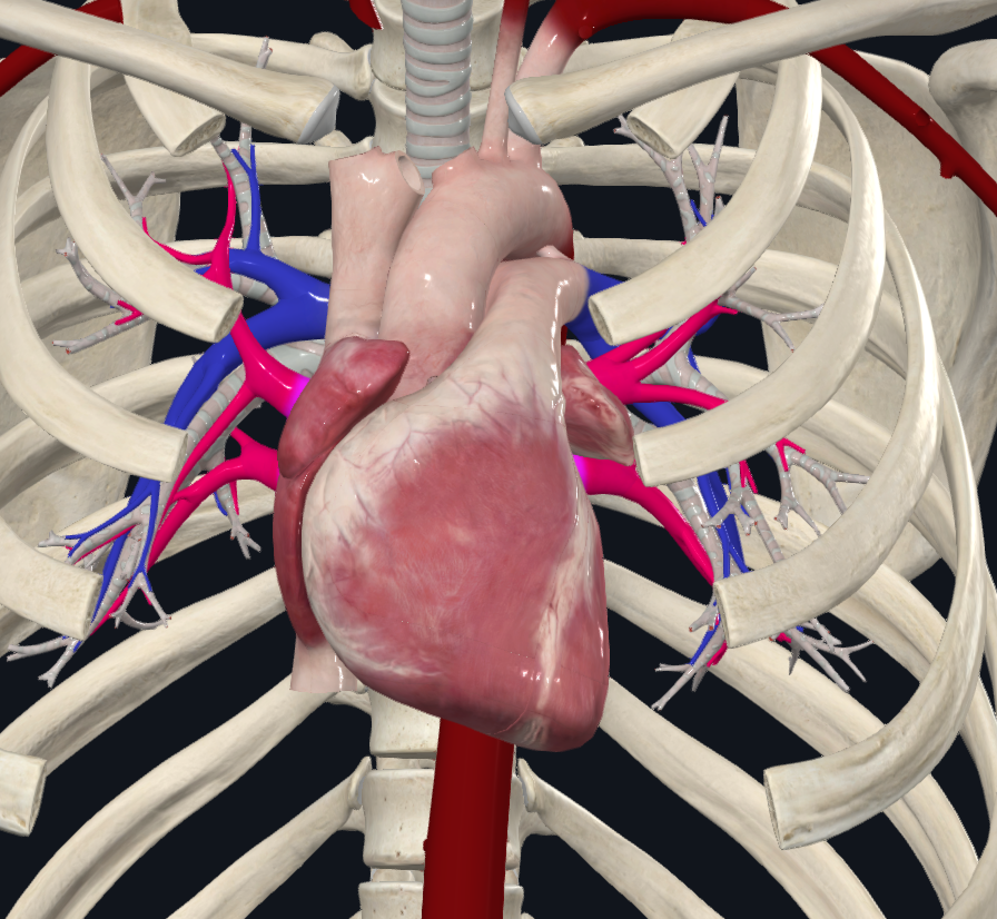
Pulmonary veins
Carry oxygenated blood from the lungs back to the heart
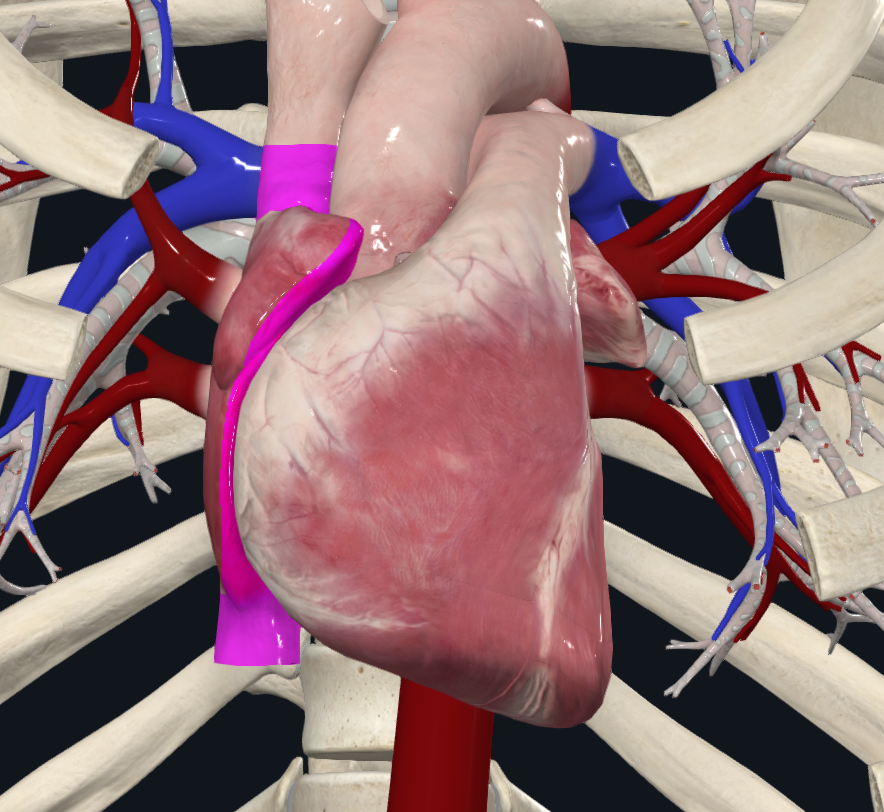
Right Atrium
receives deoxygenated blood via superior and inferior vena cava

Left Atrium
Receives oxygenated blood from the pulmonary veins

Right Ventricle
gets blood from the right atrium through the tricuspid valve

Left Ventricle
Gets blood from the left atrium through the mitral (bicuspid) valve

Tricuspid Valve
prevents backflow from the R ventricle back to the R atrium

Mitral (Bicuspid) Valve
prevents backflow from the L ventricle back to the L atrium

Pulmonary Valve
Separates R ventricle and pulmonary trunk that permits unidirectional blood flow

Aortic Valve
Permits unidirectional (oxygenated) blood flow from the left ventricle to the aorta

R Coronary Artery
Supplies R atrium, majority of R ventricle, inferior surface of L ventricle with oxygenated blood

L coronary artery
comes off of left side of aorta, supplies L atrium, majority of L ventricle, and a portion of the right ventricle with oxygenated blood

Circumflex Artery
Branch off L coronary artery - Contributes to blood supply of the L atrium and L ventricle

Great Cardiac Vein
Drains blood from the left side of the heart

Coronary sinus
Extension off of great cardiac vein and drains deoxygenated blood into the inferior vena cava

Diaphragm
Innervation: Phrenic N. (C3-C5)
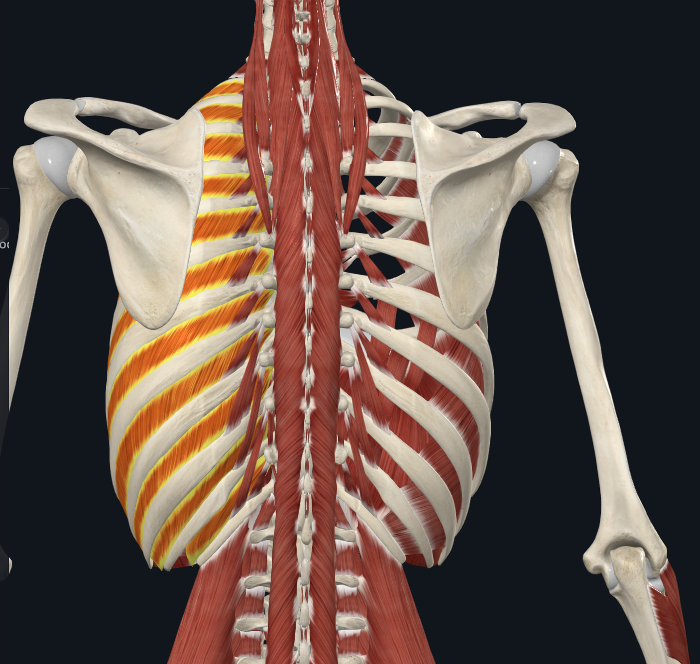
External Intercostal Muscles

Internal Intercostal Muscles

Sternum
Manubrium, body, xiphoid process
Articulates with clavicle and costal cartilages

xiphoid process
attachment site for diaphragm

Inguinal Ligament
superior iliac spine to pubic tubercle
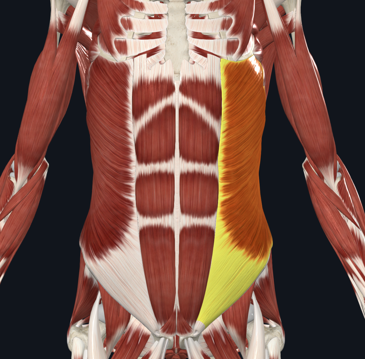
External Oblique
Origin: External surfaces and inferior borders of fifth to twelfth ribs
Insertion: Anterior superior iliac spine, anterior half of iliac crest, pubic crest, pubic tubercle, linea alba
Innervation: Anterior rami of seventh to twelfth thoracic nerves.
Action: Flexes, laterally flexes, and rotates trunk; compresses and provides structural support to adjacent abdominal structures

Internal Oblique
Action: Flexes, laterally flexes, and ipsilaterally rotates trunk

Rectus Abdominis
Action: Flexes trunk

Transverse Abdominis
Action: Compresses and provides structural support to adjacent abdominal structures.

Quadratus Lumborum
Action: Extends and laterally flexes trunk; stabilizes twelfth rib during inspiration.
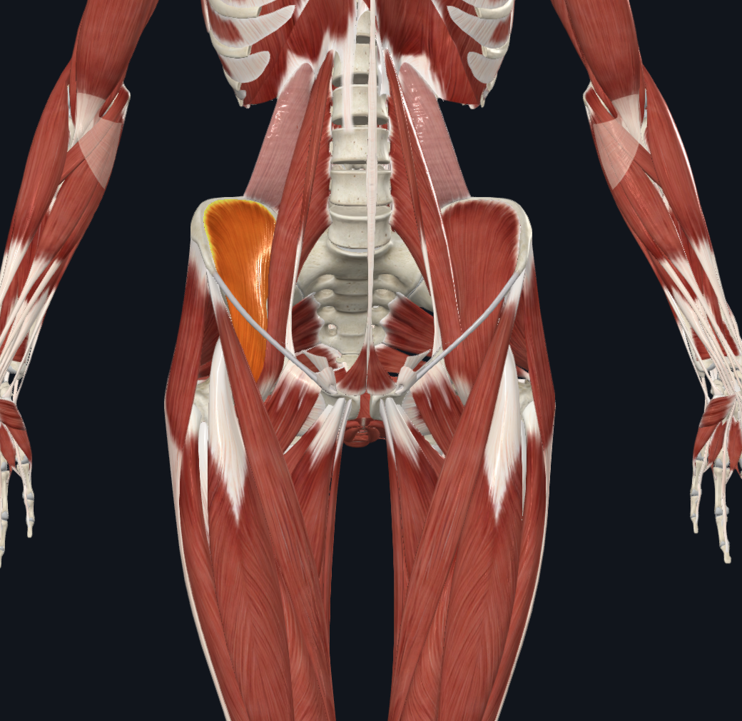
Iliacus
Origin: Iliac crest; superior two thirds of iliac fossa; ala of sacrum; anterior sacroiliac ligaments.
Insertion: Lesser trochanter of femur, via tendon of psoas major.
Action: Flexes thigh at hip joint; flexes trunk.
Innervation: Femoral nerve (L2–L3).
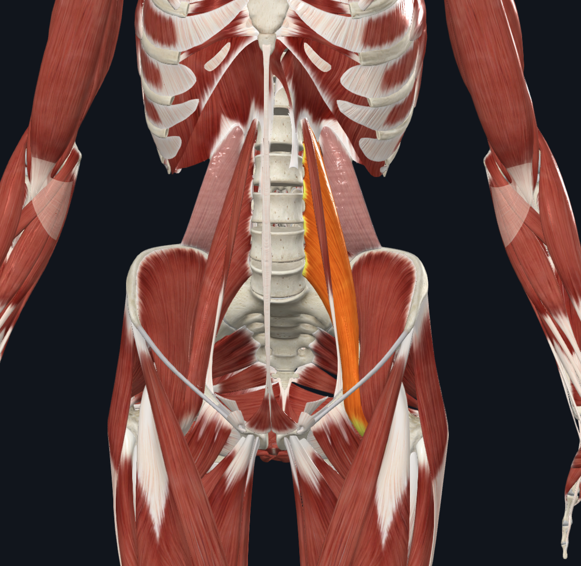
Psoas Major
Action: Flexes thigh at hip joint; flexes trunk.

Psoas Minor
**Not everyone has this**
Action: Assists in flexion of trunk.