The Cell Cycle

0.0(0)
Studied by 0 people
call kaiCall Kai
learnLearn
examPractice Test
spaced repetitionSpaced Repetition
heart puzzleMatch
flashcardsFlashcards
GameKnowt Play
Card Sorting

1/19

encourage image

There's no tags or description

Looks like no tags are added yet.

20 Terms

1
New cards

What is a genome? And how are chromosomes different in prokaryotes and eukaryotes?

Genome = all of the genetic material (DNA) in an organism. Within the genome, individual double-stranded DNA molecules are called chromosomes.

Prokaryotes generally have one circular chromosome, while eukaryotes generally have multiple linear chromosomes. There are exceptions to these general rules. Mitosis

<p>Genome = all of the genetic material (DNA) in an organism. Within the genome, individual double-stranded DNA molecules are called chromosomes. </p><p>Prokaryotes generally have one circular chromosome, while eukaryotes generally have multiple linear chromosomes. There are exceptions to these general rules. Mitosis</p>
2
New cards

What is chromatin? What is its function?

Chromatin = DNA + associated proteins, including histones.

  • Histones help organize and package DNA and can influence gene expression (which genes are used/turned on).

DNA wraps around these proteins to help organize it. Chromosomes are made of chromatin.

A chromosome is one organized DNA molecule + its proteins, whereas chromatin is the DNA-protein material making up chromosomes.


<p><strong>Chromatin = DNA + associated proteins</strong>, including <strong>histones</strong>. </p><ul><li><p>Histones help <strong>organize and package DNA</strong> and can <strong>influence gene expression</strong> (which genes are used/turned on).</p></li></ul><p>DNA wraps around these proteins to help organize it. <strong>Chromosomes are made of chromatin.</strong></p><p>A chromosome is one organized DNA molecule + its proteins, whereas chromatin is the DNA-protein material making up chromosomes.</p><p></p><p></p>
3
New cards

Explain Chromosome Structure & Sister Chromatids (all parts)

Most of the cell's life, chromosomes exist as long, thin chromatin fibres and are not easily seen individually.

After DNA replication, each chromosome has 2 genetically identical copies called sister chromatids, which remain attached.

Cohesins hold them together along their full length, especially at the centromere. The centromere is a region of highly repetitive DNA; each sister chromatid has its own centromere.

Chromatid arms extend away from the centromere. During mitosis, chromatin becomes highly condensed.

Important: 2 attached sister chromatids still count as 1 chromosome. Once they separate in anaphase, each becomes an individual chromosome.

<p>Most of the cell's life, chromosomes exist as <strong>long, thin chromatin fibres</strong> and are not easily seen individually. </p><p>After <strong>DNA replication</strong>, each chromosome has <strong>2 genetically identical copies called sister chromatids</strong>, which remain attached. </p><p><strong>Cohesins</strong> hold them together along their full length, especially at the <strong>centromere</strong>. The <strong>centromere</strong> is a region of highly repetitive DNA; each sister chromatid has its own centromere. </p><p><strong>Chromatid arms</strong> extend away from the centromere. During mitosis, chromatin becomes highly <strong>condensed</strong>. </p><p><strong>Important:</strong> 2 attached sister chromatids still count as <strong>1 chromosome</strong>. Once they separate in anaphase, each becomes an <strong>individual chromosome</strong>.</p>
4
New cards

What is the Cell Cycle? Explain interphase and the M Phase / Mitotic Phase.

Cell cycle = ordered sequence from cell birth → DNA replication → division into 2 cells.

It has Interphase + M phase.

Interphase ≈ 90% of cycle and includes

G1: cell growth + intense metabolic activity/preparation;

S: continued growth + DNA replication, making an identical genome copy;

G2: more growth + metabolic activity/preparation.

M phase: mitosis divides genetic material + cytokinesis divides cytoplasm → 2 cells with full, identical genomes
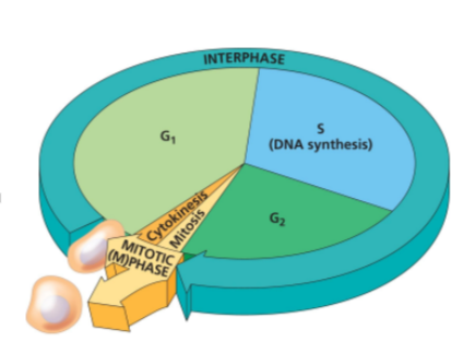
<p><strong>Cell cycle</strong> = ordered sequence from <strong>cell birth → DNA replication → division into 2 cells</strong>.</p><p>It has <strong>Interphase + M phase</strong>.</p><p><strong>Interphase</strong> ≈ 90% of cycle and includes </p><p><strong>G1:</strong> cell growth + intense metabolic activity/preparation; </p><p><strong>S:</strong> continued growth + <strong>DNA replication</strong>, making an identical genome copy; </p><p><strong>G2:</strong> more growth + metabolic activity/preparation. </p><p><strong>M phase:</strong> <strong>mitosis</strong> divides genetic material + <strong>cytokinesis</strong> divides cytoplasm → <strong>2 cells with full, identical genomes</strong></p>
5
New cards

Overall Cell Cycle process

Overall Process 

G1 → cell grows 

↓ 

S → DNA is replicated 

↓ 

G2 → cell grows + prepares for division 

↓ 

Mitosis → genetic material divides 

↓ 

Cytokinesis → cytoplasm divides 

↓ 

2 genetically identical cells 

6
New cards

Explain the structure of Mitosis & the Mitotic Spindle.

Mitosis divides the nucleus/genetic material so the 2 daughter cells receive identical genetic material.

5 stages: Prophase → Prometaphase → Metaphase → Anaphase → Telophase.

The mitotic spindle begins forming in prophase and consists of microtubules + associated proteins that move/separate chromosomes. In animal cells, spindle microtubules form from 2 centrosomes; each centrosome contains a pair of centrioles, **although centrioles are not essential for mitosis.

Asters are short microtubule arrays around each centrosome.

Mitotic spindle = centrosomes + asters + spindle microtubules.

Don't confuse centrosome (organizes spindle) with centromere (chromosome region).

<p><strong>Mitosis</strong> divides the nucleus/genetic material so the 2 daughter cells receive <strong>identical genetic material</strong>. </p><p>5 stages: <strong>Prophase → Prometaphase → Metaphase → Anaphase → Telophase</strong>. </p><p>The <strong>mitotic spindle</strong> begins forming in prophase and consists of <strong>microtubules + associated proteins</strong> that move/separate <strong>chromosomes</strong>. In animal cells, spindle microtubules form from 2 centrosomes; each <strong>centrosome </strong>contains a pair of <strong>centrioles</strong>, **although centrioles are not essential for mitosis. </p><p><strong>Asters</strong> are short microtubule arrays around each centrosome. </p><p><strong>Mitotic spindle = centrosomes + asters + spindle microtubules.</strong> </p><p>Don't confuse <strong>centrosome</strong> (organizes spindle) with <strong>centromere</strong> (chromosome region).</p>
7
New cards

What happens right before mitosis in the G2 phase?

By the end of G2, the original centrosome has duplicated → 2 centrosomes.

DNA was already replicated during S phase, so each chromosome consists of 2 genetically identical sister chromatids.

However, chromosomes are not condensed yet; the chromatin is still loose.

Flow: S phase copies DNA → G2 has 2 centrosomes + replicated, uncondensed chromosomes → mitosis begins.

<p>By the <strong>end of G2</strong>, the original centrosome has duplicated → <strong>2 centrosomes</strong>. </p><p>DNA was already replicated during S phase, so each chromosome consists of <strong>2 genetically identical sister chromatids</strong>. </p><p>However, chromosomes are <strong>not condensed yet</strong>; the chromatin is still loose. </p><p><strong>Flow:</strong> S phase copies DNA → G2 has 2 centrosomes + replicated, uncondensed chromosomes → mitosis begins.</p>
8
New cards

Explain Step 1: Prophase

Chromosomes condense, becoming tightly packed and visible. Sister chromatids remain attached by cohesins and still count as one chromosome.

The nucleolus disappears (normally responsible for rRNA transcription and ribosome assembly). The mitotic spindle begins forming, and the 2 centrosomes move toward opposite poles.

Key: chromosomes condense + nucleolus disappears + spindle forms + centrosomes move apart.

<p>Chromosomes <strong>condense</strong>, becoming tightly packed and visible. Sister chromatids remain attached by <strong>cohesins</strong> and still count as <strong>one chromosome</strong>. </p><p>The <strong>nucleolus disappears</strong> (normally responsible for rRNA transcription and ribosome assembly). The <strong>mitotic spindle begins forming</strong>, and the <strong>2 centrosomes move toward opposite poles</strong>. </p><p><strong>Key:</strong> chromosomes condense + nucleolus disappears + spindle forms + centrosomes move apart.</p>
9
New cards

Explain Step 2: Prometaphase

Chromosomes condense further and the nuclear envelope breaks down.

A kinetochore forms on the centromere of each sister chromatid. Therefore, one replicated chromosome has 2 sister chromatids → 2 centromeres → 2 kinetochores.

Kinetochore microtubules attach to kinetochores, with each sister chromatid connected to an opposite pole, preparing them to separate.

Nonkinetochore microtubules do not attach to chromosomes; they extend toward the middle and overlap/interact with microtubules from the opposite pole

<p>Chromosomes <strong>condense further</strong> and the <strong>nuclear envelope breaks down</strong>. </p><p>A <strong>kinetochore</strong> forms on the centromere of each sister chromatid. Therefore, one replicated chromosome has <strong>2 sister chromatids → 2 centromeres → 2 kinetochores</strong>. </p><p><strong>Kinetochore microtubules</strong> attach to kinetochores, with each sister chromatid connected to an <strong>opposite pole</strong>, preparing them to separate. </p><p><strong>Nonkinetochore microtubules</strong> do not attach to chromosomes; they extend toward the middle and <strong>overlap/interact with microtubules from the opposite pole</strong></p>
10
New cards

Explain Step 3: Metaphase

Kinetochore microtubules from opposite poles pull chromosomes back and forth in a tug-of-war until all chromosomes align at the metaphase plate, an imaginary plane halfway between the spindle poles.

The 2 centrosomes are at opposite poles, asters contact the plasma membrane, and each chromosome is connected to both poles through kinetochore microtubules.

Memory: Metaphase = chromosomes in the Middle.

<p>Kinetochore microtubules from opposite poles pull chromosomes back and forth in a <strong>tug-of-war</strong> until all chromosomes align at the <strong>metaphase plate</strong>, an imaginary plane halfway between the spindle poles. </p><p>The <strong>2 centrosomes are at opposite poles</strong>, asters contact the plasma membrane, and each chromosome is connected to <strong>both poles through kinetochore microtubules</strong>. </p><p><strong>Memory: Metaphase = chromosomes in the Middle.</strong></p>
11
New cards

Explain Step 4: Anaphase

Sister chromatids separate and move toward opposite poles.

Separase cleaves the cohesins holding them together. Once separated, each chromatid becomes an individual chromosome, so chromosome number temporarily doubles in the single cell.

Chromosomes move using kinetochore microtubules + motor proteins in 2 ways:

(1) motor proteins at the kinetochore “walk” chromosomes toward the pole while the microtubule depolymerizes into tubulin subunits behind them;

(2) motor proteins at the pole “reel in” the microtubule, which depolymerizes as it shortens. Meanwhile, overlapping nonkinetochore microtubules lengthen and are moved apart by motor proteins, elongating the cell.

By the end, each pole contains a complete, genetically identical chromosome set.

<p><strong>Sister chromatids separate and move toward opposite poles.</strong> </p><p><strong>Separase</strong> cleaves the <strong>cohesins</strong> holding them together. Once separated, each chromatid becomes an <strong>individual chromosome</strong>, so chromosome number temporarily doubles in the single cell. </p><p>Chromosomes move using kinetochore microtubules + motor proteins in 2 ways: </p><p><strong>(1)</strong> motor proteins at the kinetochore “walk” chromosomes toward the pole while the microtubule <strong>depolymerizes into tubulin subunits</strong> behind them; </p><p><strong>(2)</strong> motor proteins at the pole “reel in” the microtubule, which depolymerizes as it shortens. Meanwhile, overlapping <strong>nonkinetochore microtubules lengthen and are moved apart by motor proteins</strong>, elongating the cell. </p><p>By the end, each pole contains a <strong>complete, genetically identical chromosome set</strong>.</p>
12
New cards

Explain Step 5: Telophase

Two new nuclei form, one around each identical chromosome set.

The chromosomes decondense/loosen, spindle microtubules depolymerize, and telophase overlaps with cytokinesis.


13
New cards

What is cytokinesis? Explain the process and how it varies between animal and plant cells.

Cytokinesis = division of the cytoplasm into 2 daughter cells.

Animals: a cleavage furrow forms near the old metaphase plate. A contractile ring of actin + myosin tightens, pinching the plasma membrane until the cell splits. Each daughter receives a complete identical chromosome set + 1 centrosome.

Easy flow: Nuclei reform → chromosomes loosen → cell pinches in two → 2 identical daughter cells. 

Plants: the Golgi apparatus produces vesicles containing cell-wall material → vesicles fuse to form/enlarge a cell plate → cell plate joins the existing cell wall → separates the cell into 2 daughter cells.

Easy flow: Golgi vesicles → fuse → cell plate forms → reaches cell wall → 2 cells. 

<p><strong>Cytokinesis = division of the cytoplasm into 2 daughter cells.</strong> </p><p><strong>Animals:</strong> a <strong>cleavage furrow</strong> forms near the old metaphase plate. A <strong>contractile ring of actin + myosin</strong> tightens, pinching the plasma membrane until the cell splits. Each daughter receives a complete identical chromosome set + <strong>1 centrosome</strong>. </p><p><span style="background-color: inherit; line-height: 21.85px; color: rgb(48, 255, 92);"><strong>Easy flow:</strong> <strong>Nuclei reform → chromosomes loosen → cell pinches in two → 2 identical daughter cells</strong>.</span><span style="line-height: 21.85px; color: rgb(48, 255, 92);">&nbsp;</span></p><p><strong>Plants:</strong> the <strong>Golgi apparatus produces vesicles containing cell-wall material</strong> → vesicles fuse to form/enlarge a <strong>cell plate</strong> → cell plate joins the existing cell wall → separates the cell into 2 daughter cells.</p><p><span style="background-color: inherit; line-height: 21.85px; color: rgb(0, 255, 62);"><strong>Easy flow:</strong> <strong>Golgi vesicles → fuse → cell plate forms → reaches cell wall → 2 cells</strong>.</span><span style="line-height: 21.85px; color: rgb(0, 255, 62);">&nbsp;</span></p>
14
New cards

What is binary fission?

Binary fission is how prokaryotic cells divide; eukaryotic cells use mitosis. It resembles mitosis but uses different proteins/mechanisms, and mitosis may have evolved from binary fission.

Process: cell grows while DNA replicates → new chromosomes move toward opposite poles while replication is still occurring → plasma membrane pinches inward → 2 cells. Pinching involves proteins including FtsZ, not actin

Easy flow: Cell grows + DNA copies → chromosomes separate → FtsZ helps pinch cell → 2 cells. 
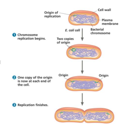
<p><strong>Binary fission</strong> is how <strong>prokaryotic cells divide</strong>; eukaryotic cells use mitosis. It resembles mitosis but uses different proteins/mechanisms, and mitosis may have evolved from binary fission. </p><p><strong>Process:</strong> cell grows while DNA replicates → new chromosomes move toward opposite poles while replication is still occurring → plasma membrane pinches inward → 2 cells. Pinching involves proteins including <strong>FtsZ, not actin</strong></p><p><span style="background-color: inherit; line-height: 21.85px; color: rgb(0, 255, 123);"><strong>Easy flow:</strong> <strong>Cell grows + DNA copies → chromosomes separate → FtsZ helps pinch cell → 2 cells</strong>.</span><span style="line-height: 21.85px; color: rgb(0, 255, 123);">&nbsp;</span></p>
15
New cards

Describe the importance of Cell Cycle Regulation & Checkpoints — function and importance of each checkpoint & phase.

Checkpoints are control points where the cell cycle can pause until the correct go-ahead signal is received, helping ensure division occurs properly.

G1 checkpoint (late G1): decides whether cell should divide. GO → enters S and likely completes cycle. STOP → may enter

G0, a nondividing state that can be reversible or permanent depending on cell type.

G2 checkpoint (end G2): checks DNA is undamaged and all chromosomes were fully replicated; problems stop mitosis.

M checkpoint (metaphase): checks that all sister chromatids are properly attached to kinetochore microtubules; if not, anaphase does not begin

<p><strong>Checkpoints</strong> are control points where the cell cycle can <strong>pause until the correct go-ahead signal</strong> is received, helping ensure division occurs properly. </p><p><strong>G1 checkpoint (late G1):</strong> decides whether cell should divide. GO → enters S and likely completes cycle. STOP → may enter </p><p><strong>G0</strong>, a nondividing state that can be reversible or permanent depending on cell type. </p><p><strong>G2 checkpoint (end G2):</strong> checks DNA is <strong>undamaged</strong> and <strong>all chromosomes were fully replicated</strong>; problems stop mitosis. </p><p><strong>M checkpoint (metaphase):</strong> checks that <strong>all sister chromatids are properly attached to kinetochore microtubules</strong>; if not, anaphase does not begin</p>
16
New cards

Explain Cyclins & Cdks and their involvement in Cell Cycle Regulation.

Cdks (cyclin-dependent kinases) are protein kinases present at a constant concentration, but they are inactive unless bound to the correct cyclin.

Cyclin concentrations rise and fall throughout the cycle. When enough cyclin accumulates → binds Cdk → Cdk becomes active → phosphorylates target proteins → promotes cell-cycle progression. Eventually cyclin is degraded → Cdk becomes inactive.

Key: Cdk amount stays constant; cyclin levels control Cdk activity. Mitosis
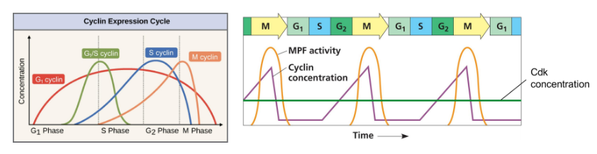
<p><strong>Cdks (cyclin-dependent kinases)</strong> are protein kinases present at a <strong>constant concentration</strong>, but they are inactive unless bound to the <strong>correct cyclin</strong>. </p><p><strong>Cyclin concentrations rise and fall</strong> throughout the cycle. When enough cyclin accumulates → binds Cdk → <strong>Cdk becomes active</strong> → phosphorylates target proteins → promotes cell-cycle progression. Eventually cyclin is <strong>degraded</strong> → Cdk becomes inactive. </p><p><strong>Key:</strong> Cdk amount stays constant; <strong>cyclin levels control Cdk activity</strong>. Mitosis</p>
17
New cards

What is MPF — Maturation Promoting Factor? Explain the process.

MPF = cyclin + Cdk complex that promotes G2 → mitosis.

How it works:

  1. Cyclin increases → binds to Cdk, forming active MPF.

  2. MPF triggers mitosis by phosphorylating target proteins.

  3. MPF helps activate a pathway that causes cyclin to be degraded.

  4. Cyclin decreases → MPF becomes inactive.

  5. Mitosis can finish, and the cell exits mitosis.

Important: Cdk concentration stays constant; MPF activity follows cyclin concentration.

<p><strong>MPF = cyclin + Cdk complex</strong> that promotes <strong>G2 → mitosis</strong>.</p><p>How it works:</p><ol><li><p><strong>Cyclin increases</strong> → binds to Cdk, forming active MPF.</p></li><li><p><strong>MPF triggers mitosis</strong> by phosphorylating target proteins.</p></li><li><p><strong>MPF helps activate a pathway</strong> that causes cyclin to be degraded.</p></li><li><p><strong>Cyclin decreases</strong> → MPF becomes inactive.</p></li><li><p><strong>Mitosis can finish</strong>, and the cell exits mitosis.</p></li></ol><p><strong>Important:</strong> Cdk concentration stays constant; MPF activity follows <strong>cyclin concentration</strong>.</p>
18
New cards

What are the three External Factors Controlling the Cell Cycle and their significance?

1. Growth factors: proteins released by certain cells that signal other cells to divide.

2. Density-dependent inhibition: when cells become crowded/touch neighbouring cells, interactions between cell-surface proteins trigger signals that stop cell division.

3. Anchorage dependence: most animal cells can only divide when attached to a solid surface. Memory: Growth factors = GO; too crowded = STOP; not attached = STOP.

19
New cards

Explain the impact of Cancer & Cell Cycle Control + what chemotherapy is

Cancer occurs when cells ignore normal cell-cycle controls and divide uncontrollably.

Cancer cells may:

  • Ignore checkpoints due to mutations in cell-cycle regulatory proteins.

  • Divide without growth factors (external signals).

  • Ignore density-dependent inhibition: Keep dividing even when crowded.

  • Ignore anchorage dependence: Divide without being attached to a surface.

Transformation: Normal cell → Cancer cell.

Types of Tumours:

  • Benign: Stays in one location and does not spread.

  • Malignant: Can invade other tissues and spread throughout the body (metastasis).

Chemotherapy

Uses drugs to kill or stop dividing cancer cells.

Side effects: Can also damage healthy rapidly dividing cells (hair follicles and intestinal lining), causing hair loss and digestive problems.

20
New cards

Who is Henrietta Lacks and why should her story remain remembered?

Henrietta Lacks was a Black woman whose tumour cells were taken in 1951 without her consent. These cells became HeLa cells, which have been widely used for important scientific research. Her medical information and genome were also shared without proper consent.



