Speech midterm
 Call Kai
 Learn
 Practice Test
 Spaced Repetition
 Match
 Flashcards
 Knowt Play
1/59

There's no tags or description
Looks like no tags are added yet.
60 Terms
Breathing vs. respiration
Breathing: inhaling (inspiration) and exhaliation (expiration)
Respiration: exchenge of oxygen and carbon dioxide at the cellular level
Speech breathing
The adjustment of normal breathing for speech production
Speech requires a controlled, steady stream of outgoing air
10% inhalation
90% exhalation
Boyle’s law
There’s an inverse relationship between volume and pressure
During inhalation (passive)
Diaphragm contracts and flattens
Thoracic cavity increases in size
Lung volume increases
Pressure in lungs drops
Air rushes in
During exhalation (passive)
Diaphragm relaxes
Thoracic cavity decreases in size
Lung volume decreases
Pressure in lungs increases
Air flows out
Pulmonary system
AKA the respiratory system
Upper respiratory: oral and nasal cavities, pharynx
Lower respiratory: trachea, broncial tree, lungs
Chest wall system
Rib cage
Sternum
Thoracic vertebrae
Abdomen
Diaphragm
Trachea
The major passage for air between larynx and lungs
Open tube with ~20 horseshoe shaped cartilages
Fibrous membrane and smooth muscle between cartilage rings holds the structure together
Goblet cells produce mucus to trap particles inside the trachea, and cilia sweep mucus up the trachea to remove them
Bronchial tree & alveoli
Primary (left and right) bronchi serve as airways to the lungs
Secondary bronchi subdivide from the primary bronchi to serve the individual lobes of the lungs
Tertiary bronchi serve the individual segments of each lobe of each lung
Terminal respiratory bronchioles supply air to the alveoli for gas exchange
Ends in alveoli that allow for gas exchange with capilaries due to a large, thin, and permeable surface
Kept constantly inflated by surfactant
Lungs
The right lung is larger
It’s shorter but wider, losing some space to the liver
3 lobes: superior, middle, and inferior
The left lung is smaller
Shares the left side of the thorax with the mediastinum (heart cavity)
2 lobes: superior and inferior
Primary inhalation muscles
Diaphragm: contracts and moves downwards
External intercostals: pulls ribs up and out
Primary exhalation muscles
Abdomen: compresses thoracic cavity
Internal intercostals: pulls ribs down
Pleural linkage
Maintains expansion of the lungs and ribcage
The inner/visceral pleural membrane encases the lungs
The outer/parietal pleural membrane lines the inside of the chest wall
The interpleural space in between is filled with a liquid surfactant that causes negative interpleural pressure, sucking them towards each other
Rib movement
The ribcage is somewhat elastic
Upper ribs move antero-posteriorly (pump)
Lower ribs move transversely (bucket)
Tidal breathing
Normal, quiet breathing while at rest
Inspiration is active
Expiration is mostly passive due to elastic recoil forces
40% inhalation
60% expiration
Lung volumes
Tidal: the volume of air we breath in and out during a normal cycle
Inspiratory reserve: the volume of air we can inhale above tidal
Expiratory reserve: the volume of air we can voluntarily exale below tidal
Residual: volume of air that can’t be exhaled voluntarily from the lungs
Lung capacities
Vital: the maximum amount of air a personal can inhale or exhale voluntarily
Functional residual: the amount of air remaining after taking a breath
Inspiratory: the amount of air that can be inhaled
Total lung: all the air the lungs can possible hold
Relaxation pressure
Created by elastic recoil trying to return the lungs and chest wall to resting position (equilibrium)
Active forces: muscles contractions
Passive forces: gravity, elastic recoil, surface tension of alveoli
Lung volume dictates the level of relaxation pressure
Resting lung volume
Outward chest expansion = inward lung recoil
Exists after we take a full breath

Above resting lung volume
Positive relaxation pressure
Elastic recoil wants lungs smaller, and eventually the ribs as well
Exhalation can be achived with passive pressure alone
Pressures above 60% VC are too much to sustain phonation
At pressures between 60% and RLV, active muscle contraction and passive pressure are used to sustain phonation

Below resting lung volume
Negative relaxation pressure
Outward chest expansion overpowers lung recoil
Exhalation can only be achieved using active muscle contraction
At pressures below RLV, only active muscle contraction is used to sustain phonation

Breath group
Number of words or syllables spoken on one exhale
Speech breathing - body type
Endomorphs (fat) use greater abdominal wall activity than rib cage activity compared to endomorphs (skinny)
Additional mass on the abdomen exerts a pulling force that tugs on the diaphragm, inhibiting rapid contraction
Speech breathing - cognitive linguistic factors
Inspiration mainly occurs during grammatical and semantic boundaries
The longer the phrase, the deeper the inhalation
When you’re actively thinking, you generally have shorter breath groups
Speech breathing - respiratory demands
Speech breathing = slight hypovention and irregular breathing
Exercise/being active = larger breaths, shorter breath groups
Hypervention
Excess of CO2
Dizziness, blurred vision
Hypovention
Lack of oxygen
Impared cell respiration, tissue damage, death
Clavicular breathing
Shoulder lifting
Uses neck muscles heavily
Less activation of diaphragm
More vertical than horizontal expansion
Least efficient
Chest breathing
Rib cage expanding a lot during inhalation
Expansion is primarily in the chest rather than the diaphragm
Less efficient
Diaphragmatic breathing
Abdomen moves outwards
Diaphragm moves freely
Most efficient pattern
Neural control of breathing
Respiratory center located in the medulla oblongata
Muscles controlled by the phrenic and intercostal nerves
Sensory receptors
Chemoreceptors monitor CO2, pH, O2, and more
Mechanoreceptors monitor tissue movement and stretch
Airflow
The volume of air moving through a given area of tube per unit of time
Airflow increases when driving pressure increases (more push, more flow)
Airflow = driving pressure/resistance
Airway resistance
Narrower airways = more resistance
Doubling airway diameter decreases resistance by a factor of 16
About half of airway resistance is in the upper airway
Lower airway resistance is mainly due to the trachea and bronchi
Rapid shallow breathing = most of work is done against airway resistance
Elastic resistance
Work required to prevent tissue from springing back once expanded
Deep slow breathing = most of work is done against elastic resistance
Viscosity
Internal friction within air/fluid
High viscosity = high friction
Higher viscosity increases breathing effort
Airflow types
Laminar: smooth, efficient, directly proportional to driving pressure
Turbulent: chaotic, eddies/whirpools, requires more driving pressure, common in trachea and large airways
Phonation
The production of speech sounds through vibration of the vocal folds
The vocal folds repeatedly open and close, creating puffs of air that generate sound waves
Larynx
Located in the front of the neck anterior to the esophogus and above the trachea
Protects the airway during swallowing
Phonation
Airflow control during breathing
Autonomic coughing
Throat clearing
Valsalva manuver: stabilization during heavy lifting (vocal folds are adducted to seal the airway, making the thoracic cavity become ridged which allows the lower body to more efficiently generate movement)
Laryngeal cartilages
Unpaired
Thyroid
Cricoid
Epiglottis
Paired
Arytenoids
Corniculates
Cuneiforms
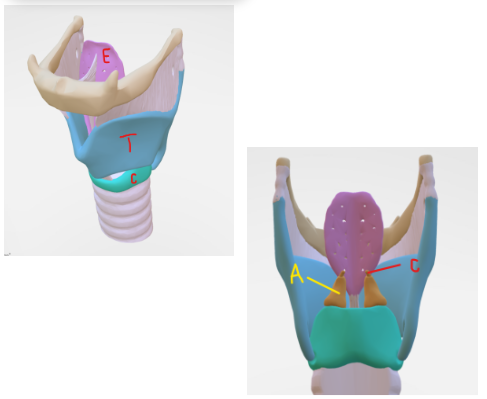
Cricothyroid joint
Allows larynx to rock/tilt
Lengthens vocal folds
Helps control pitch

Cricoarytenoid joint
Opens and closes vocal folds
Rocks and slides (undulating motion)

Aryepiglotic folds
Connects top of epiglottis to arytenoids
Just the edges
Pull the epiglottis backwards to close the larynx during swallowing
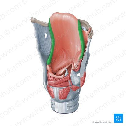
False vocal folds (ventricular folds)
Mainly protective, not sound producers
Capable of limited movement like closing during swallowing and forceful activity
True vocal folds
Complex valves that vibrate and open and close
Vibrating element of larynx
Multilayerd structure
Glottal space
Glottis: Space between the true vocal folds
Supraglottis: area above the vocal folds
Subglottis: area below the vocal folds
Intrinsic muscles of larynx
Throat muscles located entirely within the larynx
Fince control of phonation
Open and closes vocal folds
Adjusts tension (pitch)
Posterior cricoarytenoid
Lateral cricoarytenoid
Interarytenoid
Cricothyroid
Thyroarytenoid
Posterior cricoarytenoid
ONLY vocal fold abductor
Lateral cricoarytenoid
Adducts vocal folds
Interarytenoid
Adducts vocal folds
Cricothyroid
Lengthens vocal folds (raises pitch)
Thyroarytenoid
Main body of the vocal folds
Extrinsic muscles of larynx
Attaches larynx to structures outside of it
Moves entire larynx
Stablizes position
Thryohyoid
Sternothyroid
Omohyoid
Sternohyoid
Intrinisc membranes and ligaments
Connect laryngeal cartilages to each other
Conus elasticus membrane
Quadrangular membrane
Conus elasticus membrane
Below the vocal folds
Medial cricothyroid ligament and lateral cricothyroid ligament
Upper portion thickens to form the vocal ligament, the core of the true vocal fold
Quadrangular membrane
Above the vocal folds
Superior free margin forms the aryepiglotic folds
Inferiror free margin forms the vestibular ligament and false vocal folds
Extrinsic membranes
Attaches the larynx to the hyoid bone above and the trachea below
Thyrohyoid membrane: connects thyroid cartilage ↔ hyoid bone
Cricothyroid membrane: connects cricoid ↔ thyroid cartilage
Cricotracheal membrane: connects cricoid ↔ trachea
Extrinsic ligaments
Attaches the larynx to the hyoid bone above and the trachea below
Thyroepiglottic ligament: connects thyroid cartilage ↔ epiglottis
Hyoepiglottic ligament: connects hyoid bone ↔ epiglottis
Cricotracheal ligament: connects cricoid ↔ trachea
Vocal fold histology
Covered in a mucus blanket secreted from glands within the vestibular folds (false vocal folds)
Epidermal layer
Basement membrane
Lamina propia:
Superficial (gelatin)
Middle (elastic)
Deep (collagen)
Thyroarytenoid muscle
Laryngeal innervation
Vagus nerve
Reccurent branch
Motor control to all intrinsic muscles (except cricothyroid)
Superior branch
Internal: sensory
External: cricothyroid control