Boards Review (copy)
 Call Kai
 Learn
 Practice Test
 Spaced Repetition
 Match
 Flashcards
 Knowt Play
1/244

Earn XP
Description and Tags
Info for Boards Review
245 Terms
Relationship between OID and Contrast
More OID, more contrast
Which arteries supply blood to the brain?
The carotids and the verterbral arteries
Which flexure is demonstrated in an RAO?
Hepatic flexure (side down)
What does ERCP stand for?
Endoscopic Retrograde Cholangiopancreatography
What is known as the heart’s pacemaker?
SA Node
Which vessels supply our skull and face with oxygenated blood?
External Carotid
Artery Means
Blood is moving away from the lungs
SI joint degree of obliquity
25-30*
Rhese view
53*
If a pt cannot ulnar flex, we do this view
Stecher Method (20* angle on tube)
What is the Gaynor-Hart Method?
Wrist - carpal canal
Which finger is fractured in the Boxer’s fracture?
5th metacarpal bone
What is Reid’s baseline?
IOML
AEC is
1.5-2 times the correct exposure
Which are self-inductions?
Autotransformer and Choke Coil
What is the line focus principle?
A steeper anode angle will reduce the size of the effective focal spot while an anode angle greater than 45* will increase the size of the effective focal spot in comparison to the width of the electron beam
Skull
Add Mosby skull questions
What does grid cut off
do
What does ABC control in Fluoro?
KVP and mA
Anode heel impacts visibility through
SID and IR size
More KV means more
Exposure intensity
More beam restriction means more
contrast
More beam filtration means
less contrast (because it gets rid of lower wavelengths)
mA does not affect
Recorded detail
Know difference between Smith and
Colles
Coherent scattering is also known as
Unmodified or conventional or elastic scattering
Compton scattering is also known as
inelastic scattering
Anode heel effect
Is caused by x-ray absorption
Minimum response time w/modern AEC unit
.001 sec
Increasing the SID and adjust mAs to maintain exposure intensity will result in -
an increase in spatial resolution
Test accuracy of x-ray timer is called the
Spinning top
As grid ratio decreases, scale of contrast becomes
Longer
As grid ratio increases
Contrast increases
Off-focus radiation is minimized by -
Restricting the xray beam as close to its source as possible
Grid Ratios
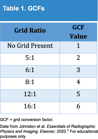
Mag fluoro does what
Increases pt does and increases spatial resolutino
What is the device used to amplify the light released by the laser scanning the CR plate before it is sent to the digitizer?
Photomultiplier
If a radiographer who stands 2 ft from an x-ray source receives an exposure of 3 mr/h, what would they receive if they stood 6 ft from the source?
Answer: .33 mR
This imaging system uses amorphous selenium to convert x-ray photons to an electronic digital signal.
Direct flat panel system
Diagnostic tube target angles range from
5 to 15*
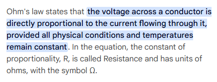
What is Ohm’s Law?
V = IR
What is the difference between voltage and current?
Voltage is the force that makes electrons flow. Current is the rate of flow of the electrons.
What is a planar joint?
They are gliding joints
Examples of joints
Include
**CR will be parallel to
intervertebral foramina
Esophagus
We see in recumbent
Obturator foramen is made up of
Ischium and pubis
Degree of obliquity for L5 and SI joints
30*
Example of a pivot joint
Proximal radioulnar and atlanto-axial view
What is a part of the tri-malleolar fx?
Lateral malleolus, medial malleolus, distal posterior aspect of the tibia
Uterine cannula is used for
HSG
Know CR plates
Study
CRs are made up of
Phosphor plate (uses a photostimulable phosphor) PSP = CR
CR: A laser is used on it to
Release stored photons
CR
Uses aluminum to absorb some x-rays
Active layers in CR uses
barium fluorohalide to store and release energy
For CR, the diameter of the laser beam affects
the spatial resolution of the imaging system
For CR, when the red laser hits the imaging plate, the plate releases
a blue visible light
This light is then captured to create the
visible image
IP (radiation detector) is erased with a
bright white light and returned to cassette
CR red laser light scans in a
Raster pattern at 2 eV
(CR) Photomultiplier tube is used to
Convert small amounts of light into an amplified electric signal (then digitized to ADC which determines the # of shades of gray in an image
CR Histogram
Graphic representation of the grays/black/whites of an x-ray exposure
Look up table (LUT)
LUT is the primary controller of digital contrast
The IP will hold the CR image for up to
8 hours before it starts to deteriorate
Flat panel director =
indirect capture
FPD - Cesium Iodide Scintillator
Converts x-rays to light
Amorphous silicon photodiode
converts light to electrons
TFT
Collects electrical signal
FPD Smaller DELs mean
increased spatial resolution
(Image Intensifier) input phosphor is made up of
cesium iodide. It converts x-rays photons into light photons
Photocathode
Converts light photons into electrons
II - Output phosphor is made of
Zinc cadmium sulfide. It converts electrons into light photons (c → z)
Hint for Image Intensifier
XLELM
Leakage radiation from the tube must not exceed
100 mr/h
What is the intensity of scattered radiation perpendicular to and 1 m from a pt compared with the useful beam at the pt’s surface?
0.1%
A controlled area has an occupancy factor of
1
Secondary radiation barriers usually require the following thickness of shielding
1/32-inch of lead
Radiation that passes through the tube housing in directions other than that of the useful beam is termed
Leakage radiation
Which of the following features of fluoroscopic equipment is designed to minimize radiation exposure to the pt and/or personnel?
Bucky slot cover; exposure switch; cumulative exposure timer
What % of x-ray attenuation does a 0.5 mm lead equivalent apron at 100 kvp provide?
75%
How is the intensity of an x-ray photon affected after each time it scatters?
It's intensity decreases 1000 times
Primary radiation barriers usually require how much thickness of shielding?
1/16 in lead
The term used to describe expectoration of blood from the bronchi is
Hemoptysis (coughing up blood)
The thoracic cavity is lined by
parietal pleura
Which bones are NOT part of the bony thorax?
Clavicles
Mediastinal structures include
heart, trachea, esophagus
The condition that results from a persistent fetal foramen ovale is
An atrial septal defect
Imperfect expansion of the lungs, often accompanied by dyspnea, is called
Atelectasis
Which of the following positions is required to demonstrate small amounts of fluid in the pleural cavity?
Lateral decubitus, affected side down
During atrial systole, blood flows into the
left ventricle via the bicuspid valve
right ventricle via the tricuspid valve
Which of the following positions will demonstrate the right axillary ribs?
LAO and RPO
Which of the following criteria are used to evaluate a PA projection of the chest?
10 posterior ribs should be visualized
Sternoclavicular joints should be symmetrical
The scapulae should be lateral to the lung fields
The sternoclavicular joints are best demonstrated with the patient PA an d
in a slight oblique position, affected side adjacent to the IR
Which of the following positions can be used to demonstrate the axillary ribs of the right thorax?
RPO and LAO
The esophagus commences at about the level of
C6
Blood is returned to the LEFT atrium, from the lungs, via the
pulmonary veins
What is described as a measure of the rate at which energy is transferred from ionizing radiation to soft tissue?
LET (linear energy transfer)
LET of occupational radiation
low energy and low LET
What minimum radiation dose is required in order for acute radiation syndrome to occur?
100 rad (1 Gy)