Bioelectrodes, Neural Prostheses, and Biosensors
 Call Kai
 Learn
 Practice Test
 Spaced Repetition
 Match
 Flashcards
 Knowt Play
1/42

There's no tags or description
Looks like no tags are added yet.
43 Terms
Bioelectrode
conducts current across the interface between the body and the electronic measuring circuit
serves as a transducer to change ionic current <→ electronic current and vice
Transducer
changes an ionic current ←> electronic current
bioelectrode acts as one
Recording Electrodes
monitor/measure electrical events in the body (reads)
ex. ECG, EEG, EMG, extracellular neural recording
ECG
electrocardiogram (heart??)
type of recording electrode
EEG
electroencephalogram
type of recording electrode
EMG
electromyogram (muscle??)
type of recording electrode
Extracellular Neural Recording
used for neural prosthesis such as “brain machine interface”
high resolution from neurons
type of recording electrode
Stimulating Electrodes
transmit electrical current into the body to influence specific physiological processes (writing)
Stimulating Electrodes Examples
cardiac pacemakers and defibrillators
neural prosthesis (deep brain stimulators, cochlear implants, and functional electrical stimulations) → activate or inhibit neurons
transcutaneous drug delivery - across the skin layer (used to open up barrier) (used with spinal cord inuries to get some function back)
stimulators for wound healing/regeneration (using body’s own bioelectricity)
Bioelectronic Medicine
instead of using chemical compounds, tap into neural system and use electronics to control it (neural, digestive system. etc.)
Electrochemical Biosensors
modulation close loop so don’t just turn things on a let it go
allows for a read out of the body’s response to a stimulator (helps to know when to shut it off etc.)

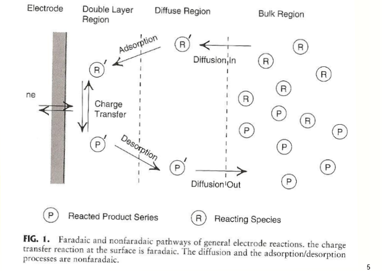
Faradaic Process
happens at the electrode-electrode interface
charge transfer governed by Faraday’s law
at interface, charge can be exchanged through redox chemical reactions bc electrons can’t just jump across

Faraday’s Law
the amount of chemical change occurring at an electrode-electrode interface is directly proportional to the current that flows through the interface
Electordes undergo mainly faradaic processes called what?
charge transfer electrodes
or
non-polarizable electrodes
Polarization at Electrode-Electrode Interface
half cell potential of electrode results in electrical double layer formation (considered as a capacitor)
another type of charge transfer
direct current can’t just cross but alternating current can cross through non-faradaic process

Non-faradaic Process
current flow can be passed by charging/discharging a capacitor
polarization at electrode-electrode interface
Perfectly Polarizable Electrodes
do not undergo faradaic process
Polarizable Electrodes
noble metal Pt, Au, or stainless steel
most bioelectrodes are
MIxed Situation
no 100% Faradaic or non-Faradaic (always a mixture but one is more dominant)
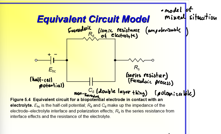
Equivalent Circuit Model - Biopoential Electrode in Contact withan Electrolyte
Ehc - half-cell potential
Rd and Cd - make up impendance of electrode-electrode interface and polarization effects
Rs - series resistance (from interface effects and resistance of the electrolyte)
circuit with half cell potentiall in series with Rd and Cd in parallel and then R
Impedance Spectroscopy
impedance is a fcn of frequency
dropping is capacitor
how to measure mixed interface

Non-polarizable Electrode
can be made by electrolytic process or sintering process
ex. Ag/AgCl → reversible rcns; silver wire/disk, put in a Cl soln., oxidize? → silver ion + chlorine togetehr to form electrode
preferred by bio stuff bc isn’t based on movement bc not conducting current through it???
Ag/AgCl
impedance increases as AgCl layer increases = more resistance (contributes to its own resistance)
Ag (silver) - Faradaic/polarizable but AgCl non-Faradaic/non-polarizable

Skin Surface Electrode
need to overcome the stratum corneum layer (top layer of skin/epidermis → layer of dead cells that makes teh surface very hydrophobic so hard for ions to pass through) → gel essentially hydrates it so ions can get through and signal is measureable (sweat too) and lowers impedance
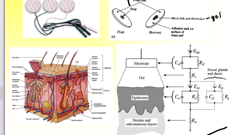
Biopotential Electrode Varieties
used fo EEGs
Myoelectric Prosthesis
for amputees
nerves are still there and tryong to find missing body part and if doesn’t it casues pain
so redirect neurons to chest instead then use EMG?? to measure what neurnon wants to do (code to control prosthesis)
Brain Machine Interface I
another application of myoelectric??
using motor neurons in the motor cortex in the brain (usually for paralyzed people)
use thoughts to control robotic arm to get food
Brain Machine Interface II
same implant as other brain one (motor neurons) just in a person
Various MEAs for Neural Recording
Utah array (sold by Blackrock) (silicon w/ doped needles??)
microwire array (manually made)

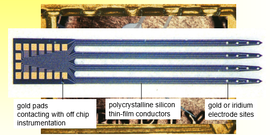
Michigan Proves
have gold pads contacting w/ off chip instrumentation
polycrystalline silicon thin-film conductors
gold or iridium electrode sites
uses same process that is used to make computer chips
Challenges at Neural Interface
unreliable single unit yield
signal fades over time
signla drift (stability of recording = not good → calibrate systems often)
stimulation threshold increase
essentially very tiny signals so electrode needs to be close to neuron to be able to pick up the signal (within 100 microns)
Problem - Unreliable Chronic Recording
?????????

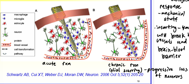
Cellular Response around the Inserted Neural Electrode
2 contributing factors (biological response and mechanical stuff)
inserting an electrode will break blood vessls and/or the blood-brain barrier
Microglia
can be resting/ramified or activated/amoeboid
Resting or Ramified Microglia
no Fc receptors or macrophage specific ones
large, dense chromatin masses in small flattened nucleus
5-10% of glial pop.
many long, thin processes
essentially sruvery environment for anything wrong
Activated or Amoeboid Microglia
upregulation of lytic enzymes
retraction of cytoplasmic procssses to stouter, thicker pseudopodia
accumulation of phagocytosed material and lipid droplets
ameboid/motile abilities
release of inflammatory factors MHC I, II expression
active proliferation
similar to monocyte → macrophage
Astrocytes
normal or reactive
Normal Astrocyte
8-10 nm diamter GFAP filaments
round nucleus
10-40 nm glucogen particles
large mitochondira
gap junctions w/ other astrocytes
maintain proper neuronal environment (structural and function support)
30-65% of glial pop.
Reactive Astrocyte
large increase in GFAP filaments, glycogen deposits
irregular nucleus
increase in mitochondria and multiple golgi complexes
hypertrophy
phagoctosis
proliferation - produce ECMs to wrap around neurons (like fibroblasts)
migration
extracellular matrix population
production of neurotrophic and inflammatory factors
GFAP
an intermediate filament that has a unique stain
Surface Chemistry Matters
antifouling zwitterionic polymers carry large amounts of positive/negative ions, are highly hydrophilic, resist protein adsorption, and cell attachment
Mechanics Matters
stimulating device inserted into materials with different mechanical moduli → how does this change the strain the tissue is experiencing

Size Matters
large device - morve invasive = more inflammation
small - electrodes the size of neuron stromas = seamles integration w/o glial response (future direction??)