Biology - Biological molecules
 Call Kai
 Learn
 Practice Test
 Spaced Repetition
 Match
 Flashcards
 Knowt Play
1/40

There's no tags or description
Looks like no tags are added yet.
41 Terms
Name 3 monosaccharide sugars (3)
Glucose
Fructose
Galactose

What is a monosaccharide? (3)
A monomer of more complex molecules containing just carbon, oxygen and hydrogen
Mainly used in respiration
Reducing sugars so positive Benedict’s test result
What are the isomers of glucose?
Alpha glucose - OH group on the bottom
Beta glucose - OH group on the top
What is a disaccharide? (3)
A disaccharide is two monosaccharides joined together by a glycosidic bond in a condensation reaction
A molecule of water is released during this reaction
They can be broken down by a hydrolysis reaction by addition of water, hydrolysed by boiling with acid or using a specific enzyme
Name 3 types of disaccharides
Maltose - glucose + glucose
Sucrose - glucose + fructose
Lactose - glucose + galactose
Describe the method to test for reducing sugars (5)
Benedict’s test - all monosaccharides and disaccharides, except for sucrose, give a positive result
The sample is places in a test tube with Benedict’s solution
The test tube is heated in a water bath for 5 minutes
Positive result - blue to brick red colour (copper (I) oxide)
Negative result - remains blue, no reducing sugar
Describe the method to test for a non-reducing sugar (5)
Sucrose - non-reducing sugar, needs to be broken down for a positive Benedict’s test result
Benedict’s test to confirm that it’s a non-reducing sugar
Hydrolyse another sample by heating with acid (dilute HCl) or using an enzyme (sucrase) at optimum temperature
Add dilute NaOH to neutralise acid
Add Benedict’s solution and heat in water bath for 5 minutes
Positive result - brick-red colour, a non-reducing sugar was initially present
Describe the structure and function of cellulose
Cellulose - a polysaccharide found in the cell wall of plants
Provides rigidity to plant cell walls and prevents bursting
Made up of many β glucose molecules joined together via condensation reactions
Forms a long, straight chain with β1-4 glycosidic bond joining the β glucose molecules so every other glucose is inverted
Hydroxyl groups form hydrogen bonds with OH groups of adjacent chains, forming a microfibril
How does the structure of plant cell walls provide rigidity?
Microfibrils group into macrofibrils which are orientated in different directions in separate layers
These layers are interwoven and embedded into a matrix, providing rigidity
Minute channels between layers of macrofibrils makes the cell wall fully permeable
Describe the structure and function of starch (6)
Starch - a storage polysaccharide found in plants
Found in starch grains (amyloplasts) in the cytoplasm
Consists of amylose and amylopectin molecules with many α glucose molecules joined together by condensation reactions
Amylose - contains α1-4 glycosidic bonds between the glucose molecules
Amylopectin - contains both α1-4 and α1-6 glycosidic bonds that form branches
Hydroxyl groups form hydrogen bonds within the same molecule which produces compact helical chains
How is starch hydrolysed? (2)
Starch is hydrolysed by the enzyme amylase to produce the disaccharide maltose
The more branched the molecule is, the faster the rate of hydrolysis
What are the properties of starch that make it an ideal storage compound? (3)
Insoluble so is osmotically inactive, prevents cell damage by osmosis
Helical compact shape so can store many glucose molecules together
Contains a large number of glucose molecules so can be used for respiration
How can you test for starch? (3)
Starch can be detected by using the iodine test:
Add a few drops of iodine solution (KI)
Positive result - blue black colour, starch is present
Negative result - remains orange
Describe the structure and function of glycogen (3)
Glycogen - the storage polysaccharide found in the cytoplasm of animal cells and fungi
It is a polymer of of α glucose and heavily branched, allowing rapid hydrolysis to release glucose for respiration
Glycogen is insoluble and osmotically inactive so can be stored largely in liver and muscle tissues

What is the structure and properties of a lipid? (3)
Lipid - contains carbon, hydrogen and oxygen but with a lower proportion of oxygen and more hydrogen compared to carboydrates
Store twice as much energy as carbohydrates
Insoluble in water so osmotically inactive but can dissolve in organic solvents (alcohols)
Describe the structure and function of a triglyceride (3)
Triglyceride - a type of lipid consisting of 3 fatty acids joined to 1 glycerol by ester bonds
Fatty acids - general formula is R-COOH where R is a long hydrocarbon chain and COOH a carboxylic acid group
Fatty acids can be saturated (no double bonds between C atoms) or unsaturated (contain one or more double bonds)

How are triglycerides formed and hydrolysed? (2)
Formation of a triglyceride - 3 condensation reactions to join 3 fatty acids with ester bonds, releasing 3 molecules of water
Hydrolysis of a triglyceride - 3 hydrolysis reactions to break 3 ester bonds between 3 fatty acids and glycerol

How can lipids be hydrolysed? (2)
Lipids can be hydrolysed into fatty acids and glycerol by:
Heating with acid or alkali
Using lipase at its optimum temp and pH
How can you test for the presence of a lipid? (4)
Lipids can be detected by using the emulsion test:
A small amount of the sample is placed into a test tube with 2cm3 of ethanol
Shake the mixture so that the fat dissolves
Add the mixture to water in another test tube and mix
Positive result - a white emulsion forms
Describe the structure of a phospholipid molecule (3)
Phospholipid - a lipid which contains 2 fatty acids, glycerol and a phosphate group joined in a condensation reaction
A phospholipid molecule contains:
A polar hydrophilic head containing the phosphate group
A non-polar hydrophobic tails consisting of fatty acids

Describe the arrangement of the phospholipid layers (3)
Phospholipids arrange themselves into spherical micelles or a sheet-like bilayer as they come into contact with water
The hydrophilic heads are attracted to water and the hydrophobic tails are water repellent, forming a single layer
A phospholipid bilayer is present in the cell membrane where the hydrophobic tails are attracted to eachother

What are proteins and amino acids? (5)
Protein - contains the same atoms found in carbohydrates plus nitrogen
Amino acid - monomer of a protein, join together to form a protein (polmer)
All amino acids have an amino group and a carboxylic acid group but differ in the structure of the R group
Amino acids can join together via condensation reactions with peptide bonds forming between them
A dipeptide contains 2 amino acids and a polypeptide contains many amino acids joined together

How can proteins be hydrolysed?
Proteins can be hydrolysed by heating with acid or by using enzymes (proteases) at their optimum temperature. These enzymes include:
Endopeptidases - hydolyse peptide bonds in proteins to produce smaller polypeptides
Exopeptidases - remove single amino acids from the ends of the polypeptide chains to produce dipeptides and amino acids
Describe the different structures of proteins (4)
Primary structure - the sequence of amino acids in the polypeptide chain, sequence determines specific shape of protein
Secondary structure - folding or coiling of the polypeptide chain
Includes alpha helices and beta pleated sheets
Weak hydrogen bonds are present in these structures
Tertiary structure - further coiling of the secondary structure
Contains many different types of bonds - ionic, hydrogen, disulfide bridges, hydrophobic interactions
Quaternary structure - consists of more than one polypeptide chain
Chains are held together by ionic bonds, hydrogen bonds (between side chains), disulfide bridges and hydrophobic interactions
May contain other groups e.g haemoglobin consists of 4 polypeptides (2 alpha chains and 2 beta) and a Haem group containing Fe2+

Describe the structures and functions of fibrous proteins (4)
Fibrous proteins - a structural protein found in keratin in nails and collagen in connective tissues
Consists of long parallel polypeptide chains with a simple tertiary/quaternary structure
Collagen - triple-helix structure, insoluble and high tensile strength
Keratin - coiled helices and cross-linking, insoluble and tough
Describe the structures and functions of globular proteins (3)
Globular proteins - a highly folded and coiled polypeptide chain, produces a complex tertiary structure and is soluble in water
Includes enzymes, antibodies receptors and hormones
Also includes hormones which act as chemical messengers which have a tertiary structure so will interact with a complementary binding site on a specific receptor
How can proteins become denatured? (5)
The denaturation of a protein is when an alteration in the tertiary structure occurs as a result of bonds breaking
It can be caused by:
High temperatures above the optimum, breaking hydrogen bonds
Change in pH breaks hydrogen and ionic bonds
Reducing agents break disulphide bridges
Heavy metal ions can bind to proteins and change its shape
How can you test for the presence of a protein? (3)
Proteins can be detected by performing a biuret test:
Add sample to test tube containing 2cm3 of biuret reagent
Positive result - blue to purple/lilac colour change
Negative result - remains blue
How do enzymes increase rate of reaction?
Enzymes can increase the rate of a reaction by lowering the activation energy:
Activation energy - the energy required for a chemical reaction to take place'
The enzyme works by lowering the activation energy so that less energy is required to fuel a reaction so rate of reaction increases
This also allows chemical reactions to occur at a normal temperature range

Describe enzyme specificity (3)
Enzymes are highly specific so only interact with a specific type of substrate
The unique tertiary structure of an enzyme determines the shape of the active site and its charges
This structure is held together by hydrogen bonds, ionic bonds and sometimes disulphide bridges
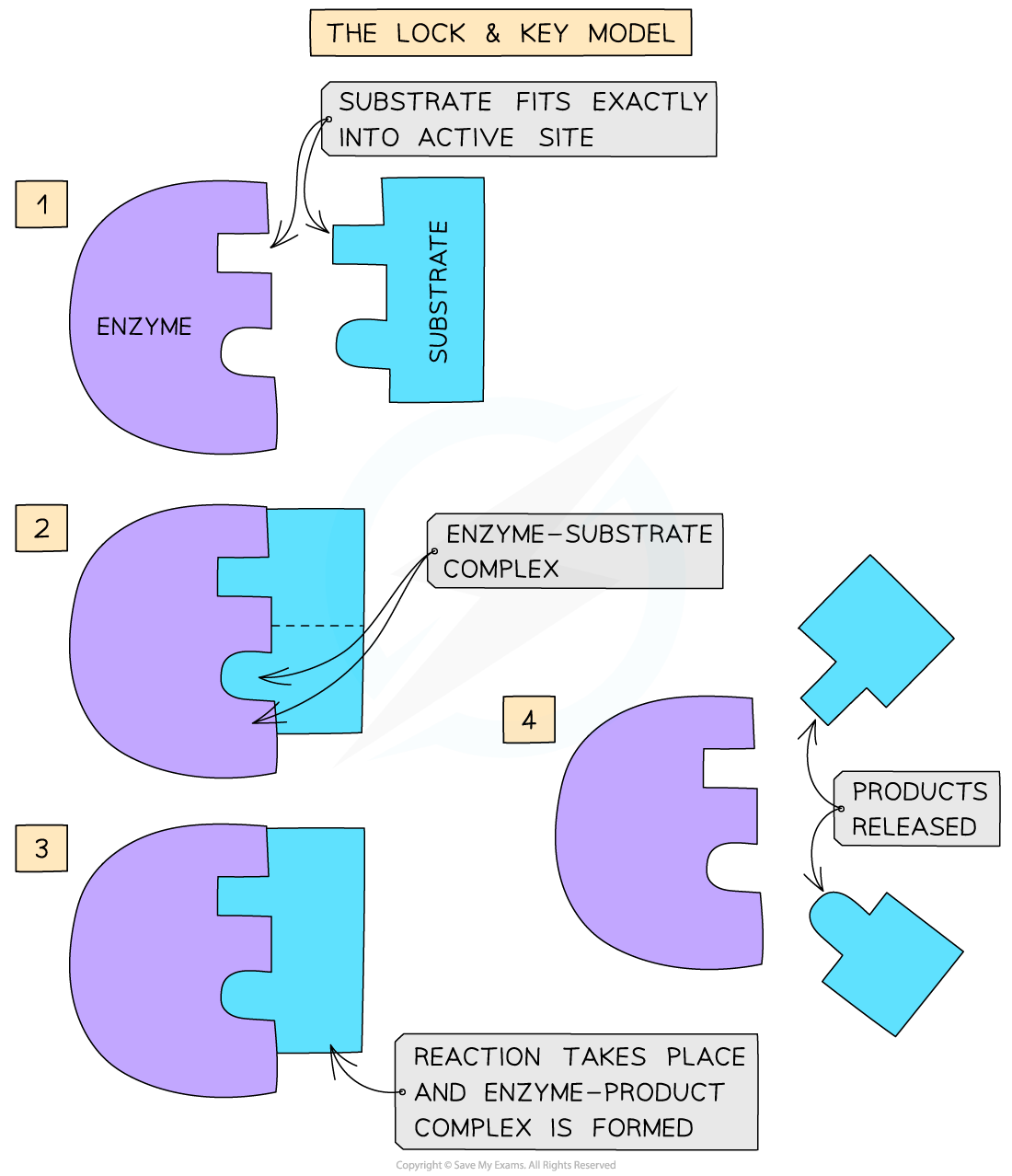
Describe the lock and key hypothesis of enzymes (2)
Only substrates with the complementary shape to the active site will bind to it forming an enzyme-substrate complex
After the substrate binds to the active site, the product is leaves the active site and is released
Describe the induced fit hypothesis of enzymes (3)
The substrate interacts with the active site and changes the shape of the enzyme so it can fit more precisely into the active site
May put a strain on bonds and make a reaction more likely e.g hydrolysis
May also allow two molecules to come close together and make a condensation reaction more likely

How can enzyme concentration affect rate of reaction? (4)
High enzyme concentration - more enzymes present so more active sites available
Increases the chances for the substrate to collide with the active site so rate of reaction increases
(This assumes that the amount of substrate does not limit the rate of reaction.)
This means that the relationship between enzyme concentration and the rate of reaction is directly proportional

How can substrate concentration affect rate of reaction? (4)
Increasing the substrate concentration will increase the rate of reaction to a particular point
As substrate concentration increases, the rate of reaction initially increases as the substrate and enzyme are more likely to collide
The rate of reaction then levels out as the active site of all the enzyme molecules are taken up by substrate molecules
At this point the rate reaches a constant value, its maximum rate Vmax

How can temperature affect rate of reaction in enzymes? (4)
As temperature increases, the molecules have more kinetic energy so are more likely to collide
This increases the rate of reaction up to the optimum temperature where the rate of reaction is at a maximum
Above the optimum temperature - tertiary structure of enzyme denatures as bonds break
The rate of reaction decreases as the shape of the active site has changed so the substrate can no longer bind to the altered active site

How do you calculate the effect of temperature on rate of reaction?
The effect of temperature on a rate of reaction can be expressed as the temperature coefficient Q10
Q10 = Rate of reaction at (x + 10°C)
rate of reaction at x °C

How does pH affect rate of reaction in enzymes? (3)
Enzymes are most active at their optimum pH and only work within a narrow pH range
If the pH change is drastic, the enzyme can become denatured as the ionic charges of acidic and basic groups are altered
This changes the shape of the active site and the substrate can no longer bind

How does a competitive inhibitor slow down rate of reaction? (3)
The inhibitor molecule has a similar shape to the normal substrate molecule and competes with it for attachment to the active site
Rate of reaction decreases as the inhibitor occupies the active site so the substrate can no longer bind to it
This effect can be reduced by the addition of more substrate

How does a non-competitive inhibitor slow down rate of reaction? (4)
The inhibitor molecule has a different shape to the substrate so does not compete for the active site
The inhibitor binds to the enzyme away from the active site and alters the tertiary structure and the shape of the active site
The substrate can no longer attach to the altered active site or can bind but no product is formed
This effect cannot be reduced by the addition of more substrate

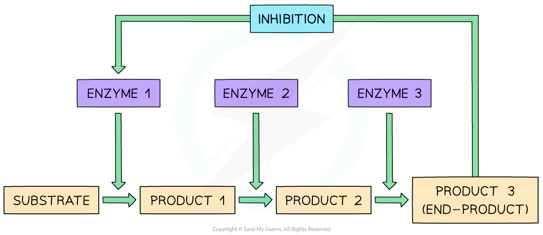
What is end-product inhibition? (negative feedback inhibition) (4)
End-product inhibition - occurs when the end product of a metabolic pathway accumulates and acts as an inhibitor
The product reduces its own production by inhibiting an enzyme in the pathway
This is a self regulating process as when product levels fall, inhibition is removed and production increases again
End-product inhibition is an example of negative feedback
What are immobilised enzymes and their advantages?
Immobilised enzymes - enzymes that are bound to or trapped in a solid support so they can be easily recovered and reused
Immobilisation reduces production costs because the enzyme can be used multiple times
Immobilised enzymes are often more stable at higher temperatures and less likely to denature
They allow continuous production by passing reactants over the enzyme and collecting products continuously
The product is not contaminated by the enzyme because the enzyme remains fixed in a matrix