619 9/28 GLA pt 2 Cellular Injury, Adaptation, and Death
 Call Kai
 Learn
 Practice Test
 Spaced Repetition
 Match
 Flashcards
 Knowt Play
1/39

There's no tags or description
Looks like no tags are added yet.
40 Terms
Apoptosis
atp dependent, enzyme mediated cell death whereby cells activate pre-programmed instructions for self destruction in response to physiologic or pathologic stimulus
Physiologic Apoptosis
involution of hormone-dependent tissues
elimination of potentially harmful self-reactive lymphocytes
death of inflammatory cells following an immune response
Pathologic Apoptosis
following fatal dna damage (radiation)
accumulation of misfolded proteins (er stress response)
cell death in certain infections (mainly viral)
Molecular Mechanisms of Apoptosis
intrinsic (mitochondiral) or extrinsic (death receptor) pathway depending on initiation, results in the activation of enzymes called caspases
2 Phases of Apoptosis
initiation phase
execution phase
Initiation Phase of Apoptosis
first step where some capsases are activated and unleash a cascade of other capsases
Execution Phase of Apoptosis
second step where terminal caspases trigger cellular fragmentation
Intrinsic (Mitochondiral) Apoptosis Pathway
activated in response to a trigger such as dna damage, infection, or loss of survival signals
pro-apoptotic proteins (bax/bak) overcome the signals of the anti-death proteins (bcl-2) initiating the apoptosis cascade
bax/bak proteins cause the mitochondrial membrane to become leaky, allowing cytochrome c to escape into the cytosol
cytochrome c (and other proteins) activate the caspase cascade inducing cell death
External (Death Receptor) Apoptosis Pathway
activated by engagement of plasma membrane death receptors such as fas/fal receptor and tnf receptor
Apoptosis as “Silent” Cellular Death
apoptotic bodies break cells into fragments for phagocyte digestion, their breakdown releases factors that recruit phagocytes, it is a controlled pathway
the end result is that apoptosis limits inflammatory reactions, even when extensive
Unfolded Protein Response and ER Stress
when the protein folding demand outweighs the protein folding capacity of the ER
can be mitigated by decreasing protein synthesis, increase production of chaperones
if the cell does not adapt, it undergoes apoptosis

Follicular Lymphoma
type of cancer caused by a genetic aberration whereby the cancer cells overexpress bcl-2 (anti-apoptotic protein), allowing cancer cells to evade apoptosis
Apoptosis Morphological Changes Diagram

Apoptosis Morphological Changes
cell shrinks with and increasing eosinophilia and nuclei become pyknotic
nucleus breaks into fragments (karyorrhexis) accompanied by dissolution of the nuclear membrane, surface membrane blebs form
eventually apoptotic bodies containing organelles and nuclear material form, apoptotic bodies are ingested by phagocytes
Unique Nuclear Features of Apoptosis
as the chromatin condenses, it accumulates peripherally and forms crescent-shapes
only on electron microscopy

Light Microscopic Features of Apoptosis
cell shrinkage (cell becomes smaller and the cytoplasm becomes dense and eosinophilic)
involves isolated cells with minimal inflammatory response

Necrosis vs Apoptosis

Cell Changes Diagram

Pathologic Calficiation
abnomal tissue deposition of calcium salts, together with smaller amounts of iron, magnesium, and other mineral salts
2 Forms of Pathologic Calcification
dystrophic
metastatic
Dystrophic Pathologic Calcification
deposition occurs locally in damaged and/or necrotic tissue, normal serum ca levels
Metastatic Pathologic Calcification
may occur in normal tissues, almost always due to hypercalcemia
Examples of Dystrophic Pathologic Calcification
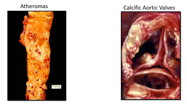
3 Reasons Metastatic Calficiation Occurs
parathyroid tumors due to excessive pth
cancer (due to pthrp secretion or bone metastasis)
vitamin-d disorders including intoxication and sarcoidosis
Where does metastatic calcification occur most often and why?
principally effects interstitial tissues of the gastric mucosa, kidneys, lungs, systemic arteries, and pulmonary veins
these tissues have an internal environment that predisposes them to form calcifications
Calcific Deposits Histology
dark basophilic character with cleft-like spaces, dystrophic and metastatic look the same histologically

4 Mechanisms fo Abnormal Intracellular Accumulations
inadequate removal of a normal substance secondary to defects in packaging and transport
accumulation of an endogenous substance, due to genetic or acquired defects, that impair its folding, packaging, transport, or secretion
failure to degrade a metabolite due to an inherited enzyme deficiency
deposition and accumulation of abnormal eogenous substance that cannot be degraded or removed becasue the cell lacks both the enzymatic machinery required for its breakdown and the means to transport it elsewhere
Example of Inadequate Removal of a Normal Substance Secondary to Defects in Packaging or Transport
hepatic steatosis (fatty change) in the liver
Hepatic Steatosis
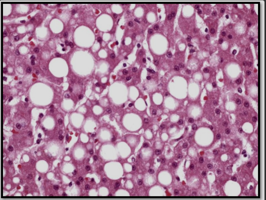
fatty change where alcohol use disorder, diabtes, and obesity cause triglycerides to accumulate within vacuoles of hepatocytes
gives liver a yellow color
Hepatic Steatosis Histology
fat vaculoes accumulate in the hepatocyte as clear circular spaces of various sized
Example of Accumulation of and Endogenous Substance, Due to Genetic or Acquired Defects, that Impair its Folding, Packaging, Transport, or Secretion
A1AT deficiency
misfolded a1at protein accumulates in the liver
A1AT Deficiency
inherited disorder where a mutation causes the aiat protein to misfold and accumulate in hepatocytes
A1AT Deficiency Histology
round-to-oval cytoplasmic globular inclusions in hepatocytes
best visualized using PAS

What does A1AT Deficiency lead to?
lung and liver disease
Example of Failure to Degrade a Metabolite due to an Inherited Enzyme Deficiency
von gierke disease
accumulation of glycogen in hepatocytes
Von Gierke Disease
mutation in enzyme that mobilizes glucose from liver glycogen, lack of enzyme leads to accumulation of glycogen within hepatocytes
Von Gierke Disease Histology
optically clear nuclei and pale cytoplasm caused by accumulation of glycogen

Example of Accumulation of an Abnormal Exogenous Substance that Cannot be Degraded or Removed Because the Cell Lacks Both the Enzymatic Machinery Required for Its Breakdown and the Means to Transport it Elsewhere
anthracosis
carbon dust in macrophages
Anthracosis of the Lung
blackening caused by inhalation of carbon dust, dust is picked up by macrophages where it remains in perpetuity due to its indigestible nature

Intracellular Accumulations vs Storage Diseases
intracellular accumulations may be reversible, but inherited storage diseases typically lead to progressive tissue injury