Nervous system flashcards
 Call Kai
 Learn
 Practice Test
 Spaced Repetition
 Match
 Flashcards
 Knowt Play
1/163

Earn XP
Description and Tags
(CH13-18)
164 Terms
Nervous system
Organ system that monitors:
stimuli
integrates information
produces responses
Three main functions of the nervous system
Monitor: Sensory receptors detect changes (stimuli) both inside and outside the body
integrate: The central nervous system (brain and spinal cord) receives, processes, and interprets the incoming sensory data.
respond: The nervous system sends signals out to effectors (muscles or glands) to produce a physical or chemical reaction.

Central nervous system (CNS)
Consists of the brain and spinal cord
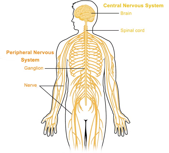
Peripheral nervous system (PNS)
Consists of nervous tissue outside the brain and spinal cord including nerves and ganglia
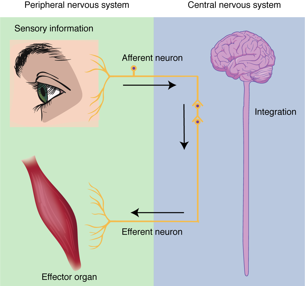
Afferent division
Sensory division that carries information through the PNS toward the CNS
Ascending pathway carries sensory information toward higher levels of the CNS
Example: You touch a hot stove.
1. Sensory receptors in your skin detect the heat.
2. Afferent neurons carry the sensory signal toward your spinal cord.
3. Your CNS receives the information and processes it.
Other examples include feeling pain, detecting pressure, and sensing temperature.

Efferent division
Motor division that carries information away from the CNS, through the PNS to effectors
Descending pathway carries motor information toward lower levels of the CNS
Example: You pull your hand away from the hot stove.
Your CNS processes the sensory information.
Efferent neurons carry a motor command away from the CNS.
Your arm muscles contract, pulling your hand away.
Efferent signals can control voluntary movements, such as raising your arm, or involuntary functions, such as changing your heart rate.
Sensory neuron
Neuron that conducts impulses toward the CNS
Sensory neurons tell your brain and spinal cord what is happening inside and outside your body. (hot stove)
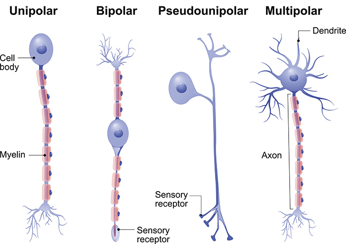
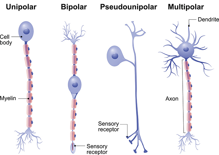
Most sensory neurons are pseudounipolar neurons
Apart of Afferent division

Motor neuron
Neuron that conducts impulses away from the CNS
Effectors include muscles and glands. Motor signals can produce skeletal muscle contraction, regulate internal muscle activity, or stimulate glandular secretion.
Most motor neurons are multipolar neurons
Apart of Efferent division
Interneuron
Association neuron located in the CNS that connects and processes information between neurons
They are important for integrating sensory information, coordinating responses, and participating in neural circuits involved in learning and other complex functions.
Most interneurons are multipolar neurons
Somatic nervous system (SNS)
Apart of Efferent division (PNS)
Primarily voluntary, controls skeletal muscles
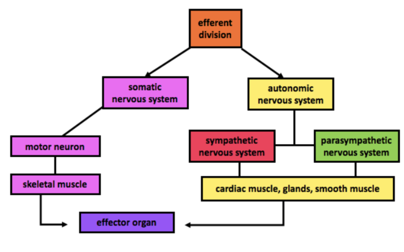
Autonomic nervous system (ANS)
Apart of Efferent division (PNS)
Primarily involuntary, regulates smooth muscle, cardiac muscle, and glands


Sympathetic nervous system
Autonomic division associated with Preparing your body for stress or activity.
fight, flight, or freeze

Parasympathetic nervous system
Autonomic division associated with Supporting recovery, digestion, and energy conservation.
rest and digest


Effectors
Muscles or glands that respond to motor signals
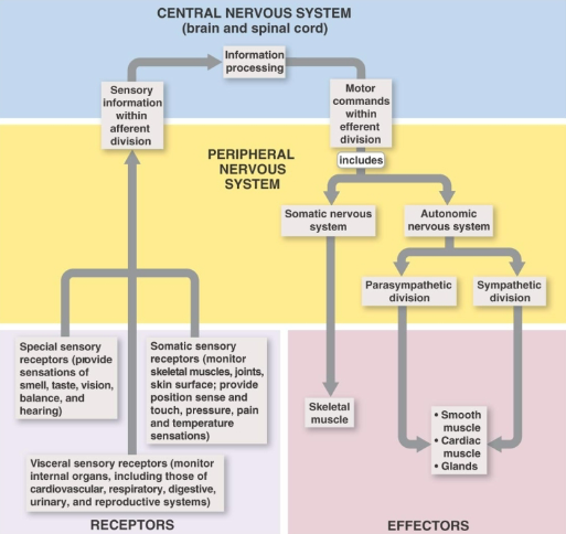
How are sensory inputs and motor outputs classified?
Somatic: Relates to the body wall and limbs, including the skin, skeletal muscles, and joints.
Visceral: Relates to the internal organs, such as the heart, lungs, stomach, and intestines.
General: Sensory information detected by receptors widely distributed throughout the body, such as touch, pressure, pain, and temperature.
Special: Sensory information detected by localized, specialized sensory organs, including vision, hearing, taste, smell, and equilibrium (balance).
Proprioception: Awareness of body position and movement through sensory information from muscles, tendons, and joints.
Branchial motor innervation: Motor nerve supply to skeletal muscles derived from the pharyngeal (brachial) muscle, including certain muscles of the face, jaw, throat, and neck.


Dorsal sensory ventral motor rule
Sensory information enters the spinal cord through dorsal roots
and motor information exits through ventral roots

Somatic vs visceral
Somatic refers to the body wall and limbs while visceral refers to internal organs
General vs special senses
General senses include touch, pressure, pain, and temperature
while special senses include vision, hearing, taste, smell, and equilibrium

Nerve
Bundle of axons in the PNS


Tract
Bundle of axons in the CNS


Ganglion
Cluster of neuron cell bodies in the PNS

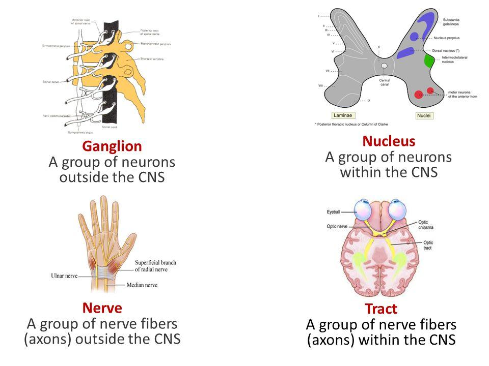
Nucleus in neuroanatomy
Collection of neuron cell bodies in the CNS

Spinal nerves
Spinal nerves are part of the PNS and are all mixed nerves,
meaning they contain both sensory (afferent) and motor (efferent) fibers.

Neuron
Functional cell of nervous tissue that receives and transmits information

Three main parts of a neuron
Dendrites
cell body or soma
axon

Soma
Neuron cell body containing the nucleus and most organelles
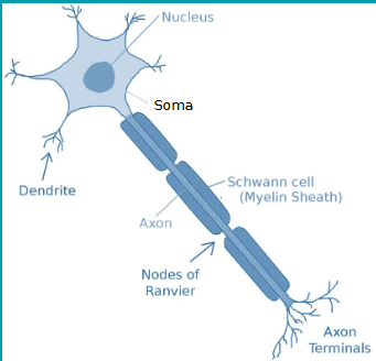
Dendrites
Branched processes that usually receive information toward the cell body

Axon
Neuron process that carries nerve impulses away from the cell body

Axon hillock (axon features)
Region where the axon emerges from the cell body
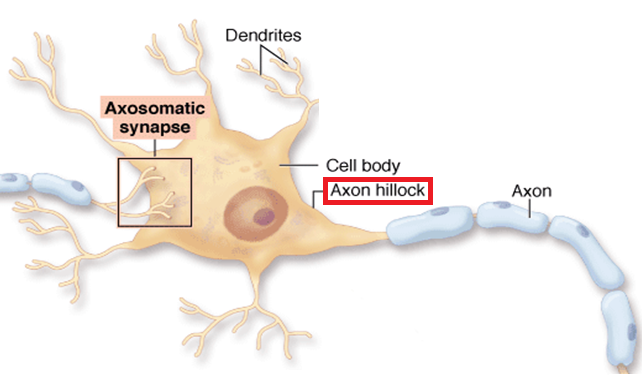

Axon terminals or terminal boutons (axon features)
Endings of an axon involved in communication with other cells

Nissl bodies (chromatophilic substance)
Rough endoplasmic reticulum and ribosomes involved in protein synthesis
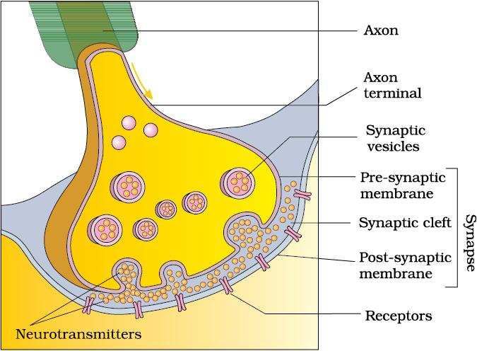
Synapse
Junction where a neuron communicates with another cell
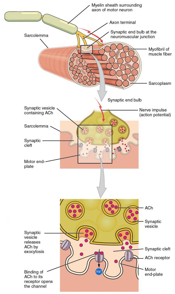

Neurotransmitter
Chemical messenger released by a neuron that binds to receptors on a target cell
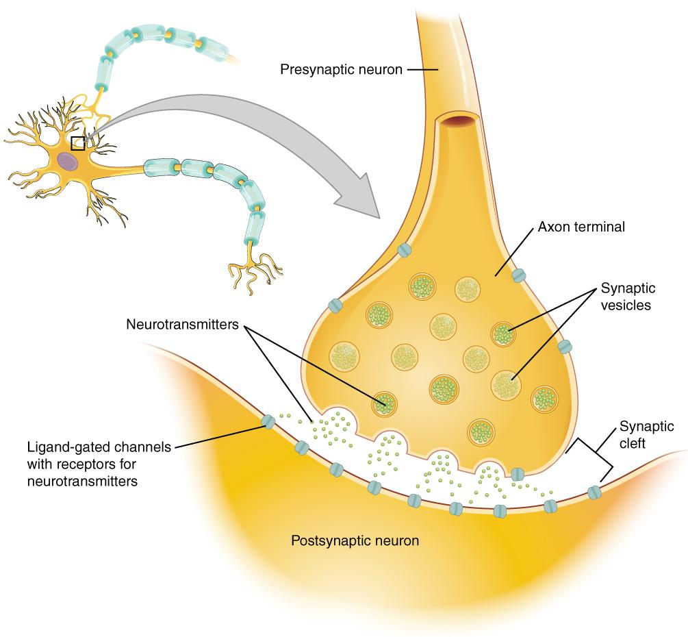
Chemical synapse
Junction where neurotransmitters cross a synaptic cleft to communicate with another cell

Electrical synapse
Junction that transmits signals directly between cells through gap junctions

Synaptic cleft
Small gap between the presynaptic and postsynaptic cells

Chemical synapse sequence
Electrical impulse reaches the axon terminal
neurotransmitter is released
target cell receives the signal through receptors
Note:
Action potential is the electrical impulse traveling along a neuron membrane
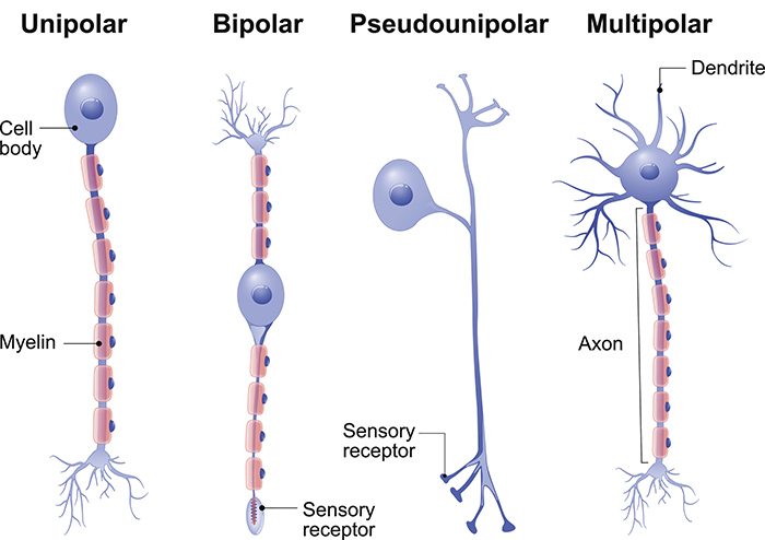
Multipolar neuron
Neuron with several dendrites and one axon
common in the brain and spinal cord
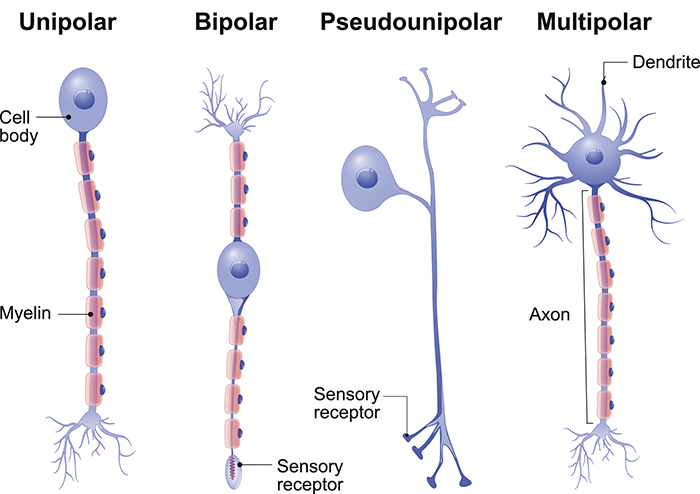
Bipolar neuron
Neuron with one dendrite and one axon
associated with specialized sensory pathways

Pseudounipolar neuron
Sensory neuron with one process that divides into two branches
Unipolar and Pseudounipolar neurons are most sensory nerves in the body

Neuroglia (glial cells)
Supporting cells that protect, nourish, regulate, and insulate neurons
Four CNS neuroglia
Astrocytes: Support neurons and help maintain the blood-brain barrier.
microglia: Remove pathogens and cellular debris by phagocytosis.
ependymal cells: Line CNS cavities and help produce and circulate cerebrospinal fluid.
oligodendrocytes: Form myelin sheaths around CNS axons.
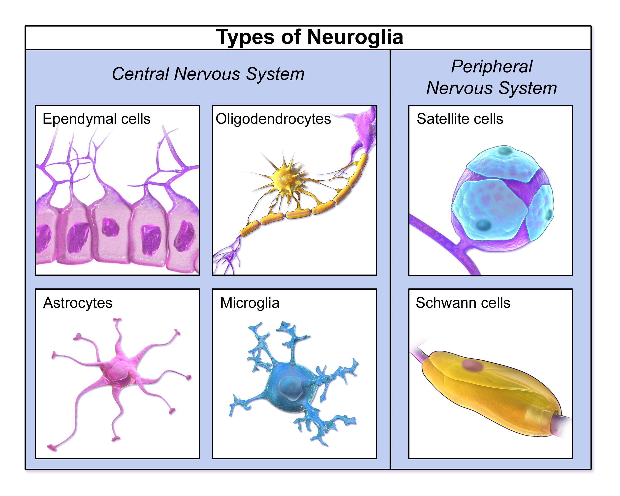
Two PNS neuroglia
Schwann cells: Myelinate PNS axons and help with nerve repair.
satellite cells: Surround neuron cell bodies in ganglia and maintain their environment.


Astrocytes
Star-shaped CNS glial cells that support neurons and help maintain the blood-brain barrier


Blood-brain barrier (BBB)
Selective barrier regulating movement of substances from blood into CNS tissue
Special feature: Tight junctions between brain capillary endothelial cells that help form barrier

Microglia
CNS immune cells that remove debris and foreign material through phagocytosis (Engulfing and digesting unwanted material)
Phagocytosis

Ependymal cells
CNS glial cells that line brain ventricles and the spinal cord central canal and help produce and circulate CSF

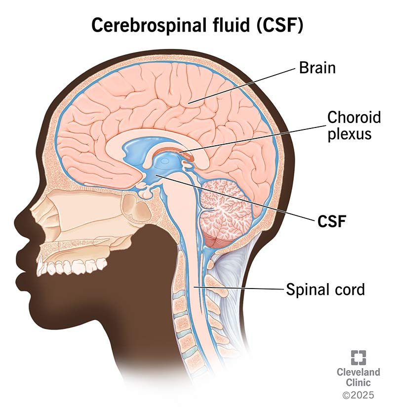
Cerebrospinal fluid (CSF)
Fluid that surrounds and cushions the brain and spinal cord
helps nourish the brain and removes wastes
transmits neurotransmitters around CNS
CSF is mostly made in the CHOROID PLEXUS

Oligodendrocytes
CNS glial cells that form myelin around segments of multiple axons


Schwann cells
PNS glial cells that form myelin around individual axon segments and assist peripheral nerve repair

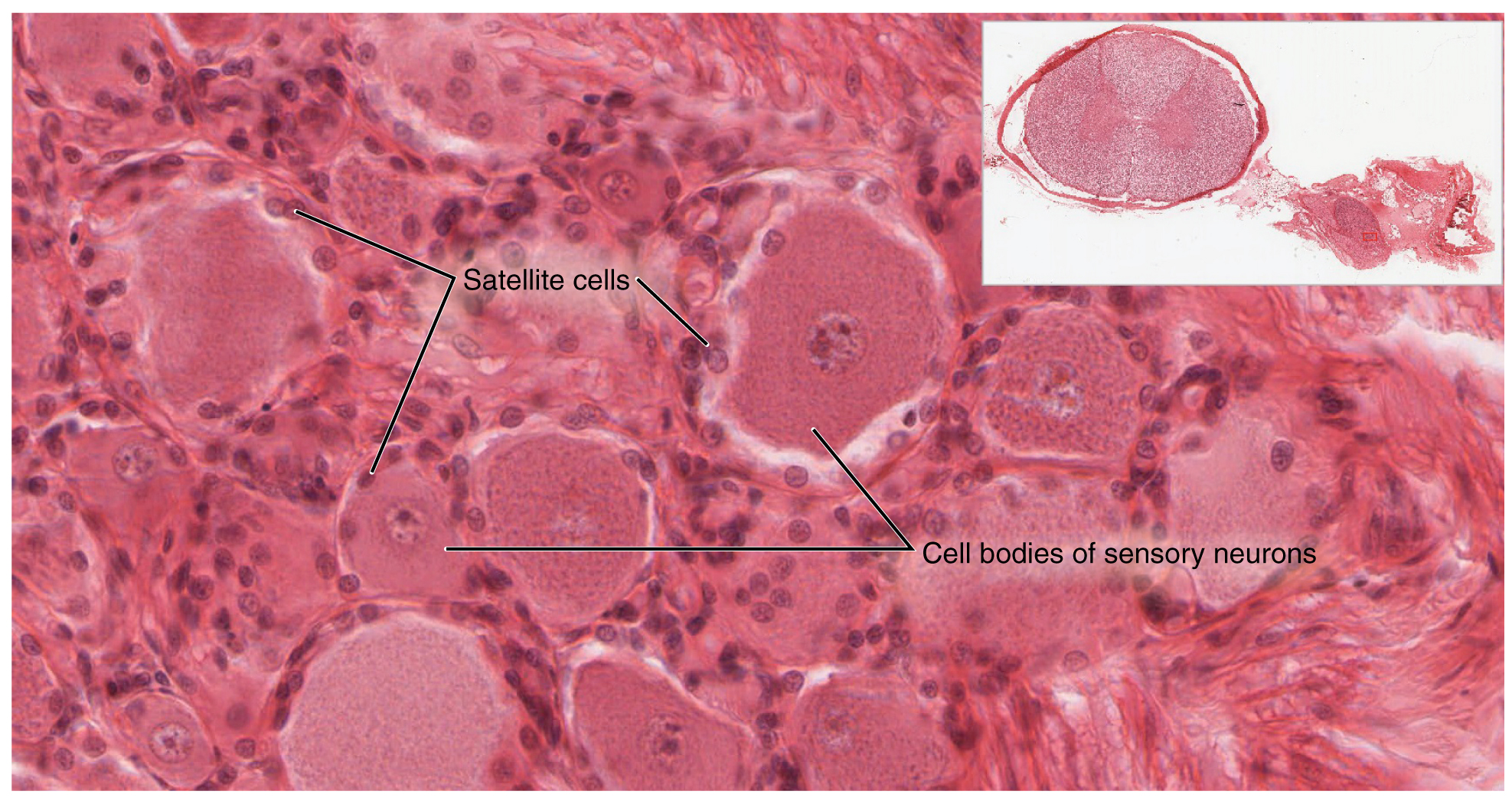
Satellite cells
PNS glial cells that surround neuron cell bodies in ganglia


Myelin sheath
Insulating layers around axons that increase conduction speed
Nodes of Ranvier
Gaps between myelin sheath segments
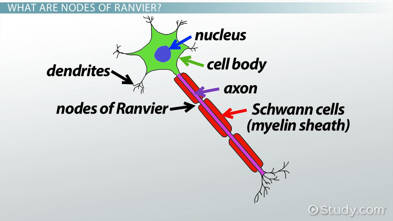
Saltatory conduction
When a nerve signal quickly jumps from one node of Ranvier to the next along a myelinated axon.

Dorsal root
Carries sensory information into the spinal cord

Ventral root
Carries motor information out of the spinal cord

Reflex
Rapid automatic response to a stimulus either somatic or visceral
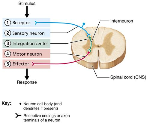
Reflex arc
Neural pathway responsible for a reflex response
There are two types:
Monosynaptic reflex arc: Reflex pathway with one synapse directly connecting a sensory neuron and a motor neuron.
Polysynaptic reflex arc: Reflex pathway with one or more interneurons between sensory and motor neurons.


Five components of a reflex arc
Receptor
sensory neuron
integration center
motor neuron
effector

Unipolar Neuron
contains one process attaching to one body.
Unipolar and Pseudounipolar neurons are most sensory nerves in the body

Anaxonic neuron
Neuron with multiple processes but no clearly distinguishable axon
found in CNS
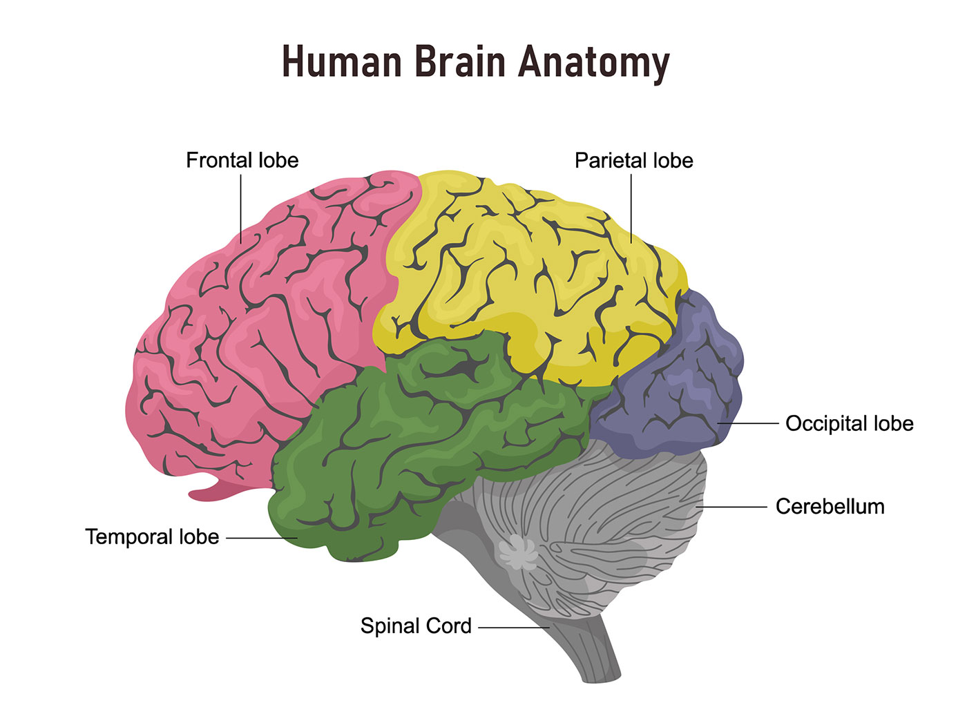
Main functions of the brain
Controls movement
interprets sensation
supports consciousness, cognition, and autonomic regulation
Four major brain regions
Cerebrum
diencephalon
brain stem
cerebellum

Cerebrum
Largest brain region with two hemispheres responsible for…
conscious sensation
movement
higher thought


Diencephalon
Region containing the…
thalamus
hypothalamus
epithalamus


Brain stem
Midbrain
pons
medulla oblongata
Contains white matter tracts with internal gray matter nuclei
produces automatic behaviors
acts as passageway for all fiber tracts running from cerebrum to spinal cord
involved with innervation of head, 10 of 12 cranial nerves attached to the brain stem


Cerebellum
Two cerebellar hemispheres connected medially by the vermis
Two primary functions:
Helps maintain posture and equilibrium
fine-tunes voluntary and involuntary movements

Gray matter vs white matter
Gray matter contains mainly neuron cell bodies
while white matter contains mainly myelinated axons and bundled into large tracts

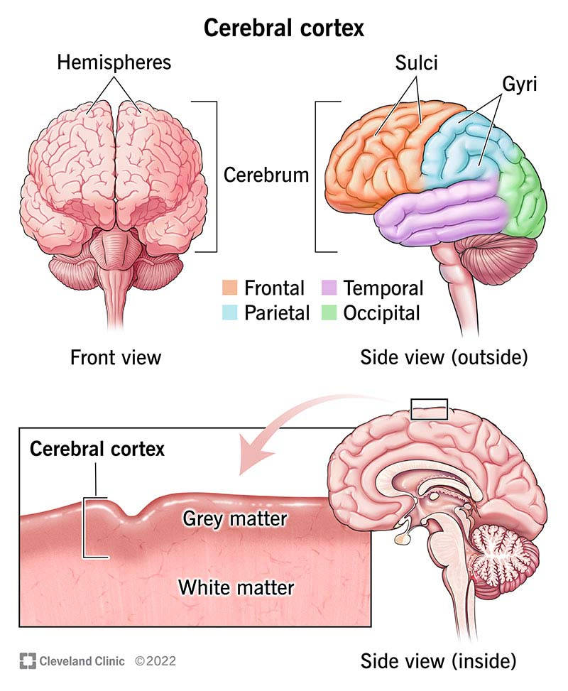
Cerebral cortex
superficial gray matter layer of the cerebrum


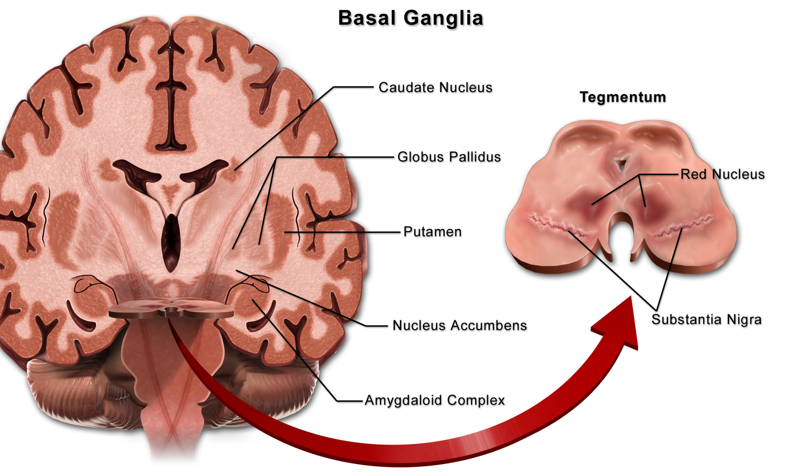
Basal nuclei
Internal structure of the cerebrum
Deep gray matter portion within the white matter involved in regulating movement
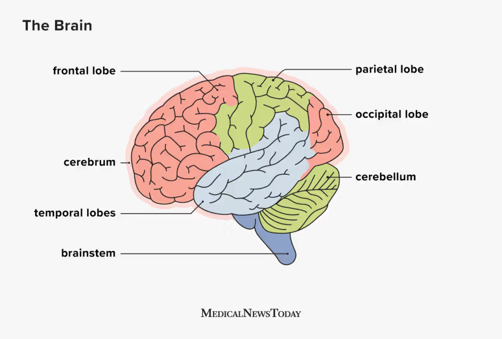
Frontal lobe
separated from the parietal lobe by the CENTRAL SULCUS
Voluntary movement and executive functions


Parietal lobe
Somatic sensation and spatial processing

Temporal lobe
Hearing, language, and memory
lateral side of the hemisphere
separated from the parietal and frontal lobes by the LATERAL SULCUS
inferiorly separated from the occipital lobe by the CALCARINE SULCUS


Occipital lobe
Vision
lies farthest posteriorly, separated from parietal lobe by the parieto-occipital sulcus occipital fissure


Insula
Deep region of the cerebral cortex within the lateral sulcus
involved in taste and visceral sensations (internal body sensations)

Central sulcus
Groove separating the frontal and parietal lobes
located between the precentral (motor) and postcentral (sensory) gyri

Lateral sulcus
Separates temporal lobe from frontal and parietal lobes
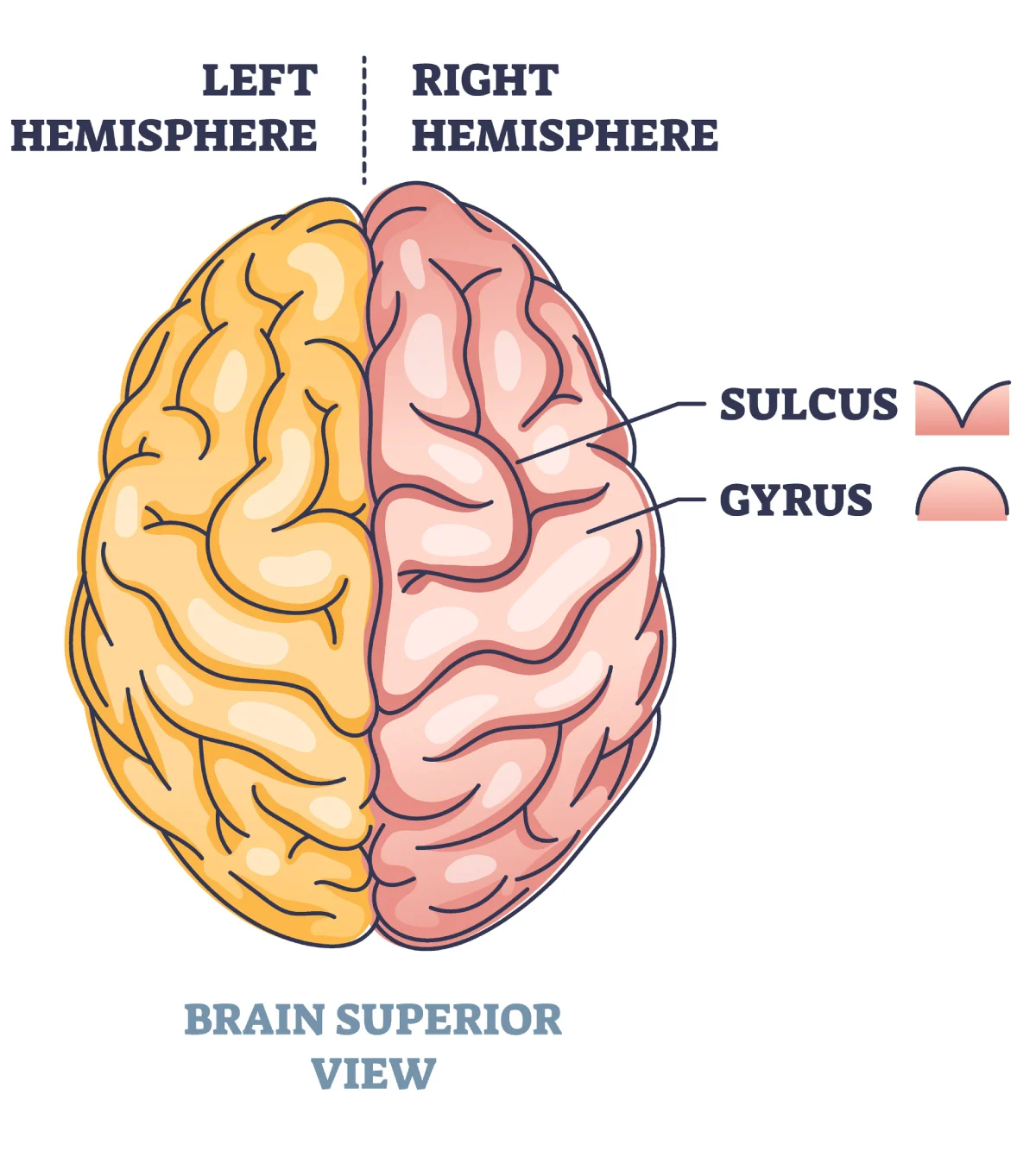
Gyrus vs sulcus
Gyrus is a raised fold, while sulcus is a groove


Primary motor cortex
Located in Precentral gyrus of frontal lobe
controls voluntary movement
especially the forearms, fingers, and facial movements

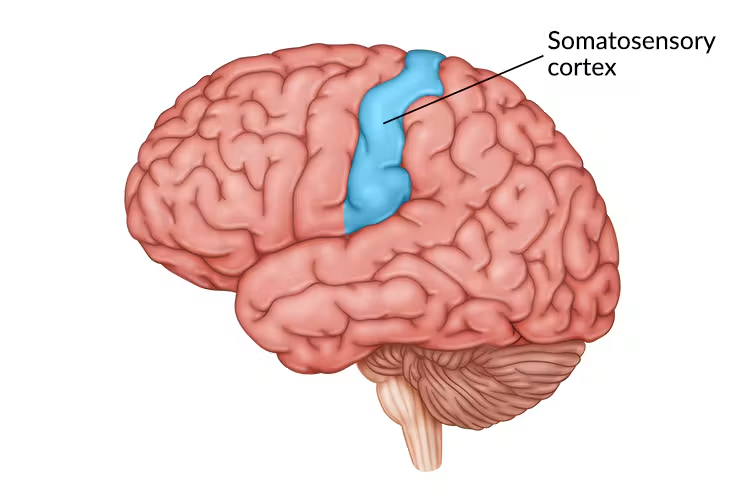
Primary somatosensory cortex
Located in the postcentral gyrus of parietal lobe
Processes body sensations
touch, pressure, pain, temperature, and body position


Contralateral cortical control
Each hemisphere primarily controls and senses the opposite body side

Motor vs sensory homunculus
Motor map is on precentral gyrus
sensory map is on postcentral gyrus
Somatotopy: the organized mapping of body parts onto the cerebral cortex.

Why are the hands and face large on the homunculi?
They require especially fine motor control or detailed sensory processing
lips and hands are the most sensitive body parts

Premotor cortex
Plans and organizes more complex movements
anterior to precentral gyrus


Frontal eye field
Controls voluntary eye movements
anterior to the premotor cortex


Broca area vs Wernicke area
Broca supports movements necessary for speech production
Wernicke supports language comprehension
Both: Usually located in the left hemisphere, which is the language-dominant hemisphere for most people.


Primary visual cortex
Largest cortical sensory area.
Receives visual information originating from the retina of the eye.
Located on the medial surface of the occipital lobe along the calcarine sulcus;

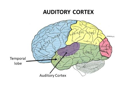
Primary auditory cortex
responsible for conscious hearing
loudness, rhythm, and pitch
Located in the superior temporal lobe primarily inside the lateral sulcus


Gustatory cortex
Taste processing, especially insular and nearby cortex
lies on roof of lateral sulcus

Olfactory cortex
Smell processing
media laspect of cerebrum in piriform lobe

Three cerebral white matter fiber types
Association fibers: Connect areas within the same cerebral hemisphere
Commissural fibers: Connect the left and right cerebral hemispheres.
Projection fibers: Connect the cerebral cortex to lower regions of the CNS


Association fibers
Connect regions within one hemisphere
run horizontally

Commissural fibers and corpus callosum
Commissural fibers connect the left and right cerebral hemispheres.
The corpus callosum is the largest bundle of commissural fibers


Projection fibers
Connect cerebral cortex with lower CNS regions
run vertically to and from brain stem and spinal cord


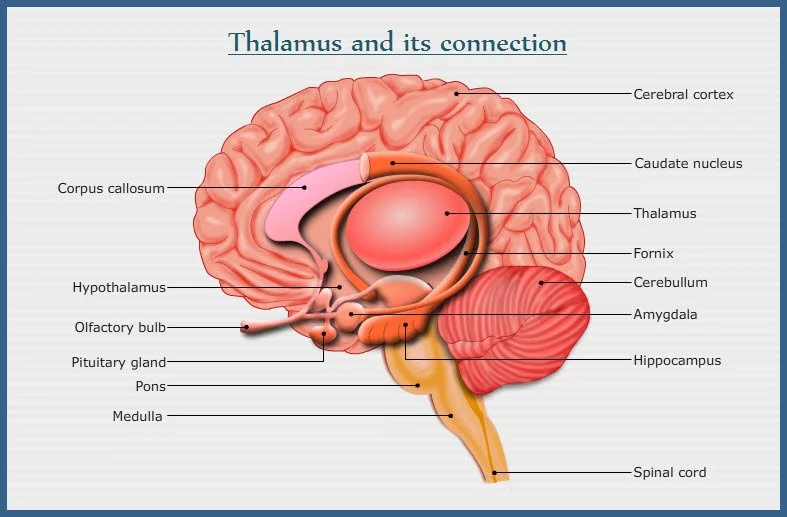
Thalamus
Major relay for sensory signals to the cortex
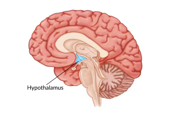
Hypothalamus
inferior portion of the diencephalon
Most important visceral control center
Regulates homeostasis, autonomic functions, and endocrine activity
forms a link between the nervous and endocrine systems


Epithalamus
most dorsal part of diencephalon and includes the pineal gland
Pineal gland secretes melatonin involved in sleep-wake timing


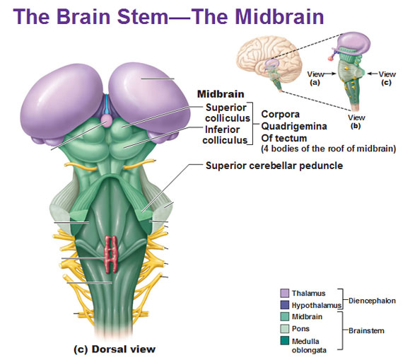
Midbrain colliculi
Superior colliculi control visual reflexes
inferior colliculi control auditory reflexes
both form the Corpora quadregemina

Pons
between midbrain and medulla oblongata
Brain stem relay region that helps…
regulate breathing
To help coordinate involuntary skeletal muscle movements and muscle tone
relaying information to and from the brain/spinal cord
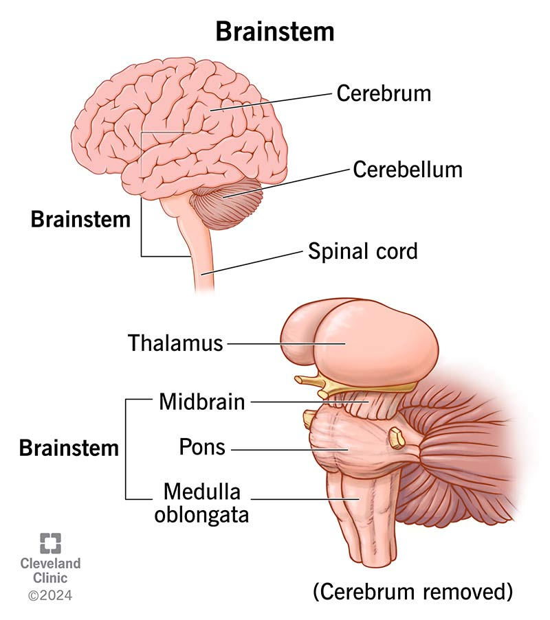

Medulla oblongata
most caudal part of brainstem, directly connects brain with spinal cord
relay station for cranial nerve nuclei
controls important visceral functions such as…
respiratory
cardiovascular (heart rate and blood pressure)
protective reflex centers
