Cell and organelles
 Call Kai
 Learn
 Practice Test
 Spaced Repetition
 Match
 Flashcards
 Knowt Play
1/53

There's no tags or description
Looks like no tags are added yet.
54 Terms
What is a cytoskeleton of a cell?
It consists of a network of fibres that extend across the cytoplasm of a cell, The cytoskeleton is the intracellular filament system that controls the shape of the cell and allows the cell to organize its internal components and to move. It also provides trafficking routes for intracellular transport.
Describe the structure of the cytoskeleton
highly dynamic structure
What techniques can we use to visualise the cytoskeleton
Use antibodies that recognise specific proteins
Using a fluorescent protein
List the three major components of cytoskeleton
Intermediate filaments
Microtubules
Actin filaments
What is the purpose of intermediate filaments?
Intermediate filaments provide tensile strength and lack the structural polarity characteristic of microtubules and actin filaments.
Which cells are abundant in intermediate filaments?
Cells that require a lot of strength (animal cells: Nerve cell axons, muscles etc..)
Why are intermediate filaments present in cells that require a lot of strength
Intermediate filaments have great tensile strength, enabling cells to resist stretching and mechanical stress
Are the toughest and most durable of the three cytoskeletal filament systems
Describe the location of intermediate filaments within the cell
Typically form networks throughout the cytoplasm, providing mechanical support
Often anchored to the plasma membrane at cell-cell junctions called desmosomes, helping neighbouring cells withstand mechanical stress
Describe the structure of the intermediate filaments
They are made up of a-helical monomers twisted together to form a coiled-coil dimer.
These dimers are staggered to form a tetromere. 8 tetromeres join together to form a lateral association, which when multiple join together form the intermediate filament
Give two take aways from the intermediate filaments structure
Take away 1: Intermediate filaments have NO polarity
Take away 2: Intermediate filaments assemble without ATP and GTP hydrolysis
What are keratins?
They represent a class of intermediate filaments
What are microtubules' purpose?
Maintain shape (e.g. axons)
Maintain cell polarity (e.g. during movement)
Transport and positioning of membrane enclosed organelles within the cell
Guiding intracellular transport of various cytosolic macromolecules
Form the mitotic spindle
Regulate cellulose deposition in plant cell walls
Describe the structure of microtubules
Microtubules are hollow tubes with structurally distinct ends
Tubes formed from dimers of two globular proteins: α and β tubulin
Microtubules have a definite structural polarity with a designated plus (+) and minus (-) end
Tubulin dimers are generally added more rapidly to the plus end than to the minus end
The polarity is crucial for microtubule assembly and function
What do microtubules display?
Dynamic instability
Describe characteristics of dynamic instability of microtubules
Dynamic instability: switching back and forth between polymerization and depolymerization
This allows microtubules to undergo rapid remodelling and is crucial for their function
Plus ends can be stabilized by binding to specific proteins (e.g. capping proteins)
Selective stabilization of microtubules can polarize a cell
Where is GTP found
Free tubulin dimers contain one GTP molecule (red)
tightly bound to β-tubulin, which hydrolyzes the GTP
to GDP (dark green) shortly after the dimer is added
to a growing microtubule
What does microtubule activity and function depend on?
Variety of accessory proteins that bind to them: microtubule associated proteins.
What can microtubule associated proteins do?
Stabilize microtubules against disassembly
Link microtubules to other cell components
Act as motor proteins that actively transport organelles, vesicles, and other macromolecules along
microtubules: kinesins move towards (+), dyneins toward (-)
What are kinesins and dyneins
Kinesins and dyneins are dimers
Are microtubules formed by tubulin monomers?
No dimers
What are actin filaments?
are thin and flexible, and allow animal cells to adopt a variety of shapes and can have
a range of functions

Give characteristics of actin filaments
Actin filaments are thin and flexible
Actin and Tubulin polymerize by similar mechanisms
Many proteins bind to actin and modify its properties
Cell crawling depends on cortical actin
Extracellular signals can alter the arrangement of actin
Filaments
Actin associated with myosin forms contractile
structures
Describe the actin filament structure
Subunit of each actin filament is an actin monomer
A cleft provides a binding site for ATP or ADP
Actin filament is a two stranded helix
Actin and Tubulin polymerize by similar mechanisms
Free actin monomers carry a tightly bound ATP
This is quickly hydrolysed to ADP soon after the actin
monomer is incorporated into the filament
This reduces the strength of binding between
monomers, decreasing the stability of the polymer
Describe treadmilling of actin filament
Occurs when ATP-actin adds to the plus end of an actin filament at the same rate as ADP-actin is lost from the minus end
Describe actin-binding proteins: Motor proteins
All actin-dependent motor proteins belong to the myosin family
They bind to and hydrolyse ATP, which provides the energy for their movement along actin filaments
Myosin-I is the simplest myosin
The tail varies among the different types of myosin-
I and determines what type of cargo the myosin
drags along
What does muscle contraction rely on?
actin and myosin filaments
Myosin-II molecules can associate with one another to form myosin filaments
• Coiled-coiled tails of myosin-II molecules associate with one another to form a bipolar myosin filament
• The bare region in the middle consists of tails only
Give the purpose of the nucleus
Contains genome;
site of DNA replication and RNA synthesis
Give the purpose of the rough endoplasmic reticulum
Synthesis of proteins destined
for secretion, insertion into the
plasma membrane, or residence
in the endomembrane system
Give the function of the golgi body
Modification, sorting
and packaging of
proteins and lipids for
either secretion or
delivery to another
organelle
Give the function of the mitochondria
ATP synthesis by
oxidative
phosphorylation
Give the function of the endosome
Sorting of
endocytosed
material
Give the function of the lysosome
Intracellular
degradation
Give the function of cytosol
Site of many
metabolic reactions
and protein
biosynthesis
Give the function of peroxisome
Oxidative
breakdown of
toxic molecules
Give the function of free ribosomes
Translate
proteins in the
cytosol
How do proteins synthesized in the cytoplasm reach their destination organelle (how do they "know" where to go)?
Proteins contain specific signal sequences that will direct them to specific organelles
Describe how proteins contain specific signal sequences that direct them to particular cellular compartments.
These signals are often short amino acid sequences, although their structure and location vary.
Some targeting signals are removed after
transport, while others remain part of the mature protein.
Both necessary and sufficient to direct a protein to a particular destination

Describe what targeting signals do
Targeting signals direct newly
synthesised proteins to specific
cellular compartments or into the
secretory pathway.
Describe exocytosis
the process of vesicles fusing with the plasma membrane,
releasing or “secreting” their contents

Describe endocytosis

Describe protein coats in endocytosis (Clathrin-coated)
Clathrin-coated vesicles transport selected cargo molecules
Surface receptors ensure specificity of transport
Each vesicle starts off as a clathrin-coated pit; clathrin molecules assemble into a basketlike network on the cytosolic surface of the membrane
When budding from the plasma membrane this process is also called clathrin-mediated endocytosis (CME)

Does the budding of clathrin-coated vesicles cost energy?
Yes
What are the three forms of endocytosis
Phagocytosis - engulf particles
Pinocytosis - engulfs liquids
Receptor mediated endocytosis - receptors will bind to specific cargo
Describe phagocytosis
A form of endocytosis in which cells engulf large particles, such as bacteria or cellular debris.
Used for feeding in some unicellular organisms and for defence (and scavenging dead and damaged cells) in animals
Performed by specialised cells including white blood cells such as neutrophils and macrophages
Recognition of a particle triggers actin-dependent membrane extensions (pseudopods) that surround and engulf it
The resulting phagosome matures and can fuse with lysosomes, where the contents are degraded
Describe pinocytosis
Is performed by most cells and occurs continuously
This process of cellular drinking is not molecule specific
Relatively small quantities of extracellular fluid, containing salts, water, amino acids etc. is taken up
Whatever happens to be around the invaginating region will end up being engulfed into the cell
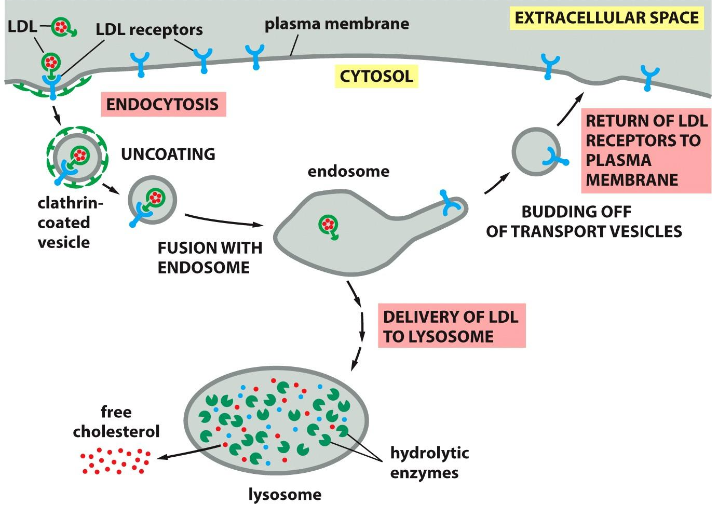
Describe receptor mediated endocytosis (RME)
Cells need to take up cholesterol to make new membrane
It is extremely water insoluble
Cholesterol bound to Low-density lipoproteins (LDL)
LDL-cholesterol bind to receptors → RME
Naked vesicle fuses with the endosome
Acidic environment triggers dissociation of LDL with its receptor
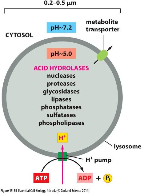
What is the pH of lysosomes?
pH 5 - Acidic
How is the pH of the lysosomes kept low?
The presence of many protons, by using a proton pump
Following exocytosis glycosylated domains of a protein can end up where?
Outside the cell
Describe protein targeting Specific signal sequences direct proteins to the appropriate
cellular compartments.
Describe Vesicle transport
Transport vesicles move proteins (and lipids) between
compartments. Exocytosis releases material outside the cell, while endocytosis brings material into the cell.
Describe Vesicle formation and fusion
Coat and adaptor proteins help select cargo
and form vesicles, while Rab proteins, tethering factors and SNAREs ensure
correct targeting and membrane fusion