Exam Two - Intro to Cognitive Neuroscience
 Call Kai
 Learn
 Practice Test
 Spaced Repetition
 Match
 Flashcards
 Knowt Play
1/136

There's no tags or description
Looks like no tags are added yet.
137 Terms
Overview: Visual cortical hierarchy
Two pathways:
Ventral/temporal: “What” = recognition
Dorsal/parietal: “Where/how” = space & guiding motor planning
Reciprocal interactions
Hierarchy ⇒ moving from V1 to downstream targets
More complex selectivity
Larger receptive fields
Visual Object Recognition: Ventral pathway
What/ventral pathway: occipito-temporal ⇒ V1 → inferior longitudinal fasciculus [white matter tract] → inferior temporal cortex.
Lateral occipital cortex/complex & posterior inferior temporal cortex: shape processing
Anterior inferior temporal cortex: object recognition
Superior temporal sulcus: Biological processing (hands, faces, body parts, interpreting emotions)
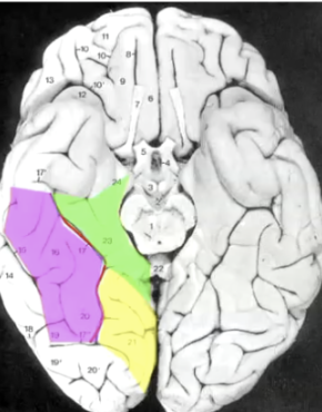
Ventral temporal cortex
Ventral temporal cortex
Fusiform gyrus (magenta): face
Separated from others by collateral sulcus (red line)
Parahippocampal area (green)
Lingual gyrus (yellow)
Visual agnosia
Failure to visually identify objects
Other perceptual modalities intact.
E.g., see object on table = can’t identify; hold object in hand (feel) or hear object = can identify.
DIFFERENT THAN MEMORY LOSS.
Shape Processing in Humans: Healthy fMRI
Healthy: activation in LOC in response to shape processing; visual agnosia = no activation.
fMRI study:
Control: random motion OR directional motion (w/o showing object) ⇒ little activation
Experimental: Motion object: form items/objects in dot movement patterns ⇒ activation
Control: luminance object (clearly defined) ⇒ activation

Shape Processing in Humans: Healthy object recognition
Familiar [feature extraction, shape description, memory matching] & novel objects [feature extraction, shape description] drive LOC, but scrambled objects [indistinguishable lines, no shape description] do not.
Bilateral temporal lobe damage: Explicit matching task
Task paradigm: explicit matching task (matching card’s orientation to wall’s slit), action task, memory task.
Patient w/ visual agnosia cannot match the card's orientation to the slit, but can reach for it and place the card in it.
Can do reaching task based on memory of slit orientation.
TAKEAWAY: Dorsal stream (“how”/ “where”) intact, even if the ventral stream is damaged.
Object constancy
Same object should be recognized independent of context & viewing conditions (viewing distance, angle, shadows, illumination, occlusions)
Strengths/abilities of human object recognition
Extremely rapid processing
~150 ms processing time = sufficient
Flexible: different views/contexts
Generality: recognize new category members
Identification: can identify individuals
Mental modules
Jerry Fodor: certain mental processes are self-contained or MODULAR.
Visual object recognition = highly specialized neural circuitry = mental module.
Requires vast amount of stored knowledge from past experiences.
Mental modules could, but do not need to correspond to brain modules ⇒ could be networks.
Properties of Modules
Domain-specific: only work on 1 type of information (e.g., visual information)
Little conscious awareness
Evolutionary mind modules.
VERSUS Domain-general processing: general-purpose processing → tends to be slow & attentionally demanding.
Beliefs cannot affect operations
Fast processing
Informationally encapsulated
Outputs are very basic or “shallow”
Experiment: Specificity of inferior temporal cortex
Single neuron in monkey’s inferotemporal cortex responds specifically to complex images (e.g., strongest response to bare hands; no response to face)
Hierarchical coding hypothesis
Gnostic unit: neuron that can recognize complex object.
Primary visual cortex (features) → secondary visual cortex (conjunction of features: e.g. corners, lengths) → occipital complex (shape) → higher up: object recognition.
Firing of specific higher-up neurons to objects = based on memory.
Computational models of visual recognition:
Increased complexity/selectivity from layer I → VI
Grandmother Cell Hypothesis
Extreme form of hierarchy: single cells proposed to sit at top of hierarchy w/ job of recognizing specific objects (e.g. recognizing ur grandma)
PROBLEMS:
What if a cell dies?
How do we recognize novel objects?
How does a neuron adapt over time?
Evidence for Grandmother Cell: Jennifer Aniston Neurons
Jennifer Aniston Neurons in Human Medial Temporal Lobe:
Showed famous faces to patients & conducted neurosurgical recordings ⇒ neurons respond specifically to Jennifer’s face.
Contrasting evidence for grandmother cell
MTL recognition cells not limited to objects ⇒ strongest response to text phrase “Halle Berry” rather than her face.
Violates “Grandmother Cell” Hypothesis as text is not person ⇒ representations can be more conceptual than perceptual.
therefore: MTL = memory region rather than visual region
Ensemble coding hypothesis
Proposes that:
A group of neurons codes high level features (e.g. mouth, face shape)
Together form an ensemble that flexibly codes objects.
Types of feedback in rapid object recognition
Bottom-up & feedforward: rapid speed of visual object recognition demands that most recognition occurs through this process.
Top-down & feedback
Low spatial frequency info “races ahead” ⇒ possibly to guide search for a match to memory.
E.g. sends a “blurred” shape of an umbrella (LSFs) → fusiform gyrus to identify potential matches.
Face inversion effect
Face recognition/detection is sensitive to orientation changes.
Objects do not experience the same effects → face perception is a different class of stimuli.
Case studies: Dr. P
Could recognize features but could not integrate them to recognize objects; no problems with other modalities/no dementia.
Tried to lift off his wife’s head = mistakes wife for hat.
Face recognition deficits: Prosopagnosia
Deficit in recognizing faces (including their own)
Unlike Dr. P, pure prosopagnosiacs can recognize standard objects:
Basic-level categorization
Ventral temporal lobe damage (more anterior) ⇒ fusiform face area (FFA)
Other visual recognition conditions
Double dissociation experiments show that agnosiacs w/o face deficit: damage to LOC
Congenital prosopagnosia (no lesion/stroke): “Faceblind”
~2% suck at facial recognition
Super recognizers: 1-2%
Some people are advanced at facial recognition ⇒ >80% of faces they see.
Substantial individual differences.
Face recognition areas
Monkey: Superior temporal sulcus
Human:
Specificity for faces in the right fusiform gyrus → more fMRI activation for faces > scrambled faces, objects, etc.
Causal stimulation of FFA: induce percepts of faces in a surgical patient w/ electrodes along fusiform gyri.
Face-specific ERP: N170
Faces produce a stronger EEG signal than other visual categories ⇒ negativity at 170 ms detected in the occipitotemporal cortex.
Overview of human recognition system & areas
One generalized “all-pupose” recognition system:
Lateral occipital cortex/complex: surfaces, shapes, general objects
Inferotemporal cortex
A few category-specific recognition modules
Faces: face network, including fusiform face
Body parts: extrastriate body area
Hands, torso/body-shape
Words: visual word form area → Left fusiform gyrus
Places: parahippocampal cortex
Visual word form area
Left fusiform gyrus supports sight reading of words.
Literacy is likely not driven by brain evolution
Experiment: trained monkeys to learn alphabets ⇒ LFG developed specificity for language, whereas untrained monkeys did not.
Face processing network
Fusiform (ventral temporal) face area + occipital face area + superior temporal sulcus (movement of face parts = emotions)
Conserved across species: marmosets, macaques, humans.
Categorization of stimuli
Basic Level Categorization: between different stimuli.
Subordinate level categorization: stimuli are similar to each other, but w/ finer-scale distinction (e.g. different species of penguins) → requires expertise/learning.
Face recognition may be an advanced form of subordinate-level categorization.
Visual expertise (e.g. w/ cars, birds) involves FFA recruitment ⇒ therefore, FFA is recruited for more than just faces.
Machine learning applications to object recognition
Encoding model: generates predictions about brain activity (stimuli → brain)
Decoding model: generates predictions based on brain activity (brain → stimuli)
Multivoxel pattern analysis (MVPA)
Decoding pattern from ROI → brain region contains specific category information
Model can be built by machine learning: train model on a set of patterns + stimuli AND then test it on unknowns.
Brain plasticity study
Early blind individuals who learn braille ⇒ recruit a large amount of visual cortex.
Tactile + auditory systems takes over visual machinery.
Someone who became blind later in life = shows less.
Key anatomy of attention
Posterior parietal
Temporoparietal junction
Lateral parietal cortex:
Intraparietal sulcus (IPS) separates superior & inferior parietal lobule.
Inferior parietal lobule: angular gyrus & supramarginal gyrus
Superior frontal cortex
Ventral prefrontal
Superior colliculus: midbrain → eye movement
Pulvinar of thalamus

Properties of attention
Engage mechanisms = allow continued cognitive processing of attended things.
Limited: Cannot attend to everything at once
Competition for cognitive resources
Stimuli & tasks compete for access
Selective: attention is a filter
→ Implications:
Some processing w/o attention
Most cognition requires attention → enables OR makes processing better.
Arousal states
Global effects: wakefulness (inattentiveness or attentiveness) OR sleep ⇒ arousal effects
Selective states: ignoring or attending⇒ attention effects
Cocktail party effect & Dichotic listening
Auditory selective attention
Limited/selective attention ⇒ competition of stimuli
Dichotic listening (one speaker in one ear, one in the other):
Dichotic: two ears = separate presentation
Subjects listened to one of two spoken passages that are simultaneously presented & asked to repeat one stream aloud (shadowing)
Results:
Ability to focus attention on one speaker among many distractors
Undetected changes: semantic content, language, backward speech
Detected changes: speaker volume, intonation, speaker gender.
Unattended speech appears to go unprocessed EXCEPT when hearing your own name.
Stages that attention act
Early selection: registration, perceptual analysis
Late selection: semantic encoding/analysis; executive functions & decision/memory.
ERP evidence: Observe difference between attended & unattended stimulus to left or right ear.
Early selection (between auditory channels): N1 effect
Within auditory channel (late selection): P300 effect
Exogenous vs. endogenous attention
Exogenous attention: reflexive
What you attend to may not always be in your control → stimuli may “capture” attention
Alerts of changes in the world
Endogenous attention: voluntary
Directed by mind not stimuli
Attention system failures: Inattentional blindness
“Looking but not seeing”: salient stimuli can go undetected. ⇒ highly focused (endogenous) attention blocks out other salient (exogenous stimuli)
Endogenous: self-directed, top-down
Exogenous: stimulus-driven, bottom-up
Implications:
To see object = must attend to it.
Most failures of attentional system occur w/o awareness.
Change blindness
Exogenous distractors mask other exogenous cues
One change = attention cued to location; widespread change = attention not cued.
Demo: film loop w/ images (original & change image) interspersed by blank screen. ⇒ forces memory to hold information about the original image.
Dan Simons & Levins: Experiment on Cornell campus.
Experimenter holds map & stops to ask a passerby for directions on campus
2 workmen walk between them with door, and experimenters will switch out.
With work outfit = can get away with race changes (e.g. Asian-Caucasian)
Implications of change blindness error
Fallibility of eyewitness testimony to strangers: limited long-term memory of the person/details.
We rely heavily on exogenous attention to detect changes:
Object changes usually produce a transient signal that attracts attention = permits us to detect change.
Distractor flashes and transients mask exogenous cues to attention = overwhelming the system.
Perception encodes less than we think:
We rely on the external world to hold most stimulus information = counting on things to not change too rapidly.
Sensory working memory capacity is tiny (2-4 objects)
Sites of attentional action
Where attentional selection acts: Reveals representations of selected information → attention alters/modulates these representations
Widespread sites: e.g., sensory cortex & thalamus
Dr. Somers’ lab: spatial attention to stimulus
Retinotopic mapping of V1-4 and visual field
Result: visual spatial attention modulates retinotopically specific regions of primary and other early visual cortex.
Earliest visual selection: LGN of thalamus ⇒ low-down: attention modulation can happen.
Attention experiments: Overt vs. covert attention
Overt attention: look directly at objects that we attend to
Increases attentional resources
Increased visual resolution: due to photoreceptor density in eye
Covert attention: eye position & attention are separable (e.g. “no-look pass”: directing attention improves performance)
Spatial attention & Paradigms
Spatial attention: direct to location
Paradigms:
Point of fixation differs from location of covert spatial attention
Increased processing in that location/ “spotlight” of location
Spatial attention: spatial cuing task
Central fixation
Must respond as soon as possible to the stimulus appearing on either left or right.
Cue participant (valid trial or invalid trial that misdirects )
Result:
Directing attention to a target (valid cue) = shorter reaction time than attending to wrong side.
EEG ⇒ ERP responses: component P1 (~100 ms after onset)
Mechanisms of spatial attention in monkey
Recorded in neuron from cortical area V4 (w/ fairly large receptive field) [later also seen in V1 & 2]
Monkey fixated at specific field
Presented one at a time: Maximum response to stimulus w/ correct color & orientation; poor response to incorrect color & orientation.
When both stimuli presented = averaging out of response levels.
Monkey can then attend to stimulus 1 or 2
Effective stimulus attended: response as though it was the only stimulus presented = great response ⇒ ignores the other stimulus.
Ineffective stimulus attended: response as though it was the only stimulus presented = poor response.
FINDING: Spatial attention shrinks the receptive field to the extent of the attended stimuli.
Feature-based attention: ERP
Different neural signatures for different forms of selection
Space-based = sooner; waveforms different drastically. ⇒ support argument that space-based and feature-based attention are distinct.
Feature based attention: Motion & color dot experiment
Number of dots that were colored or moving ⇒ participants instructed to pay attention to either color/movement on each trial.
Motion ⇒ increased activity in MT/V5
Color ⇒ increased activity in V4
Monkey Experiment: Object selection effect on similar objects
Selecting an object can attentionally enhance other objects that share key features with selected object (e.g. color, motion direction, orientation)
Preferred stimuli for neuron: Cloud of dots that move in the same direction (C) ⇒ up.
Experiment 1: Monkey pays attention to either blue dots moving up (A; preferred attention) or dots moving down (B) outside of neuron’s receptive field.
If pays attention to downward dots = downregulation of neuron response.
If pays attention to upward dots = upregulation of neuron response.
Experiment 2: if attending to both correct area of space + feature (up, inside receptive field) = stronger response.
Object-based attention: Experiment (fMRI)
Spreads to entire person/object
Experiment: subject shown transparent overlays of faces, house, etc. ⇒ some can also be moving.
fMRI findings:
FFA more activated for face attention & especially when stimulus is static.
PPA (parahippocampal place area) more activated for house attention & especially when stimulus is static.
MT/MST: Motion attended
Object-based attention: paradigm
Two objects with two locations on each side of the object.
Valid: dot shown up where cued ⇒ fastest reaction time
fMRI activity spread effect more strongly in V4 → weaker towards V1.
Invalid: uncued
Different side of same object
People are faster at detecting this than when the object is uncued at different location.
Different object
Attention & Rhythms
Attention mechanisms rely on networks & integration across many brain regions
Oscillation & synchrony:
Performance fluctuations at 4-6 Hz (theta band): performance (e.g. attentional strength) oscillates over time = likely due to synchronization of signals across brain regions.
Neural evidence for other frequency bands w/ other effects.
Sources of attentional control: Dorsal
Dorsal: Directed attention network
Supports attentional focus = maintains attention
“Endogenous” attention
Dorsal parietal regions: IPs (intraparietal sulcus); SPL (superior parietal lobule) & FEF (frontal eye field)
Anatomically similar to dorsal pathway to visual network: projections from occipital → parietal (dorsally)
Sources of attentional control: Ventral
Orienting/reorienting system
Alerts to salient stimuli outside of attentional focus => “Exogenous” attention
Disengages from current attentional focus & reorients
Brain region:
TPJ (temporal-parietal junction): Inferior parietal lobule (IPL) and superior temporal gyrus (STG)
VFC (ventral frontal cortex): Inferior frontal gyrus (IFG) & medial frontal gyrus (MFG)
Anatomically different ventral visual network (“What”)
TPJ → more frontal regions vs. occipital cortex to more inferior temporal cortex.
Invalid conditions of spatial cue paradigm = more strongly activated.
TPJ asymmetric activation in hemispheres
Asymmetric: stronger activation in right hemisphere > LH when stimuli appear in unexpected locations ⇒ reorienting attention
Circuit breaker that seems to happen in right temporal-parietal junction.
Superior longitudinal fasciculus
Fronto-parietal connections
SLFI: dorsal attention network
SLFIII: ventral attention network.
Default mode network
Deactivated during directed attention ⇒ NOT THE SAME AS VENTRAL ATTENTION NETWORK.
No task (resting state) & scanner = time course ⇒ see opposition in activation (default mode network deactivated when dorsal network is activated)
Implicated in some functions: Social cognition, memory functions, language, etc.
Reorientation to stimuli
Depends on bottom-up, non-conscious pathway (e.g. eye movements)
Eyes move in “saccades” ⇒ eye position dwells on the most interesting/informative parts of the scene (e.g. faces).
Saliency map: bottom-up map of relative interest of scene regions -> predicting direction of eye movements & attention
Neural locus: superior colliculus (in midbrain), pulvinar
Low-level visual processing = extract salient parts of image
Winner-take-all: competition across the map determines where to make the next eye movement, as some parts are more “attractive”
Priority maps
Neural locus: intraparietal sulcus.
Where we choose to move our eyes/covertly direct our attention:
Stimulus salience
Our goals/values (e.g. if ur hungry -> food)
Where we have already looked
More cognitive than saliency map → combines bottom-up & top-down
Premotor theory of attention
Shifts of attention & preparation of goal-directed actions are closely linked ⇒ share similar neural structures.
Covert shift of attention = similar to saccadic eye movement but w/ suppression of eye movement to location. {Wrong assumption}
Frontal eye field stimulation & V4 in covert attention: Exp. 1
FEF: region that plans eye movements
Experiment 1:
Researchers put stimulating electrode in monkey’s FEF & recording electrode in V4 ⇒ stimulate FEF site that matches V4 neuron’s RF.
Then, gave FEF a weak, subthreshold pulse → too small to cause eye movement
Visual stimulus in V4’s RF
Nothing in RF
Results: Modulation only when visual stimulation in receptive field (RF) of V4 neuron.
Weak FEF changed neuron’s response w/ sth in RF but not when there was nothing ⇒ therefore, FEF isn’t directly driving V4
Frontal eye field stimulation & V4 in covert attention: Exp. 2
Monkey did a task w/ target among distractors, where target is positioned in FEF’s site movement field. → weakly stimulate FEF & measured how well monkey detected targets.
FEF stimulation made monkey better at detecting target at stimulated location
FEF stimulated → V4 responses at location boosted → monkey’s perceptual threshold shifts.
Takeaway from FEF & V4 experiments
Subthreshold FEF stimulation may not cause eye movement, but monkey behaves as if its attention has been shifted
Signal preparing eye signal may serve as top-down attention signal ⇒ Supports idea that attention + eye-movement planning share circuitry
Evidence against premotor theory of attention
Separation at individual neuron level:
FEF: contains both “eye movement” and “visual” neurons
An attention task that does not require eye movements may modulate visual but not movement neurons.
Separation = argues against Premotor Theory of Attention
Hemispatial neglect
Can see (no sensory loss, not visual deficit) but not attend to stimuli in one hemisphere due to unihemispheric damage.
Disorder of spatial attention: treat neglected side of space as though it does not exist.
Confusing findings:
Hemispheric asymmetry: Observed in right hemisphere damage (e.g. stroke) almost exclusively ⇒ “Left neglect”
Key damage in ventral attention network (exogeneous stimuli) rather than areas w/ visual maps.
⅔ of subjects recover from neglect
Hemispatial neglect: Brain region
Primarily damage in the ventral region (temporoparietal junction) → superior temporal gyrus & temporal lobe
Not in dorsal network
Hemispatial neglect: Line bisection test
Test paper w/ horizontal lines on it ⇒ patient must draw bisecting vertical lines to evenly divide horizontal
Types of errors in patients w/ neglect:
Neglect to bisect lines all the way on the left
Poor bisection => not centered.
Hemispatial neglect: Mental imagery
Ask patients to imagine themselves facing door of Piazza standing at North & facing South -> describe to researcher
Could only describe things that are on the right, even when shifting perspective
Hemispatial neglect: Multiple reference frames
Ego-centric: body’s coordinates
Retino-centric: eye-centered
Head-centered
Body-centered
Allocentric → Object-centered:
Patients can only draw half an object, whether on the left or right.
Brain regions & coordinate reference frames
Task A: Patients given figure & asked to make copy.; Task B: Circle all the C & O (where some Cs were opened on the left)
More dorsal regions (inferior parietal lobule IPL/TPJ): Ego-centric neglect of space ⇒ only drew what was on the right & neglected letters on the left.
In contrast, patient with damage in superior temporal gyrus (STG) ⇒ object-centered coordinate loss
Drew objects throughout the visual space, but only drew their right-halves.
When opening of C on the right = detected; when opening of C on the left = cannot detect it well.
Recovery from neglect
Acute spatial neglect: Reduced activation in right IPS/SPL; increased activation in left IPS/SPL
Over time (poststroke) ⇒ rebalancing of hemispheres
Note: Structural damage to IPL/TPJ leads to hypoactivity in IPS/SPL
Theories behind hemispatial neglect
Representational model:
LH encodes right side of space
RH encodes both side of space
However, no evidence for asymmetry in parietal visual maps of healthy subjects.
Attentional bias model
Each hemisphere has contralateral bias
Stronger bias for LH
Normally, competition keeps them in check.
Extinction
Weaker form of neglect ⇒ attention drawn preferentially to one field (typically right).
Can also be a recovery stage of neglect
Experiment:
Stimuli presented either left or right: hemispatial subject cannot detect left, but patient with extinction can direct attention to the left.
Simultaneous stimuli presented left & right ⇒ patient w/ extinction only pick up right side of space.
Reveals competition btwn hemispheres
Balint’s syndrome
Bilateral damage to posterior parietal-occipital cortex
Simultanagnosia: Patients perceive only one object/time. ⇒ difficulty in perceiving multiple objects in the same visual space.
Test: How many colors do you see ⇒ Seeing scene = may only report seeing one object/color.
Optic ataxia: Impaired visually-guided movements ⇒ difficulty reaching or grasping objects.
Ocular apraxia: Difficulty voluntarily moving the eyes. They will display an inability to guide their eye movements to smoothly scan a visual scene.
Motor areas of cerebral cortex
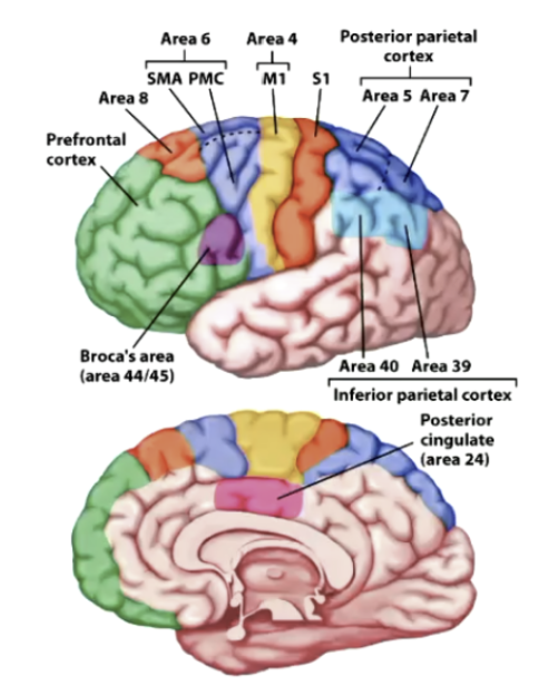
Primary motor cortex: BA 4 → voluntary movements to muscles
Anterior to primary motor cortex:
Supplementary motor area (SMA): dorsal BA 6 → planning internally guided movements.
Premotor Cortex: ventral BA 6 → Planning externally-guided movements + mirror neurons
Dorsolateral PFC: goal-directed actions
Frontal eye field: Area 8
Parietal eye field/LIP: BA7
Parietal reach region: BA 5 & 7
Broca’s area: includes motor aspects of speech production (BA 44/45)
Muscle fibers
Activated by alpha motor neurons
Acetylcholine: makes muscle fibers contract
Alpha-MN cell bodies in spinal cord (mostly) & brainstem (controlling head & neck)
1 motor unit: 1 motor neuron + muscle fibers that it innervates.
Agonist/antagonist muscle pairs at control joints: 1 flexes & 1 extends.
Electromyogram (EMG)
Electrical activity contracts muscles = produces electrical changes
EMG measures changes in membrane potential of muscles
Intramuscular electrodes or surface electrodes
Expected recording:
Recording from cortical motor neuron: Spike first
Spike from motor neuron follows after (from muscle activity)
Reverse pattern seen for sensory neurons (must receive signal from body first before brain)
Spinal cord
At spinal cord, lower motor neurons are organized into somatotopic maps
Long-axis: anatomical segment of spinal cord. ⇒ clustering of motor neurons that control specific muscle group clusters.
Proximal & distal mapping: Where muscles are located on body (e.g. abdomen = proximal; fingers = distal.)
Stretch reflex: Knee-jerk
Spinal cord reflect
Muscle spindles sense stretching in muscle
Sensory signal transmitted to dorsal root of spinal cord
Directly activates alpha motor neuron
Muscle contracts
Experiment: severed spinal cords in cats
Cat with severed spinal cord placed on treadmill ⇒ hind-legs started to move passively independent of supraspinal signals
Spinal circuits can be activated by tonic inputs = stepping rates increases w/ stimulus intensity
Even when dorsal roots are severed, leaving only motor neurons ⇒ agonist/antagonist muscles & left/right limbs are still coordinating.
Central pattern generator: small, autonomous neural networks in spinal cord = generate rhythmic motor behavior.
Central pattern generators: Clinical applications
Spinal cord damage: Long-axis of spinal cord.
Paraplegia: loss of control of lower limbs
Tetraplegia or quadriplegia: loss of control of all 4 limbs
Epidural stimulation in paraplegics = can produce rhythmic movements consistent w/ walking.
Fine control not yet achieved
Amyotrophic lateral sclerosis (Lou Gehrig’s disease)
Motor neuron disease:
Degeneration of neurons in lateral horn of spinal cord
Specific to neurons governing voluntary movements
Progressive degeneration, fatal ⇒ muscles atrophy as motor neurons decay
Cognitive functions mostly spared.
Motor pathways to spinal cord
Most motor neurons decussate on the way to the spinal cord (EXCEPT: cerebellum)
Corticospinal tract (CST) or pyramidal tract neurons: originate in motor cortex
Extrapyramidal tract neurons: originate in midbrain or medulla.
Primary motor cortex: Primates
Rostral subdivision:
Conserved across mammalian species
CST neurons terminate on spinal interneurons
Caudal subdivision:
Humans & some other primates
CST terminate on alpha motor neurons
Implicated in tool use, dexterous hand control
Disorders of PMC
Hemiplegia: loss of voluntary movement ⇒ contralateral to lesion in primary motor cortex.
Hemiparesis: motor weakness
Motor homunculus
Along pre-central gyrus.
Area proportional to degree of fine motor control.
Refined homunculus model:
Effector (effector=body part being moved)-specific & effector independent zones
Hand, foot, mouth are key effector-specific zones.
Multi-effector integration zone:
Co-activation seen in inter-effector zone = coordination between body parts
Behavioral code: Endpoint control
Deafferented monkeys: cut dorsal root sensory signals ⇒ does not know where limb is.
Experiment: apply external torque to oppose movement, then stop
Hypothesis: If the brain only sent a pre-programmed command for a specific amount of force or duration, an unexpected external resistance would disrupt the trajectory = monkey would not reach as far once the resistance was removed.
Result: reach the same endpoint, just delayed in movement. ⇒ Therefore, motor circuit code endpoint, not just force/torque
Coding both agonist & antagonist muscle motor signals (find equilibrium point) = can derive position
Motor cortex versus alpha motor neurons
Movement direction coded, independent of starting position.
Monkey starts hand at certain direction and must return to middle:
Each individual neuron respond more strongly to certain directions.
Alpha motor neuron: muscle movement ⇒ cares about postural movement
Population vector
Summation of preferred direction movement of individual neurons (0, 90, 180, 270) = determine probable direction of movement.
Motor cortex codes movement direction before movement is initiated (during motor planning)
Superior colliculus: eye movements
Compared to control, inactivation of part of direction map can alter motor readout
Cerebellum
Adaptive control: “On the fly” adjustments to load/obstacles based on predictions
No direct synaptic connections between cortex & cerebellum (pass through other structures)
Inputs from frontal motor & parietal cortices received at pons → cerebellar cortex (through middle cerebellar peduncle)
Cerebellar cortex also receives input from: inferior olive, spinal cord, vestibular nucleus (through inferior cerebellar peduncle)
Output: Cerebellar cortex → Deep cerebellar nuclei → ventrolateral complex in thalamus (through superior cerebellar peduncle) → primary motor & premotor cortices.
Cerebellar ataxia & Hypermetric arm movements
Loss of fine control & coordination.
Maintaining posture stability is difficult
Integration of feedback signals is disrupted.
Hypermetric arm movements:
Cerebellum have a special role in timing of agonist & antagonist muscles for smooth movement.
In cerebellar patients: antagonist muscles are activated (EMG oscillate) without waiting for feedback ⇒ causes arm position deviates from target.
Cerebellum: Internal model
Predicts sensory feedback to motor command.
Motor command deviates to two paths:
Efference copy: parallel signal from motor signal → predictor → predicted sensory feedback
Sensorimotor system (external influences: no delay/trajectory perturbation) → actual sensory feedback
When actual sensory feedback = predicted sensory feedback ⇒ no sensory discrepancy.
If error/discrepancy = correction signal quickly
If error persists = body will continue to adapt
Basal ganglia
Proceduralization: practice changes ⇒ structure of motor knowledge, requires less attention & cognition.
Striatum: Receives inputs from M1, S1, frontal cortex
Caudate nucleus:
Putamen:
Subthalamic nucleus (STN):
Globus pallidus: Gpi and Gpe (external)
Outputs: To ventral lateral nucleus of thalamus
Internal segment of globus pallidus (Gpi)
Part of substantia nigra: Source of dopamine → “go” signal
Output is inhibitory

Direct motor pathway
Cortex sends excitatory signals to striatum
Role of dopamine: Dopamine from SNc binds & excites D1 receptors in striatum ⇒ strengthen subsequent steps = increase in movement output
Striatum inhibits Gpi
Gpi disinhibits the thalamus → able to send excitatory signals to cortex ⇒ facilitates movement (“Go signal”)

Indirect motor pathway
Cortex sends excitatory signals to striatum
Striatum inhibits GPe
Role of dopamine signaling (SNc): Binds & inhibits D2 receptors in striatum ⇒ striatum inhibits GPe less = “silence” indirect pathway to facilitate movement.
Inhibition of GPe disinhibits the subthalamic nucleus (STN)
STN sends excitatory signals to GPi
GPi inhibits thalamus → cannot excite cortex ⇒ inhibits movement.
Winner-take-all: Motor go signal
When preparing to move = multiple potential action choices are competing ⇒ Winner-take-all where network amplifies strongest signal while mutually suppressing weaker alternatives.
Frozen addicts case study
Chem grad students synthesized MPPP (demerol-like opioid) → MPTP by-product & other consumed MPTP in synthetic heroin.
Destroyed dopamine producing cells in substantia nigra ⇒ Akinesia (inability to produce volitional movement) = extreme Parkinson’s syndrome.
Parkinson’s research: MPTP induces PD in monkeys, but not rats.
Parkinson’s Symptoms
Positive symptoms:
Resting tremor
Rigidity
Agonist & antagonist muscles simultaneously activated
Negative symptoms:
Postural instability
Hypokinesia: reduction of voluntary movement
Freezing: difficulty in initiating voluntary movements
Bradykinesia: slowed movements
Parkinson’s: Dopamine
Symptoms appear when SNc has lost many dopamine producing neurons
Treatments: L-DOPA (increases gambling addiction); deep brain stimulation (surgical implant into structures like subthalamic nucleus)
Experiment: PET scan = visualize dopamine uptake in striatum ⇒ observe reduction in Parkinsonian subjects.
Model of Parkinsons: Increased inhibition of thalamus
Loss of direct pathway ⇒ stronger GPi inhibition of thalamus
Indirect pathway no longer inhibited by dopamine ⇒ GPE inhibited more by striatum = weak inhibition of STN = more excitation of GPi = inhibits thalamus.
Deep brain stimulation placed in STN helps restore some function ⇒ reduces inhibition of thalamus = easier to facilitate movement.

Huntington’s Disease
Gene: huntingtin ⇒ but no cure/few treatments
Neurodegenerative genetic disorder:
Symptoms typically emerge ages 35-50 ⇒ 50% inheritance
Chorea/hyperkinesia: involuntary movements
Early symptoms: clumsiness, balance problems, restlessness
Dementia of subcortical type: deficits in cognitive executive function & memory.
Pathology of Huntington’s: Motor pathway
Greatest pathology: striatum
Loss of inhibitory neurons in “indirect” pathway
Less inhibition of striatum on GPe → so greater inhibition on STN & GPi = decreasining overall inhibition on thalamus.
Also affects substantia nigra, hippocampus & cortical layers (3, 5 & 6)

External vs. internal loop
External loop: Dominates visually-guided movements
Cerebellum, posterior parietal cortex, lateral premotor cortex
Internal loop: self-guided, well-learned movements
Basal ganglia, supplementary motor area