Exam 1 unit 1 patho
 Call Kai
 Learn
Learn Practice Test
 Spaced Repetition
 Match
Match Flashcards
 Knowt Play
1/82

Earn XP
Description and Tags
Name | Mastery | Learn | Test | Matching | Spaced | Call with Kai | Chat |
|---|
No analytics yet
Send a link to your students to track their progress
83 Terms
Physiologic hyperplasia
Occurs due to a normal stressor (e.g., increase
in the size of the breast during pregnancy)
Pathologic hyperplasia
Occurs due to an abnormal stressor (e.g., proliferation of the endometrium due to prolonged estrogen stimulus)
Can the heart and neurons undergo hyperplasia?
No, they cannot undergo hyperplasia due to their post-mitotic nature; instead, they may undergo hypertrophy.
Physiologic hypertrophy:
Occurs due to a normal stressor (enlargement
of skeletal muscle with exercise)
Pathologic hypertrophy:
Occurs due to an abnormal stressor (e.g., an increase in the heart due to aortic stenosis)
Can you distinguish hyperplasia and hypertrophy grossly
no, and microscopic examination is required to distinguish the two.
Physiologic atrophy
Occurs due to a normal stressor (e.g., decrease in
the size of the uterus after pregnancy)
Pathologic atrophy
Occurs due to an abnormal stressor. In general, atrophy is due to the loss of stimulus to the organ. Specific types of loss of stimulus include loss of blood supply or innervation, loss of endocrine stimulus, disuse, mechanical compression, decreased workload, or aging.

idk just know

Myostatin Mutations
Metaplasia
Change of epithelium at a site, or location, from one type of
epithelium to another type.
Mech of metaplasia
The epithelium normally present cannot handle the new environment it is in, so it converts to a type that can adapt
Is metaplasia reversible
yes, metaplasia can be reversible if the underlying cause is removed.
Barrett esophagus
a type of metaplasia where the normal squamous epithelium of the esophagus is replaced by columnar epithelium due to chronic gastroesophageal reflux disease (GERD).
squamous metaplasia in the respiratory tract epi of the trachea and the bronchi in response to prolonged exposure to cig smoke
Metaplasia occurs when normal ciliated columnar epithelium is replaced by stratified squamous epithelium, providing increased protection in the airway.
Causes of cell injury
include hypoxia, ischemia, chemical exposure, infections, and physical trauma, genetic or acquired metabolic abnormalities, aging.
Hypoxia
a condition where there is inadequate oxygen supply to tissues, leading to cell injury and dysfunction.

Hypoxia pathway
Decreased oxygen results in decreased production of
ATP.
• Na/K+ and Ca2+ pumps are ATP-dependent.
• When ATP levels decrease, these pumps fail and Na+
enters the cell (along with H2O), causing swelling.
• Ca2+ enters the cell and activates proteases,
endonucleases, phospholipases, and DNAses, which
damage the cell.
• Cells switch to anaerobic respiration to produce ATP,
which results in accumulation of lactic acid,
decreasing cellular pH.
• Decreased pH causes disaggregation of ribosomes
from the ER (Na+/H+ ion exchanger is also activated,
bringing in more Na+).
Free radicals?
Highly reactive molecules with unpaired electrons that can damage cells by reacting with lipids, proteins, and DNA.
Oxygen-derived free radicals
ROS
How are free radicals generated?
by normal physiologic reduction-oxidation reactions, ultraviolet light, x-rays and ionizing radiation, and transitive metals.
Metabolism of exogenous chemicals, such as carbon tetrachloride induces formation of what?
formation of ROS
Damage by free radicals
Lipid peroxidation (damages cell membranes), DNA fragmentation, and protein cross-linking which result in increased degradation and decreased activity.
Antioxidant Defenses
Detoxifying Enzymes and Scavengers of ROS
Detoxifying Enzymes
Superoxide Dismutase (SOD) - is the first line of defense against O2- (superoxide), converting it to hydrogen peroxide (H2O2).
Catalase - mainly located in peroxisomes, is one of two enzymes that complete the detoxification of O2- by converting H2O2 to water.
Glutathione peroxidase (GPX), catalyzes the reduction of H2O2 and lipid peroxides in mitochondria and the cytosol.
Scavengers of ROS
– Vitamin E (𝛂-tocopherol)
– Vitamin C (ascorbic acid)
– Retinoids (precursors to vitamin A)
– Nitric Oxide (NO)
Chemical Injury
Damage caused to cells due to exposure to toxic agents, leading to cell dysfunction or death. Common sources include drugs, heavy metals, and radiation, which disrupt cellular structures and metabolic processes. Some require conversion to a toxic metabolite
Ethylene glycol → metabolite oxalic acid
Chemical Injury
Cyanide inactivating complex IV impairing ATP synthesis
Direct chemical injury
Altered mitochondrial membrane potential
When the cell experiences stress or high calcium levels in the cytoplasm, more calcium flows into the mitochondria
What happens when Ca2+ increases in the mitochondrial matrix?
↑ROS, ↓low pH) lead to lipid peroxidation and the formation of the mitochondrial permeability transition pore (MPTP). This promotes cytochrome c release, which activates the apoptotic pathway.

Altered mitochondrial membrane potential KNOW THIS
What are the types of cell injury
Reversible/Irreversible
Define reversible cell injury
Decreased ATP production causes sodium to enter the cell, bringing water and swelling the cell and organelles. The conversion from aerobic to anaerobic respiration decreases cellular pH. These changes are reversible. If ATP is once again produced by the cell, the Na+/K+ ratio and pH will be corrected.
Define irreversible cellular injury
This type of injury occurs with damage to the plasma or lysosomal membranes, loss of DNA, or loss of mitochondria. In these cases, the damage cannot be reversed. The two most important factors determining irreversible damage are membrane disturbances and the inability to reverse mitochondrial dysfunction
When thinking about Reversible injury
Cellular swelling (hydropic) and fatty change
Necrosis
uncontrolled cell death due to one of the various causes of cell injury (swelling, breakdown of membrane and organelles)
Two types of necrosis
Coagulative necrosis and Liquefactive (others do exist but thanks Canela)
Coagulative necrosis
Coagulative necrosis is the type of necrosis in which protein denaturation is more prominent than enzymatic breakdown. There is increased eosinophilia of the cytoplasm and decreased basophilia of the nucleus; both are associated with preservation of the general cellular architecture (the organ type is identifiable).
What organs are affected by coagulative necrosis
Can occur in any organ but organs with high fat content like the brain is followed by liquefactive necrosis. (also spinal cord)
Liquefactive Necrosis
Occurs in situations in which enzymatic breakdown is more prominent than protein denaturation or in organs that lack a substantial protein-rich matrix (e.g., lipid-rich organs such as the brain). Histology impressions: Loss of organ cell architecture. In the brain, there are sheets of lipid- laden macrophages that replace dead tissue.
What organs are affected by liquefactive necrosis
most commonly associated with organs with high fat and low protein content (brain) or high enzymatic content (pancreas) also can be in skin
Fat necrosis
A change in adipose tissue due to trauma or release of enzymes from adjacent organs (pancreas) the trauma → breakdown of lipids/ release of fatty acids → combine to form chalky deposits
Caseous Necrosis
A form of tissue necrosis characterized by the presence of cheese-like (caseous) necrotic tissue, often associated with tuberculosis infections. It features a granular collection of macrophages and necrotic cells surrounded by a distinct inflammatory border.
Cell death by necrosis
1. Ischemia reduces O2 and glucose
2. Anaerobic glycolysis produces lactate/lactic acid
a) Reduced pH (increase in H+)
b) Reduced ATP
3. Reduce plasma membrane ion pump function→ ionic
imbalances
4. Ca2+ accumulates in cell
5. Ca2+ activates phospholipase A2
a) Plasma membrane disrupted
b) Cell swelling
6. Impaired mitochondrial electron transport
a) Reduced ATP
b) ROS formation
7. Cell dies

Apoptosis
Programmed cell death
Patterns of occurrence of apoptosis
An organized process that occurs in specific physiological or pathological contexts, such as during development, tissue homeostasis, and in response to cellular stress or damage. Apoptosis may be triggered by signals from within or outside the cell to eliminate damaged or unwanted cells.
Phases of apoptosis
Initiation - the phase in which caspases (cysteine aspartic acid proteases) become catalytically active.
Execution - the phase in which the action of caspases causes the death of the cell
Initiation of extracellular pathway (apoptosis)
The Fas ligand (FasL) binds to a member of the tumor necrosis factor (Fas receptor) → activated receptor activates FADD → activated caspases
Initiation of intracellular pathway (apoptosis)
The mitochondria release cytochrome c → combines with Apaf-1 → activates caspases → cleave DNA (looks like a ladder)
does apoptosis generate inflammation?
No lol Apoptosis is a non-inflammatory process, designed to eliminate cells without triggering an immune response.
Extrinsic pathway of apoptosis
The extrinsic pathway of apoptosis is initiated when death ligands bind to their corresponding death receptors on the cell surface, triggering a cascade that activates caspases and leads to programmed cell death.

Necrosis vs. Apoptosis


Know this silly goose
Lipofuscin
Wear and tear pigment
Lipofuscin mech of formation
Lipofuscin is a product of lipid peroxidation, which accumulated in the lysosomes as the cell ages. The cell CANNOT rid itself of these lipofuscin-laden lysosomes.
Organs with lipofuscin accumulation
Heart and liver
Gross morphology of lipofuscin accumulation
Lipofuscin accumulation can impart brown discoloration to organs. Such organs may also be atrophic, giving rise to the term “brown atrophy.”
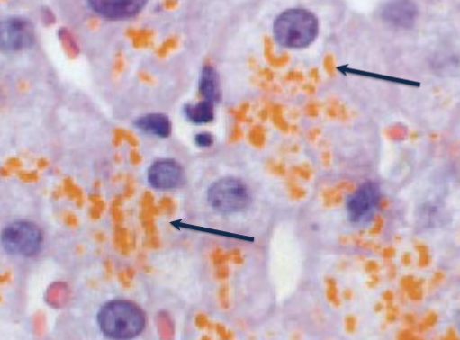
Microscopic (Histology) morphology of lipofuscin accumulation
Finely granular, yellow-brown pigment which often surrounds the nucleus
Forms of calcium deposition?
Metastatic and dystrophic
Mech of dystrophic calcification
Patients who have normal levels of calcium have deposition of calcium only within abnormal tissue, such as necrotic tissue.
Mech of metastatic calcification
Patients who have hypercalcemia have deposition of calcium within normal or abnormal tissue.
Some causes of hypercalcemia
Increased PTH by parathyroid adenoma or parathyroid gland hyperplasia - destruction of bone via tumors - Vitamin D intoxication - renal failure - sarcoidosis
Organs most commonly affected by calcium accumulation
Vasculature, kidneys, and lungs
Gross morphology of calcium accumulation
Hard yellow nodules
Protein accumulation involvement
Intermediate filaments (Mallory hyaline in the liver and neurofibrillary tangles in Alzheimer’s disease.
Forms of iron accumulation
Hemosiderosis and hemochromatosis
Hemosiderosis
Accumulation of iron in organs without resultant side effects. The iron pigment is frequently within macrophages. Hemosiderin is a term used for aggregates of ferritin micelles
Hemochromatosis
Accumulation of iron in parenchymal cells resulting in side effects, including congestive heart failure, diabetes mellitus (from damage to the pancreas), and cirrhosis. Hemochromatosis can be acquired or hereditary.
Organs affected by hemochromatosis
liver, skin, heart, pancreas
Microscopic morphology of iron accumulation
Chunky, yellow-brown granules.
(iron stains positive with a Prussian blue stain).
Fat accumulation (steatosis) organs affected
Liver, kidney, heart, skeletal muscle
Steatosis gross morphology
Enlarged, fatty liver with a yellow appearance and potential for a smooth surface.
Steatosis microscopic morphology
One or several clear vacuoles within the cell.
What can steatosis indicate?
Reversible damage or may be the sign of an intrinsic abnormality in fat metabolism.
Cellular aging
Tissue cells have a fixed number of divisions Telomer gets shorter if too short DNA is interpreted as broken leading to cellular senescence or apoptosis.
Immortal cells have what?
Telomerase
rare mutations in telomerase leads to?
shortened telomeres and aplastic anemia, skin and nail defects, infertility, pulmonary fibrosis, and cancer.
Shortened telomeres are associated with what diseases
cirrhosis, ulcerative colitis
Disease resembling ageing
is known as progeria. It features symptoms similar to accelerated aging, including growth delays and hair loss.
Werner syndrome
caused by recessive mutations in the WRN gene (a DNA helicase involved in replication and telomere maintenance). Succumb to MI or cancer by their 40s or 50s.
Hutchinson-Gilford progeria
is a rare genetic disorder caused by a mutation in the LMNA gene, leading to accelerated aging and associated with a variety of health issues, including cardiovascular problems and skeletal abnormalities. Live, on average 13 years