Nervous System
 Call Kai
 Learn
 Practice Test
 Spaced Repetition
 Match
 Flashcards
 Knowt Play
1/171

There's no tags or description
Looks like no tags are added yet.
172 Terms
Dendrite
protrusions from the cell body where signals are recieved
Cell body
the large central part of the neuron where the nucleus and other organelles are housed
Initial segment
top of the axon where action potentials are triggered
Axon
long protrusion from the cell body that carries electrical signals
Axon termini
small protrusions from the bottom of the axon where signals are sent from
What are special features about the cell cycle of neurons?
amitotic, high metabolic rate
amitotic
do not divide
Clusters of cell bodies in the CNS
nuclei
Clusters of cell bodies in the PNS
ganglia
Function of a microtubule in an axon
make tracks that transport proteins can walk upon
Central nervous system (CNS)
brain and spinal cord
Peripheral nervous system (PNS)
everything other than the CNS
Afferent
carry information from the periphery to the brain
Efferent
carry information from the brain to the periphery
Interneurons
found only in the CNS, connect sensory and motor neurons to the brain
Oligodendrocyte
myelinated CNS glial cell that insulates axons so that electrical signals can propagate
Microglia
CNS glial cell that acts as the immune system
Ependymal cells
ciliated CNS glial cells that produce cerebrospinal fluid
Astrocytes
CNS glial cells that establish the blood-brain barrier
Schwann cells
surround and form myelin sheaths around larger nerve fibers in the PNS, important for regeneration and proper nerve signal conduction
Synapse
space between a sending axon and a receiving dendrite that can use electrical or chemical stimuli to inhibit or excite action
Cerebral cortex
the main functional portion of the brain split into different lobes
Parts of the forebrain
cerebrum and diencephalon
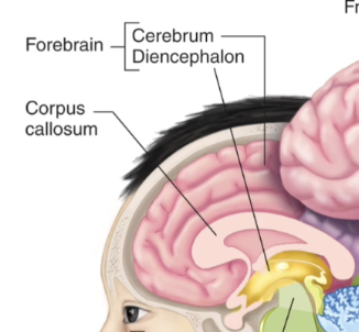
Parts of the brainstem
midbrain, pons, medulla oblongata
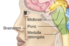
Parts of the hindbrain
pons, medulla oblongata
Cerebellum
packed with neurons, in charge of complex motor functions

Corpus callosum
separates ledt and right brain
Parts of the cortex
motor cortex and somatosensory cortex
Motor cortex function
specific, voluntary movements
Somatosensory cortex function
processing environmental sensory input
Hypothalamus
below the thalamus, master command center for neural and endocrine coordination, in charge of preservation of the individual, preservation of the species
Brainstem
nerve fibers relaying signals between forebrain, cerebellum, and spinal cord pass through the brainstem
Reticular formation
loosely arranged cell bodies in the brainstem intermingled with axons
Function of the reticular formation
absolutely essential for life, controls motor functions, cardiovascular, respiratory, sleep, focus, integrates CNS info
Brainstem nuclei
clusters of neurons creating integrating centers
Function of brainstem nuclei
integrating info from cardiovascular, respiratory, swallowing, eye-movement, reflex orientation
Cranial nerve function
10 of 12 pairs connect directly with the brain and innervate muscles, glands, sensory receptors of the head and thoracic and abdominal cavities
Gray matter
contains interneurons’ cell bodies, dendrites of efferent, axons of afferent, glial cells
Dorsal root
brings sensory info to the dorsal horn

Ventral root
brings motor information from the ventral horn to the periphery

White matter
myelinated axons
Path of descending fibers
brain to periphery
Path of ascending fibers
periphery to brain
Cervical spinal nerves
8 nerve pairs, control muscles and glands, receive input from neck, shoulders, arms, hands
Thoracic spinal nerves
12 nerve pairs, chest and upper abdomen
Lumbar spinal nerves
5 nerve pairs, lower abdomen, hips, legs
Sacral spinal nerves
5 nerve pairs, genitals, lower digestive tract
Coccygeal spinal nerve
1 nerve pair, tailbone
Subclasses of efferent nerves
somatic and autonomic
Somatic nerves
single neuron between CNS and skeletal muscle cells, innervates skeletal muscle cells and only leads to excitation

Autonomic nerves
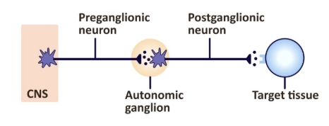
two-neuron chain between CNS and organ, innervates smooth and cardiac muscle, glands, GI neurons, can be excitatory or inhibitory

Subclasses of autonomic nerves
sympathetic and parasympathetic
Sympathetic/thoracolumbar nerves
short pre-ganglionic, long post-ganglionic synapses, control fight or flight responses

What neurotransmitters are used in the sympathetic nervous system
ACh pre-ganglionic, NE/Epi post-ganglionic
Parasympathetic/craniosacral nerves
long pre-ganglionic, short post-ganglionic, control rest and digest responses

What neurotransmitters are used in the parasympathetic nervous system
ACh pre- and post-ganglionic
Ganglion
cluster of cell bodies that connect the body to the CNS
Cholinergic receptors
receptors on skeletal muscles innervated by somatic neurons, nicotinic and can cause contraction
What is special about the neurons in the adrenal medulla
postganglionic sympathetic but never develop axons
Function of the adrenal medulla
release and 80/20 mixture of Epi/NE into the blood
Neuron
individual nerve cell
Nerve
many neurons working together in a network
Neurotransmitter
signal passed from neuron to neuron
Hormones
signal released into the blood
Catecholamines
neurotransmitters/hormones that activate the fight or flight response
What structures receive both sympathetic and parasympathetic input?
heart and glandsW
What are the effects of sympathetic and parasympathetic divisions on the heart and glands?
whatever effect one division has, the other has the opposite and are usually activated reciprocally
Meninges
membranes between skull and brain that support and protect
Cerebrospinal fluid
generated in the choroid plexus and goes around the brain and spinal cord to protect and cushion the CNS
Cranium and vertebrae
bones that support and protect the CNS
Layers of the meninges
dura mater, arachnoid mater, pia mater
Dura mater
outermost layer, thick membrane
Arachnoid mater
middle layer, web-like membrane
Pia mater
clings to brain, contains blood vessels
Function of the meninges
cover and protect the CNS, protect blood vessels and enclose venous sinuses, contain cerebrospinal fluid
Subarachnoid space
in between the arachnoid mater and pia mater, filled with CSF, contains largest blood vessels serving the brain
Superior sagittal sinus
found in the subarachnoid space, arachnoid villi absorb CSF into venous blood to recycle it
Blood-brain barrier
highly selective protective mechanism to maintain a stable environment for the brain where substances in the brain’s capillaries are separated from extracellular space by continuous endothelium and a thick basal lamina surrounding it
What kinds of substances cross the blood-brain barrier easily?
highly lipid-soluble
Resting membrane potential
-70 mV for the average neuron, results from the separation of charge across membranes
What causes changes in membrane potential?
movement of ions
What is the use of the Nernst equation
can tell the potential where there is no movement of a given ion, what the potential would be if permeable to that ion only/equilibrium potential
Nernst equation

Depolarization
potential goes from RMP to less negative
Repolarization
potential returns to RMP
Hyperpolarization
potential goes away from RMP in a more negative direction
Graded potentials
changes in potential confined to a small region
How does depolarization work?
a stimulus causes ligand gated ion channels to open and allow ions to flow
Excitability
feature of neurons and muscle cells that is their ability to generate action potential
What is special about the Na channel as compared to the K channel?
Na channel has an inactivation gate that is separate from the closed state
Is Na more highly concentrated inside or outside of the cell?
almost 10x more outside
Is Cl more concentrated inside or outside of the cell?
about 14x more outside
Is K more concentrated inside our outside of the cell?
30x more inside
Which side of the cellular membrane is more negative and which is more positive?
Outside is more positive, inside is more negative
Mechanism of action potential
cell recieves input through graded potentials and the sum of those meets the threshold
sodium channels open and move into the cell
at +30 mV, sodium channels inactivate and potassium channels open
repolarization of the membrane brings it back to resting potential
hyperpolarization causes Na channels to go from inactivated to closed
closure of K channels returns potentual to resting value

Refractory period
neuron cannot fire a second action potential to allow individual signals to pass down the axon and not get mixed up
Action potential propagation
signal moving down the axons
Nodes of ranvier
non myelinated portions where voltage gated channels are found, action potentials jump from one to the next as they propagate
Excitation
making the inside of the cell more positive by moving cations into it
How does inhibition work
inhibitors open ligand channels for Cl t0 move through, making the cell more negative