BILD 2 Final All Untested LOs
 Call Kai
 Learn
Learn Practice Test
 Spaced Repetition
 Match
Match Flashcards
 Knowt Play
1/79

There's no tags or description
Looks like no tags are added yet.
Name | Mastery | Learn | Test | Matching | Spaced | Call with Kai | Chat |
|---|
No analytics yet
Send a link to your students to track their progress
80 Terms
What are the differences between asexual & sexual reproduction?
Asexual reproduction
Occurs without the fusion of gametes
Doesn’t involve the exchange of genetic information
Offspring are genetically identical to parent
Sexual reproduction
Occurs through the fusion of gametes (fertilization)
Offspring are genetically unique
How do the environment, sex chromosomes, sex-determination genes, & hormone levels contribute to sex determination?
Environment
In some species, environmental conditions determine sex
Temperature during embryonic development can determine whether offspring develops as male or female
Sex chromosomes
XX is female (in humans)
XY is male (in humans)
Sex determination genes
SRY gene on the Y chromosome
If SRY gene is present:
Gonads develop into testes
Testes produce testosterone
Male reproductive anatomy develops
If SRY gene is absent:
Gonads develop into ovaries
Higher estrogen/progesterone levels dominate
Female reproductive anatomy develops
Hormone levels
High testosterone levels promote the development of male internal/external reproductive structures
High estrogen & progesterone levels promote the development of female reproductive structures
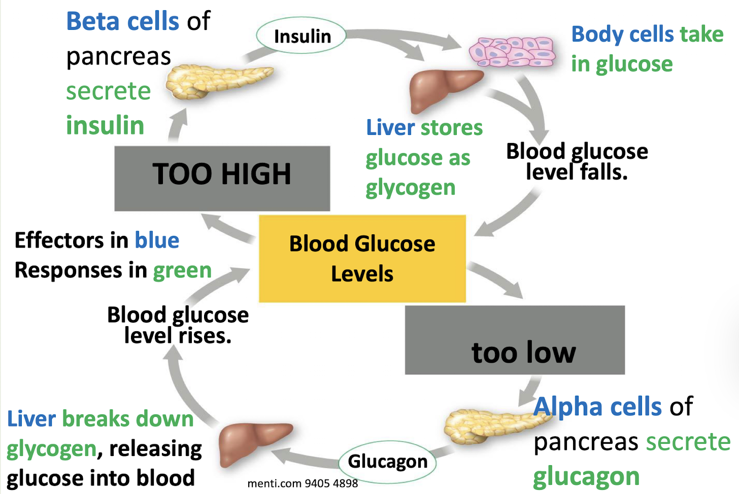
How do changes in levels of blood sugar, insulin, or glucagon affect the homeostatic control of blood glucose levels?
Increase in insulin levels
Blood glucose levels decrease
Glucose uptake by cells increases
Glycogen formation increases
Decrease in insulin levels
Blood glucose levels remain elevated
Cells take in less glucose
Increase in glucagon levels
Increase in blood glucose levels
Increase in glycogen breakdown in the liver
More glucose released into the blood
Decrease in glucagon levels
Reduced ability to raise blood sugar during fasting
Risk of low blood sugar
How would changes in neurotransmitter release, receptor activity, or neurotransmitter clearance from the synapse affect signaling between neurons & their targets?
Increase in neurotransmitter release → Increased signaling because more neurotransmitter binds receptors
Decrease in neurotransmitter release → Decreased signaling because fewer receptors are activated
Increased receptor activity → Stronger signaling response
Decrease receptor activity → Weaker signaling response
Faster neurotransmitter clearance → Decreased signaling because neurotransmitter spends less time in the synapse
Slower neurotransmitter clearance → Prolonged or increased signaling because neurotransmitter remains in the synapse longer
What are the two ways the hypothalamus & pituitary gland work together to release hormones?
Anterior pituitary pathway: Hypothalamus releases hormones into blood vessels that travel to the anterior pituitary → Stimulates or inhibits the release of anterior pituitary hormones
Posterior pituitary pathway: Hypothalamus directly produces hormones → Hormones travel down axons of hypothalamic neurons to the posterior pituitary → Hormones are stored in the posterior pituitary & released into the bloodstream
What are the two methods of long-distance communication in animals? What are their differences?
Endocrine system
Uses hormones
Endocrine glands secrete hormones into surrounding fluid, including blood
Targets cells throughout the body because all cells have a blood supply
Reaches target cells more slowly
Effects tend to last longer
Nervous system
Uses signaling by nerve cells (neurons)
Signals are electrical or chemicals carried through extracellular fluid
Targets neurons, muscles, & some glands only
Reaches target cells more quickly because electrical signals are quicker than chemical signals
Effects tend to last a shorter amount of time
What are the different types of glia? What are their functions? Where are they found?
Central nervous system
Oligodendrocytes
Insulate neurons with myelin
Can myelinate multiple neurons at once
Astrocytes
Main support cells for the brain
Transport nutrients from the blood to neurons
Balance ionic & chemical environment
Involved in care & maintenance of the brain, like healing
Microglia
“White blood cells” of the brain
Fight micro-organisms
Scavenge & clean up debris
Peripheral nervous system
Schwann cells
Insulate cells of the PNS with myelin
How does the flow of ions during the action potential change the voltage inside the cell?
Na+ flowing in will make the cell more positive
K+ flowing out will make the cell more negative
What do the terms depolarization & hyperpolarization mean?
Depolarization: Membrane potential gets more positive (less negative)
Hyperpolarization: Membrane potential gets more negative (less positive)
How does an action potential occur?
1.) A neuron is at resting potential, then is stimulated, causing it’s membrane potential to rise above threshold
2.) Rising Phase: When a neuron reaches threshold, all voltage-gated sodium channels open & sodium flows into the cell due to the concentration gradient
3.) Peak: Right when the rising phase changes to the falling phase; Na+ channels inactivate due to time, meaning the sodium ions stop passing through even though the voltage is above threshold; Voltage-gated K+ channels open due to the voltage reaching threshold, they are just slow to respond
4.) Falling Phase: Because the K+ channels have just opened, K+ flows out of the cell, causing hyperpolarization; Because Na+ channels have just become inactive, Na+ stops flowing in & stops depolarization
5.) Undershoot Phase: So much K+ leaves the cell that the membrane potential sometimes hyperpolarizes below the resting potential; Then, the K+ channels close, the Na+ channels go back to rest, & the cell returns to resting membrane potential; The neuron can now make another action potential & Na+/K+ pumps restore ion concentrations
How does the synapse function?
1.) High voltage causes voltage-gated Ca2+ channels to open → Ca2+ enters the cell
2.) Ca2+ causes vesicles to fuse with the membrane → That fusion releases neurotransmitters into the synapse
3.) Neurotransmitter binding causes the receptor to have an effect in the receiving cell
What causes neurotransmitter signaling to stop?
Diffusion away from the synapse
Reuptake into the presynaptic terminal
Enzymatic degradation
What happens to the graph of voltage vs. time of an action potential if Na+ channels are blocked?
You do not get an action potential
No sharp rising phase
At most a small subthreshold bump, but not enough to stimulate an action potential
Because rising phase depends on Na+ influx
What happens to the graph of voltage vs. time of an action potential if the function of Na+ channels is reduced?
Harder to reach threshold
If it fires, the peak may be reduced or the action potential may fail to fully develop
What happens to the graph of voltage vs. time of an action potential if Na+ channels fail to inactivate?
The action potential stays depolarized longer
Repolarization is delayed
Neuron may be prone to repeated firing/seizures
What happens to the graph of voltage vs. time of an action potential if K+ channels are blocked?
Falling phase is slower
Repolarization is delayed
Undershoot is reduced or missing
Action potential is wider
Takes longer to return to rest
Action potential propagation can still occur because propagation depends on Na+ driven depolarization to trigger the next segment
What happens to the graph of voltage vs. time of an action potential if K+ channels open more easily?
Faster repolarization
Bigger undershoot (more hyperpolarization)
Peak might be cut short
Because K+ “fights” Na+ sooner
What do the hypothalamus & the pituitary do?
Hypothalamus
Receives information from the rest of the brain & initiates responses
Pituitary
Works with the hypothalamus to secrete hormones into the blood
How do the hypothalamus & posterior pituitary work together to release hormones?
Posterior pituitary is made of axons from hypothalamic neurons
The cell bodies are located in the hypothalamus & make the hormone
The hormone is physically moved down the axon & released into the pituitary blood vessels
How do the hypothalamus & anterior pituitary work together to release hormones?
Hypothalamic neurons secrete the first hormone
That hormone acts on the anterior pituitary to release the second hormone into the bloodstream
What is the sensory receptor’s function?
To turn an outside stimulus into a change in membrane potential
What type of channels in sensory neurons respond to touch signals? Smell & taste?
Touch: Stretch-sensitive channels
Smell, taste: Receptors that detect chemicals & indirectly open channels
Map out how how light, retinal, opsins, & cation channels affect a photoreceptor’s membrane potential & neurotransmitter release.
Light (photons) enter the eye & hit the retina
The retina’s photoreceptor cells (rods & cones) detect light
When more light is detected:
The molecule retinal that is in the photoreceptor cells is activated, and it changes shape
Opsin is bound to retinal, and detects retinal’s change in shape, activating the opsin
The activation of opsin activates a pathway that decreases cGMP and closes Na+ channels
K+ channels are always open, so without Na+ entering the neuron, K+ continues to flow out, causing hyperpolarization
Neurotransmitter release decreases
When less light is detected:
Retinal in the photoreceptor cells remains inactive
Opsin is not activated
cGMP levels remain high, so Na+ channels remain open
With both K+ & Na+ channels open, K+ flows out & Na+ flows in, causing depolarization
Neurotransmitter release increases
How does information flow in the nervous system?
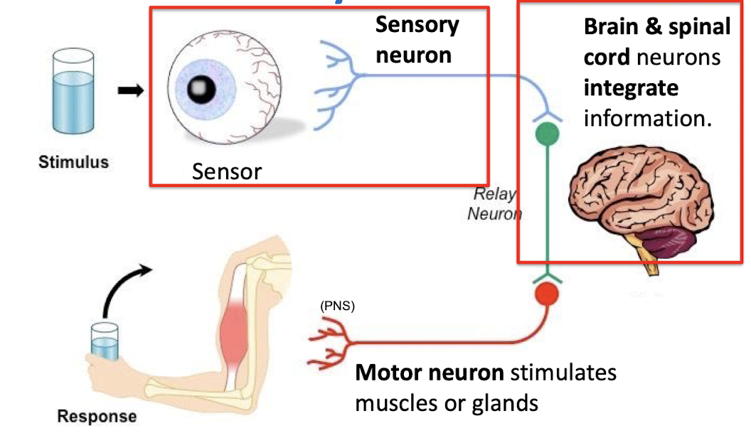
How does the sensory pathway flow?
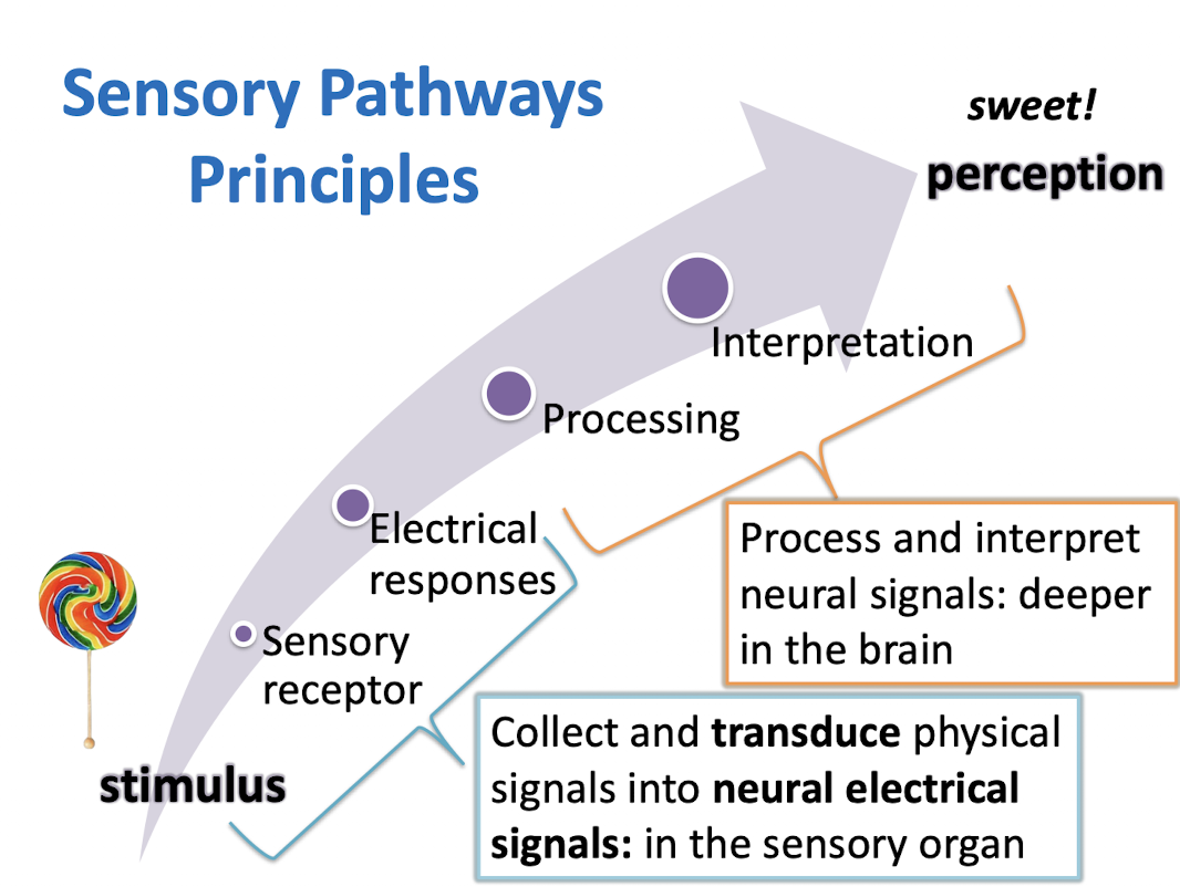
How does light turn into neurotransmitter signals?
External stimulus (photons in light) → Change in membrane potential → Neuron releases a certain amount of neurotransmitter
Given a neuron’s responses to stimuli, how can you hypothesize its receptive field?
A neuron’s receptive field is the stimulus & location in space that causes the strongest response
To identify the receptive field:
Present stimuli at different locations
Observe when the neuron’s firing rate increases
The stimulus & location that produce the strongest response are the neuron’s receptive field
How do you distinguish between innate & learned behavior?
Innate
All individuals in a population behave alike
Instinctive
Not modified & unaffected by environment
Low variation in population
Evolved through natural selection
Often have to do with important tasks for survival or reproduction
Cannot seem to adjust the behavior when the situation changes
Learned
Allows individuals to change their behavior in response to their specific environment
Based on experience
Modified via trial & error
Affected by environment
High variation in population
Capacity to learn is a product of natural selection
How could an experiment using optogenetics demonstrate the concept of “fire together, wire together”?
Principle: If two neurons that are connected to each other fire at the same time, it will cause stronger or more connections between them, so in the future if one neuron is activated, it will cause the other to fire too.
Optogenetics: Genetically inserting Channelrhodopsin (ChR2) into mouse’s brain
Blue light inserted in the mouse’s brain will activate ChR2 → Na+ influx → Depolarization
Experiment:
Part 1:
Put mouse in Location A → Location A neurons will fire
Scientists also force location A neurons to express ChR2 → Neurons will “fire together” that usually wouldn’t
Part 2:
Put mouse in footshock location B & turn on blue light → Location B neurons will fire & ChR2 in location A neurons will be activated → Location A ChR2-activated neurons will be linked with Location B footshock neurons
Test if neurons wired together:
Put mouse back in Location A → Location A channels activated because mouse is in Location A → Because Location A neurons are connected to (or wired to) Location B footshock neurons, then Location B neurons will also be activated → Mouse will freeze
What does it mean to say that a synapse is strengthened or weakened?
A synapse is strengthened when an AP in the presynaptic neuron will cause a bigger response in the postsynaptic neuron.
A synapse is weakened when an AP in the presynaptic neuron will cause a smaller response in the postsynaptic neuron.
Distinguish between the innate & adaptive immune systems in terms of cell types, pathogen recognition, & protection from infection.
Innate immune system
Characteristics:
Fast, immediate response
Present at birth
Can attack pathogens never encountered before
Found in all plants & animals
Recognizes broad classes of pathogens
Recognition:
Detects PAMPs (Pathogen-Associated Molecular Patterns) such as:
Bacterial flagella
Bacterial cell wall
Viral RNA
Use Toll-Like Receptors (TLRs) to detect PAMPs
TLRs on innate immune cells detect PAMPs → Secrete cytokines, recruit more immune cells → Phagocytose pathogens, etc
Major cell types:
Macrophages
Large phagocytic cells that engulf pathogens
Functions:
Phagocytosis
Cytokine secretion
Talks to adaptive immune system
Dendritic cells
Functions:
Detect pathogens
Talks to adaptive immune system
Neutrophils
Functions:
Phagocytoses pathogens
First responder
Secretes cytokines
Eosinophils
Attacks larger pathogens like parasites & cancer cells
Protection:
The skin & other membranes
Some bodily fluids
Sweat
Tears
Mucus
Inflammation
Antimicrobial peptides: Molecules that directly attack invading microbes
Cytokines: Molecules that signal to other cells to do something
Adaptive immune system
Characteristics:
Acquired
Prior exposure needed
Slower response (days-weeks)
Very specific signals recognized by countless receptors
Only found in vertebrates
Recognition:
Recognizes specific antigens
B & T cells respond to unique pathogen antigens
Major Cell Types:
B & T cells
Produce antibodies
Antigens are the particular outside shape of each pathogen
Antigens can be detected by antibodies
After infection, you make memory B & T cells
When you are exposed to the pathogen again, the memory cells react to the antigens of the pathogen directly & quickly
Antibodies
Tag the pathogen as being “bad” as a signal to the innate immune system
Block their ability to infect
Big variety possible
What are the two arms of the adaptive immune system?
Cell-mediated arm
Cytotoxic T-cells infect body cells & cancer cells
When pathogens are inside body cells (viruses & some bacteria), the infected body cells need to be killed
Humoral arm
B-cells divide to make plasma cells, which secrete antibodies
When pathogens are in blood & body fluids, antibodies can reach them
Map out the path of the adaptive immune system.
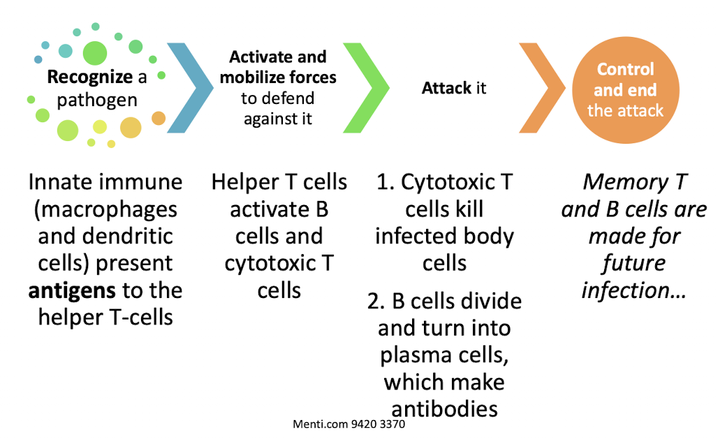
Map out the path of the innate immune system.
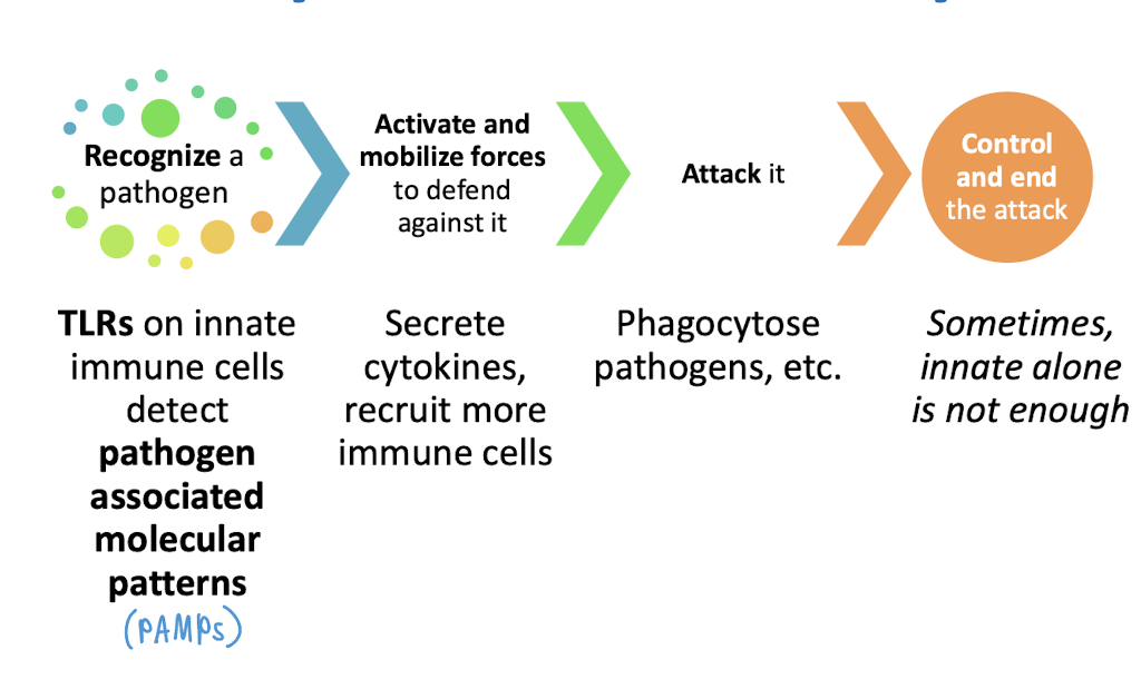
How would the innate immune system function be affected by a loss of macrophages? Defective TLR signaling? Loss of dendritic cells?
Loss of macrophages
Effects:
Reduced phagocytosis
Fewer cytokines
Weaker activation of adaptive immunity
Result: Higher pathogen load early in infection
Defective TLR signaling
Effects:
Pathogens not detected efficiently
Delayed immune response
Result: Pathogens replicate before immune response begins
Loss of dendritic cells
Effects:
Adaptive immunity not properly activated
How would the adaptive immune system function be affected by a loss a B cells? A loss of T cells? Loss of memory cells?
Loss of B cells
Antibodies not produced
Extracellular pathogens persist
Loss of T cells
B cells not activated
Cytotoxic T cells poorly activated
Antibodies reduced
Loss of memory
Repeated infections cause the same severity of illness
No enhanced secondary immune response
How do traditional vaccines work? How do mRNA vaccines work?
Traditional vaccines
Types include:
Weakened pathogen
Killed pathogen
Purified pathogen protein
How they work:
Vaccine introduces antigen
Antigen-presenting cells ingest antigen
Helper T cells recognize antigen
Helper T cells activate B cells
B cells becomes plasma cells
Plasma cells produce antibodies
Some B cells become memory cells
Result: Future infection triggers rapid antibody production
mRNA vaccines
Deliver genetic instructions for making a pathogen protein
How they work:
Vaccine introduces mRNA from pathogen (antigen source)
mRNA codes for pathogen protein
Presented to immune cells
Helper T cells activate B cells
Plasma cells produce antibodies
Memory B cells form
Predict whether an infection or changes in B- & T-cell development might predispose someone to develop an autoimmune disease.
Normal Self-Tolerance
During B- & T-cell development:
Many different receptors are generated
Developing lymphocytes are exposed to self-antigens
Cells that strongly recognize self-antigens are eliminated.
This prevents the immune system from attacking the body’s own tissues
Defective B- or T-cell Development
Self-reactive B or T cells are not eliminated
They mature & attack self-antigens
Increased risk of autoimmune disease
Infection with Cross-Reactive Antigens
A pathogen has antigens similar to a body’s own antigens
Immune cells attack the pathogen
The same immune cells may accidentally attack the body’s tissues
Increased risk of autoimmune disease
How is pressure involved in the movement of water & nutrients in plant xylem & phloem?
Xylem
Water moves from roots → leaves
No pumps in the roots
Transpiration at the leaves lowers pressure, so water evaporates through stomata
Very low pressure in the leaves causes water to be pulled upward from higher pressure in the roots
Movement occurs because of a pressure gradient created by evaporation at the top
Phloem
Sugar is actively loaded into phloem at source cells in the leaves
High sugar concentration draws in water by osmosis, increasing pressure
At sink tissues (roots/storage), sugar is unloaded
Water leaves, lowering pressure
Sugar moves from high pressure (source) to low pressure (sink)
How is pressure involved in the movement of blood in vertebrates?
The heart acts as a pump
During ventricular systole, contraction increases pressure in arteries
Blood flows from high pressure (ventricles/arteries) to lower pressure (capillaries → veins → atria)
Pressure gradually decreases along pathway: Arteries → Arterioles → Capillaries → Venules → Veins
Ventricles generate the most pressure because they have thicker muscle walls
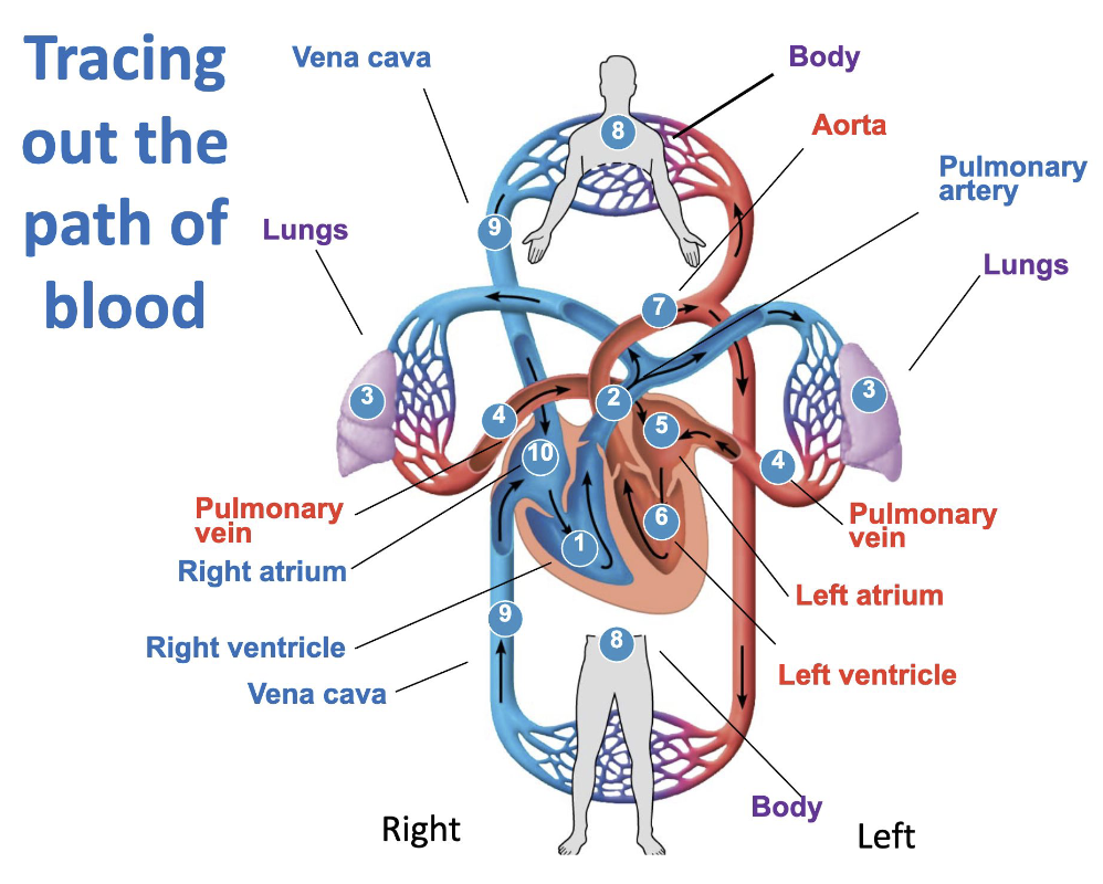
How does blood flow through the four chambers of the heart, the great vessels, lungs, and the blood vessels?
Blood passes the heart twice
Body to heart (Systemic circuit) (deoxygenated)
Body capillaries → Venules → Veins → Vena cava → Right atrium
In heart to lungs (Pulmonary circuit)
Right atrium → Right ventricle → Pulmonary artery → Lung capillaries → Pulmonary veins → Left atrium → Left ventricle
Heart to body (System circuit) (oxygenated)
Left ventricle → Aorta → Arteries → Arterioles → Capillaries
Arteries → Arterioles (Away from heart)
Veins → Venules (Toward heart)
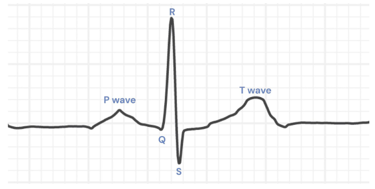
Draw out the ECG associated with heart beat and describe what is occuring during each section of the graph.
P wave
Atrial contraction
Generated by the SA node
Atrial depolarization occurs
QRS
Ventricular contraction
T wave
Ventricular relaxation
What are the components of the circulatory system?
Fluid in which materials are transported (blood)
A pump to move the fluid around (heart)
Vessels to provide controlled paths (veins, arteries, capillaries)
What are the roles of the veins, venules, capillaries, arterioles, & arteries?
Veins: Carry blood toward the heart
Venules: Connect capillaries to veins
Capillaries: Allow for exchange of O2/CO2, nutrients, & waste
Arterioles: Control blood flow into capillaries & help regulate blood pressure
Arteries: Carry blood away from the heart
What is the cardiac cycle, & what it its order?
Cardiac cycle: One complete phase of pumping & filling
Contraction phase is systole
Relaxation phase is diastole
Order:
Atrial & ventricular diastole
Atria & ventricles are relaxed, & blood is returning to the heart
Atrial systole (ventricular diastole)
Atria contract, ventricles are still relaxed
Ventricular systole (atrial diastole)
Ventricles contract
Pushes blood to the next structure
What happens if the SA node is destroyed?
The AV node becomes the pacemaker, and there are no P waves
What happens if the atria do not depolarize normally?
Atrial contraction is abnormal or absent
Ventricles still contract, but rhythm may be slower
How does the electrical signal travel in the heart?
Sinoatrial node: Pacemaker
Spreads to atria
Spreads to atrioventricular node
Spreads down the septum
Spreads out to both ventricles
What are the differences between systolic & diastolic blood pressure?
Systolic blood pressure
Arterial blood pressure during ventricular contraction
It is the higher number in a blood pressure reading because ventricular contraction generates the greatest pressure
Pumping pressure
Diastolic pressure
Arterial blood pressure during ventricular relaxation
It is the lower number because the heart is not actively contracting, so arterial pressure falls
Resting pressure
What are cross-sectional & total cross-sectional area, & what is the pattern in the body?
Cross-sectional area: The area of a vessel if you slice it & look at the opening
Total cross-sectional area: The sum of all vessels at that level
Pattern in the body:
Small in aorta (one large vessel)
Larger in arteries
Largest in capillaries (because there are millions of them)
Decreases again in veins
Capillaries have the greatest total cross-sectional area
How is velocity related to total cross-sectional area? What is the pattern of blood velocity & why?
Total cross sectional area & velocity are inversely related
If total cross-sectional area increases → velocity decreases
If total cross-sectional area decreases → velocity increases
Pattern:
Highest near heart (aorta, arteries)
Decreases dramatically in capillaries
Increases somewhat again in veins (but not as high as arteries)
Cause:
Pressure (higher pressure → faster velocity)
Total cross-sectional area (lower area → Faster velocity)
How is pressure related to total cross-sectional area? What is the pattern of blood pressure?
Pattern:
Highest in aorta & arteries
Gradually decreases through arterioles
Much lower in capillaries
Lowest in veins & vena cavae
Cause:
Pressure is generated by ventricular contraction
As blood moves through vessels, energy is lost due to:
Stretching of vessel walls
Friction within vessels
Pressure steadily declines with distance from heart
What are pressure, total area, & velocity like in the aorta, capillaries, & veins?
Aorta
High pressure
Low total area
High velocity
Capillaries
Lower pressure
Highest total area
Lowest velocity
Veins
Very low pressure
Lower area than capillaries
Moderate velocity
How is blood pressure controlled homeostatically?
If blood pressure is low, heart rate increases & arteries & arterioles constrict, making blood pressure rise
If blood pressure is high, heart rate decreases & arteries & arterioles relax, making blood pressure fall
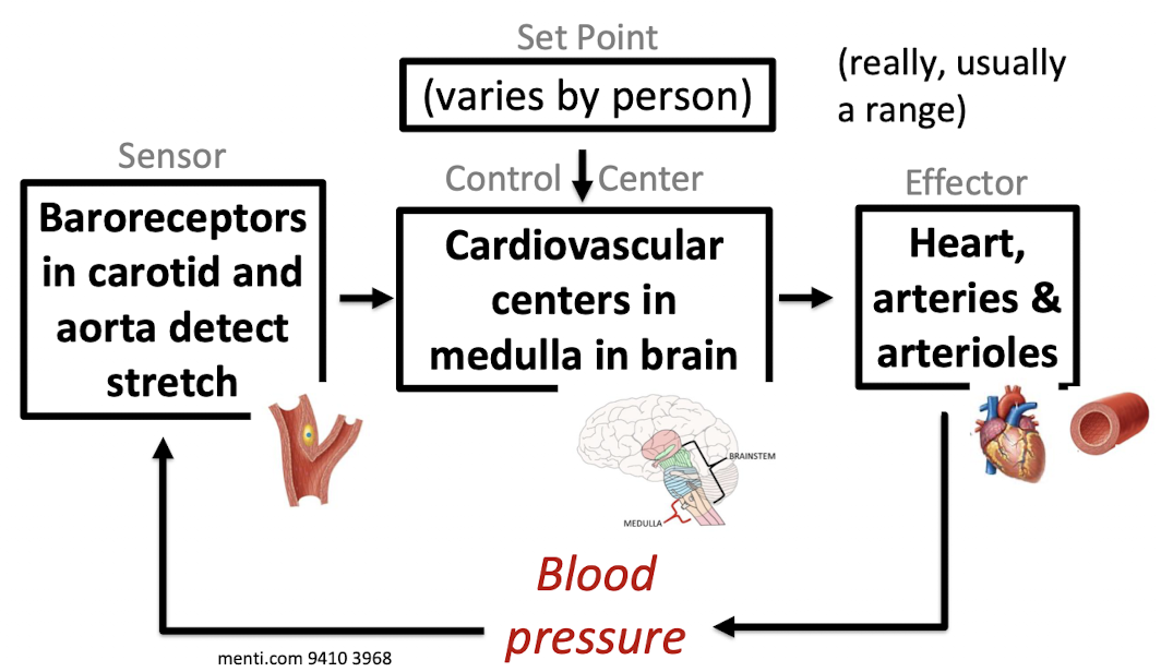
How do changes in body posture affect homeostatic control of blood pressure?
When a person stands up suddenly:
Gravity pulls blood downward
Less blood returns to the heart
Arterial blood pressure falls
Homeostatic response:
Baroreceptors detect less stretch
Signal sent to medulla
Effectors respond:
Heart rate increases
Arteries & arterioles contrict
Result:
Blood pressure rises back toward the set point
If this reflex did not occur → Dizziness or fainting
How do changes in baroreceptor function affect homeostatic control of blood pressure?
Typically, when blood pressure increases:
Baroreceptors detect increased stretch
Medulla decreases heart rate
Arteries relax (vasodilation)
Blood pressure falls toward normal
If baroreceptors cannot detect stretch:
Changes in blood pressure are not sensed properly
The medulla does not adjust heart rate or vessel diameter appropriately
Blood pressure becomes unstable
Standing up could cause prolonged drops in pressure
If baroreceptors reset to a higher set point, like in chronic hypertension:
High blood pressure is treated as acceptable
Homeostatic correction does not occur
Hypertension (high blood pressure) persists
How will blood pH levels change when the kidney reabsorbs bicarbonate ions? When bicarbonate ions are secreted?
Reabsorption
From filtrate back to blood
Adds bicarbonate back to the blood
Blood pH increases (becomes more basic)
Occurs when body needs to correct low blood pH
Secretion
From blood to filtrate
Removes bicarbonate from the blood
Blood pH decreases (becomes more acidic)
Occurs when body needs to correct high blood pH
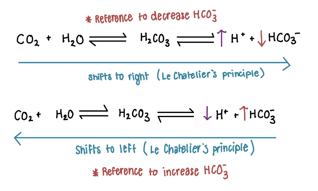
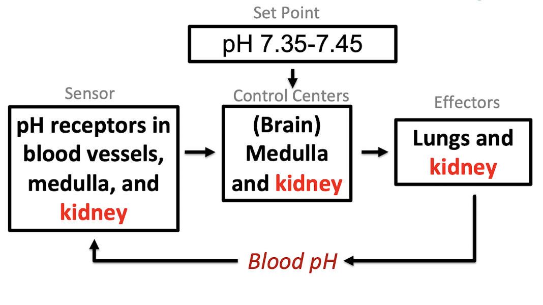
Map out how blood pH is homeostatically controlled.
Map out the creation, processing, & path of filtrate in the human kidney.
Glomerulus: Blood pressure forces arteriole blood through slits to make the filtrate. Small things like water, ions, & sugars can go through the filter, but not cells or proteins.
Proximal tubule
Filtrate has been made, so things can go back into the blood
Glucose, amino acids, water, sodium, & bicarbonate are reabsorbed by blood
Most of the reabsorption is done here
The filtrate ends isosmotic
Descending nephron loop
Water leaves filtrate & is reabsorbed into the blood
Filtrate ends hyperosmotic
Ascending nephron loop
Sodium is reabsorbed into the blood
Extracellular fluid in medulla is salty
Actively pumps salt out
Filtrate ends hyposmotic
Distal tubule
Water is reabsorbed if there is ADH, and sometimes bicarbonate is reabsorbed
Filtrate ends hyposmotic without ADH, isosmotic with ADH
Collecting duct
Water is reabsorbed if there is ADH because the ADH inserts aquaporins into the membrane, which allow water to flow out because the medulla fluid is saltier
Sometimes sodium & bicarbonate are reabsorbed into the blood
Bicarbonate is sometimes secreted into the filtrate
Filtrate ends hyposmotic without ADH or hyperosmotic with ADH
What happens when glomerular filtrate rate is low? High?
Low GFR
Waste products stay in tubule too long & move back into the body
Too much fluid is retained in blood
Filtration stops & wastes/excess fluids remain in blood
High GFR
Important materials flushed out with urine before they’re recovered
Too much fluid loss
Damage to glomerular capsule & kidney failure
How do the kidneys control GFR?
Myogenic mechanism
Smooth muscle around the arteriole detects stretch & controls the arteriole’s size
High blood pressure stretches the arteriole → arteriole constricts → less blood in kidney → GFR lower
Low blood pressure → arteriole is not stretched → more blood in kidney → GFR higher
Local; only affects the glomeruli
Controls how much blood reaches the glomeruli
More blood flow → higher GFR
Controlling the renin-angiotensin-aldosterone system
Global; affects the whole body
Controls the blood pressure of all the body’s blood
Higher blood pressure → higher GFR
How do changes in the renin-angiotensin-aldosterone system affect blood volume & pressure?
Low GFR causes the release of the hormone renin
Renin causes the formation of angiotensin I
Angiotensin I turns into hormone angiotensin II
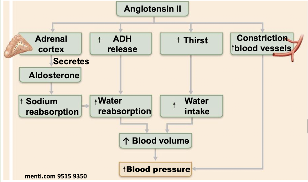
Angiotensin II causes the adrenal glands to release the hormone aldosterone
Aldosterone increases the reabsorption of sodium in the distal tubule & collecting duct, making the blood saltier
Blood pressure & volume increase because the increased salt concentration in the blood attracts more water through osmosis
What are the different types of osmolarity?
Hyperosmotic → Concentrated
Isosmotic
Hyposmotic → Dilute
How do the functions of the distal tubule & collecting duct change with & without ADH?
With ADH: Water is reabsorbed from the filtrate into the blood
In the distal tubule, filtrate stays hypotonic & ends isotonic
Fluid around the collecting duct is very salty, so water leaves the filtrate
Filtrate goes from isotonic to hypertonic, & hyperosmotic urine is produced
Without ADH: Water is not reabsorbed & stays in the filtrate
In the distal tubule, filtrate remains hypotonic
In collecting duct, water cannot leave the filtrate, so the filtrate remains hypotonic
A large volume of hypotonic, dilute urine is produced
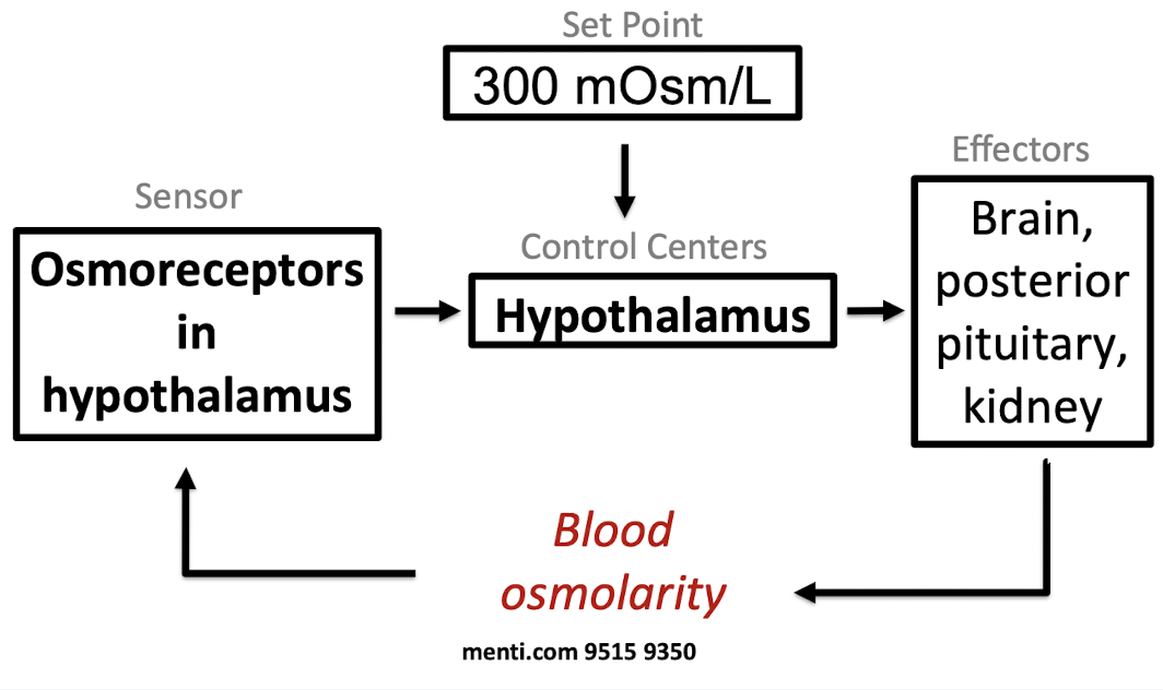
How is blood osmolarity homeostatically controlled? Map it out.
When osmolarity is high:
Hypothalamus contacts other parts of the brain, to create a feeling of thirst
Hypothalamus tells the posterior pituitary to release more of the hormone anti-diuretic hormone (ADH)
When osmolarity is low:
Hypothalamus tells the posterior pituitary to release less of anti-diuretic hormone (ADH)
Map out the effects of angiotensin II.
Map out the flow of information in the nervous system.
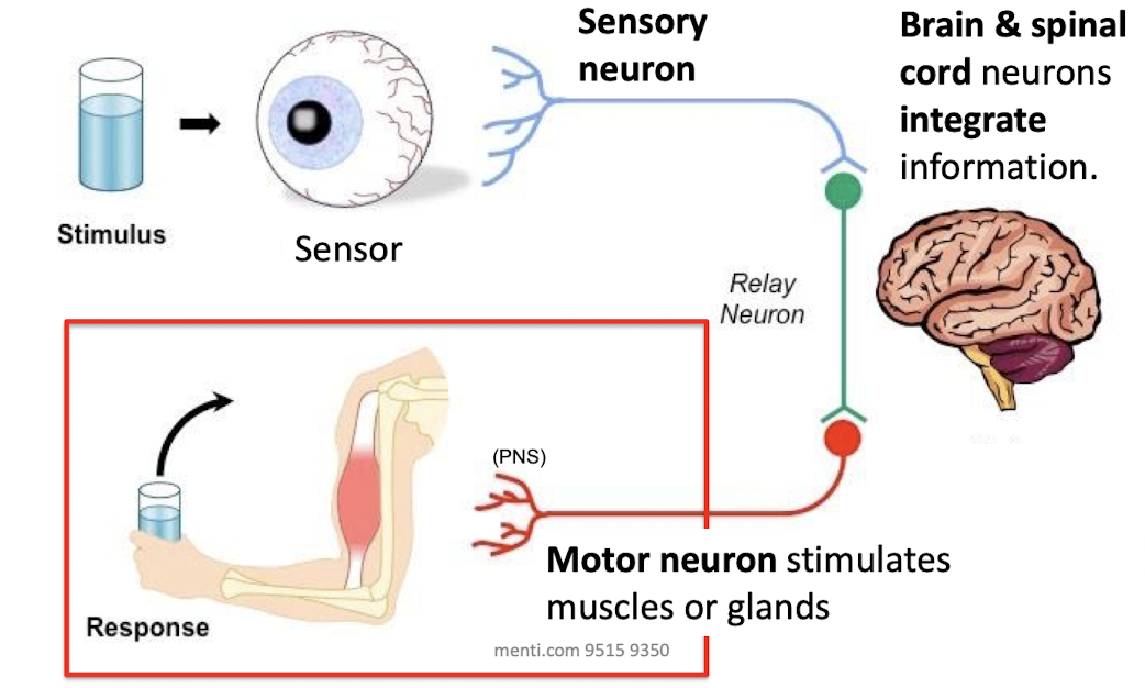
What are the different types of muscle? What are sarcomeres & do they have sarcomeres?
Skeletal
Attached to bones
Voluntary movement
Have sarcomeres
Cardiac
Walls of the heart
Involuntary movement
Have sarcomeres
Smooth
Walls of hollow, visceral organs
Involuntary movement
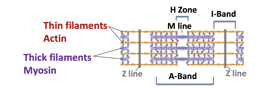
How do thin filaments, thick filaments, and Z lines move during muscle contraction?
During contraction, thick & thin filaments slide together lengthwise
Thin filaments (actin) slide toward the center of the sarcomere
Thick filaments (myosin) stay in the same position
Z lines move closer together as the sarcomere shortens
What are the steps of skeletal muscle fiber contraction?
Neuron has an action potential, which activates the neuromuscular junction
In the neuromuscular junction:
The action potential in the neuron opens voltage-gated calcium channels
Calcium influx into the neuron causes the release of acetylcholine
Acetylcholine binds to its receptor on the muscle fiber, opening a channel that lets in sodium
Acetylcholine is broken down in the cleft by acetylcholinesterase
The muscle has an action potential, and the signal propagates to the rest of the muscle
Muscle contracts using cross bridge cycling
How is muscle contraction influenced by calcium?
Without calcium
Myosin heads cannot bind to actin because tropomyosin is bound to actin and is in the way
With calcium
Ca2+ binds to troponin
Troponin moves tropomyosin off the actin binding sites so the actin & myosin can form cross bridges
Myosin head turns & pulls thin filaments to contract
ATP binding to the myosin detaches the cross bridges
Energy from ATP hydrolysis moves myosin back into the initial state
What are the differences between the sympathetic & parasympathetic nervous system?
Parasympathetic
Rest & digest
Promotes maintenance functions & conserves body energy
Promotes low blood pressure, low heart rate, & digestion
Sympathetic
Fight or flight
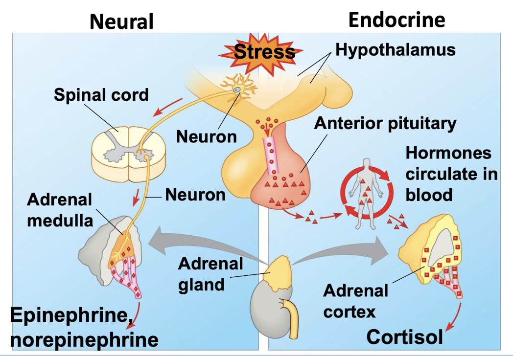
Release epinephrine & norepinephrine
Increase heart rate
Increase blood pressure
Vasoconstriction
Increase blood sugar
What are the two parallel stress pathways?
Would a larger animal or a smaller animal have more trouble keeping its body temperature at its set point & why?
A smaller animal
Because smaller animals have a larger surface area to volume ratio
This means that it has a lot of surface area compared to its volume, so there are lots of places for the heat to leave and not much heat stored inside, meaning that heat leaves quickly compared to how much heat it has
A larger animal has less surface area compared to its volume, so there are fewer places for heat to leave and a lot of heat stored inside, so heat leaves slowly compared to how much heat it has
How can you become big while still having a high SA/V ratio?
Be made up of many smaller units
Shape
Have a folded, elongated, or branched shape instead of a compact sphere to increase surface area without greatly increasing volume
What does this arrow mean? ⊣
Block, inhibit, or decrease
What is a negative feedback loop?
Occurs when a change in the level of something causes an effect that counteracts that change
Stabilization
What is a positive feedback loop?
Occurs when a change in the level of something causes an effect that increases that change
Amplifies an effect or pushes the organism into a new state
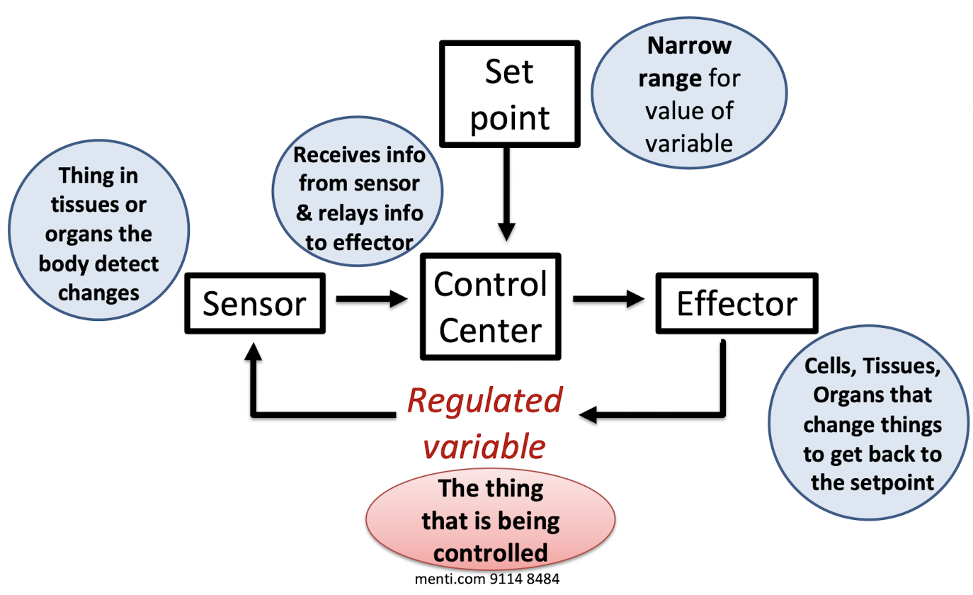
What are the parts of a basic homeostatic circuit? Describe each part.
Set point
Narrow range for value of variable
Control center
Receives information from the sensor & relays info to the effector
Effector
Cells, tissues, & organs that change things to get back to the set point
Sensor
Thing in tissues or organs that detects body changes
How is the exchange of stuff with the environment related to the SA/V ratio?
High SA/V allows for more exchange with the environment
Because there is more surface area relative to volume, so there is more space for diffusion of heat, gases, nutrients, and waste to occur
Low SA/V allows less exchange with the environment
Because there is less surface area relative to volume, so there is less space for diffusion of heat, gases, nutrients, and waste to occur