CP.09 Nerves
 Call Kai
 Learn
 Practice Test
 Spaced Repetition
 Match
 Flashcards
 Knowt Play
1/35

Earn XP
Description and Tags
• Define Neuron, Nerve and Nervous system • Characterise the types of Neuronal Cells, Neurons and Nervous Systems • Describe reflex arc • Describe Synaptic transmission • Outline Excitatory and Inhibitory Neurotransmitters
36 Terms
Define neuron vs nerve vs nervous system.
Neuron is an electrically excitable cell that receives, processes, and transmits information through electrical and chemical signals.
Nerve is a bundle of fibres that conduct impulses between the brain or spinal cord and another part of the body. Nerves include fragments of neurons (axons) and non—
Nervous system is the network of nerve cells and fibres which transmits nerve impulses between parts of the body and coordinates its action.

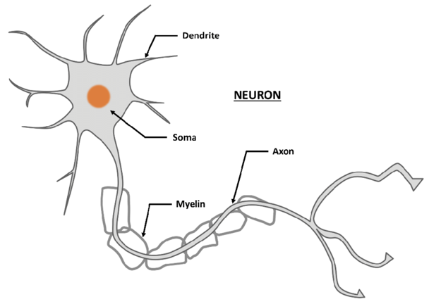
What are the structural components of a neuron?
Soma (Greek σῶμα "body”) or perikaryon is the spherical portion of a neuron containing the cell nucleus.
Dendrite is a short, branched extension of a nerve cell, along which impulses received from other cells at synapses and transmitted to the cell body.
Axon the long thread like part of a nerve cell along which impulses are conducted from the cell body to the other cells
+ Axon hillock is the last site in the soma where membrane potentials propagated from synaptic inputs are summated before being transmitted to the axon
Neurons can be classified according to their function. Define an afferent, efferent, interneuron and neurosecretory neuron. Which NS are they within (PNS/CNS/autonomic).
Sensory (afferent) input neurons convey information from tissues and organs in the PNS (peripheral NS) into the CNS (central NS).
Motor (efferent) output neurons transmit signals from the CNS to the effector cells (muscles and glands). Found in Central Nervous System (spinal cord and brain stem), and Autonomic Nervous System.
Interneurons (relay or association neurons) - most common type of neurons, connects sensory and motor neurons to each other via dendrites, also connect neurons within specific regions of the Central Nervous System. Functions are, information processing and decision making.
Neurosecretory cells secrete hormones and other signal substances. They are located in hypothalamus, adrenal medulla, thyroid gland and other hormone and hormone-like substances secretory organs.

Neurons can be classified according to their structure. Define unipolar, pseudounipola r, bipolar, multipolar and anaxonic neurons.
Unipolar neuron = only one neurite extends from the cell body. Most of the unipolar neurons are sensory.
Pseudounipolar neuron = dendrites and axons have fused in a single process, which divides into two processes: axon and dendrite. Often, it is an intermediate state in the development of unipolar or bipolar neurons.
Bipolar neuron = two neurite extensions. Bipolar neurons are specialized sensory neurons for the transmission of special senses, e.g.: olfactory cells, inner ear, retina.
Multipolar neuron is a type of neuron that possesses a single axon and many dendrites. There are about 99% of human neurons are multipolar.
Anaxonic neuron is a neuron type in which the axon cannot be differentiated from the dendrites. Often, it is an intermediate state in the development of multipolar neurons.

What are the cell types with the nervous system which do not transmit action potentials? And what are their roles?
Glia or Neuroglia (Greek γλία which means "glue “) non-neuronal cells that maintain homeostasis: form myelin and provide support and protection for neurons in the Central and Peripheral Nervous Systems.
What are the 4 functions of glial cells?
To surround neurons and hold them in place
To supply nutrients and oxygen to neurons
To insulate one neuron from another.
To destroy pathogens and remove dead neurons
What is the difference between macroglia and microglia?
Macroglia is generic term for cells in Central and Peripheral Nervous Systems. Umbrella term for astrocytes, oligodendrocytes and Schwann cellsà together provide nutrition, physical support and synthesise myelin.
Microglia are the immune cells tissue-based macrophages (perform phagocytosis and protect neurons of the Central Nervous System)

Macroglia is generic term for cells in Central and Peripheral Nervous Systems. What are the 4 types of macroglia within the CNS and 3 types within the PNS?
Macroglia within the CNS: Astrocytes, oligodendrocytes, ependymocytes and radial glia.
Macroglia within the PNS: Schwann cells, satellite cells, enteric glial cells.

What is the function of the macroglia within the CNS? Astrocytes, oligodendrocytes, ependymocytes and radial glia.
Astrocytes (Astroglia): These are Star-Like Cells (hence the name Astro) and link neurons to their blood supply by forming the blood-brain barrier (BBB). They also help regulate the external chemical environment of neurons by removing excess potassium ions, as well as recycling neurotransmitters released during synaptic transmission.
Oligodendrocytes: These cells coat the neuronal axons with their cell membrane, squeezing the glial cytoplasm out and wrapping around it, forming myelin. These are similar to Schwann Cells apart from the fact that they are located in the CNS, whereas Schwann cells are located in the PNS.
Ependymocytes: These line the Spinal Cord and the Ventricular System of the brain, secrete Cerebrospinal Fluid, and act as Neural Stem Cells in the brain repair mechanism,
Radial Glia: These participate in Neurogenesis (the production of neurones), by functioning as Neural Progenitors (meaning that every neuron in the cerebral cortex is a descendant of these). In addition to this, it acts as a scaffold for the neurons during neuronal migration (in order to move into the position needed).

What is the function of the macroglia within the PNS? Schwann cells, satellite cells, enteric glial cells.
Schwann Cells: These coat axons with their cell membrane and produce Myelin (like CNS Oligodendrocytes). Schwann cells are in the PNS.
Satellite Cells: These regulate the external chemical environment in PNS (like CNS Astrocytes),
Enteric Glial Cells: These regulate Homeostasis in Intrinsic Ganglia of the digestive system.

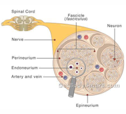
A nerve is not a cellular structure but rather a bundle of fibres that conduct impulses between the brain (or spinal cord) and another part of the body. Nerves include fragments of neurons (Axons) and non-neuronal cells (Glial Cells/Neuroglia), as well as some other supplemental cells such as Blood Vessels. On the picture to the right we can see a nerve that projects from the spinal cord. Within the cross-section provided, there are many microscopic elements.
What are the four key components labelled in the nerve structure below?
(1) Epineurium: This is the external layer of the nerve, covering it in a Dense Sheath of Connective Tissue (it is very rich in collagen),
(2) Perineurium: This is a layer that forms a complete sleeve around a bundle of axons. In the larger nerves, there can be many bundles of Nerve Fibres surrounded by Perineural Septa.
(3) Endoneurium: This is a layer that forms a cover surrounding each fibre. Within the Endoneurium, the individual nerve fibres are surrounded by Endoneurial Fluid (similar to the Cerebrospinal Fluid in the central nervous system).
(4) Fascicle (or Fasciculus). This is the structure that is enclosed by the Perineurium, with the term fascicle referring to the nerves in the PNS (if the nerve shown was in the CNS, this would be called a Tract instead.

How are each of the subsections of the nervous system related?
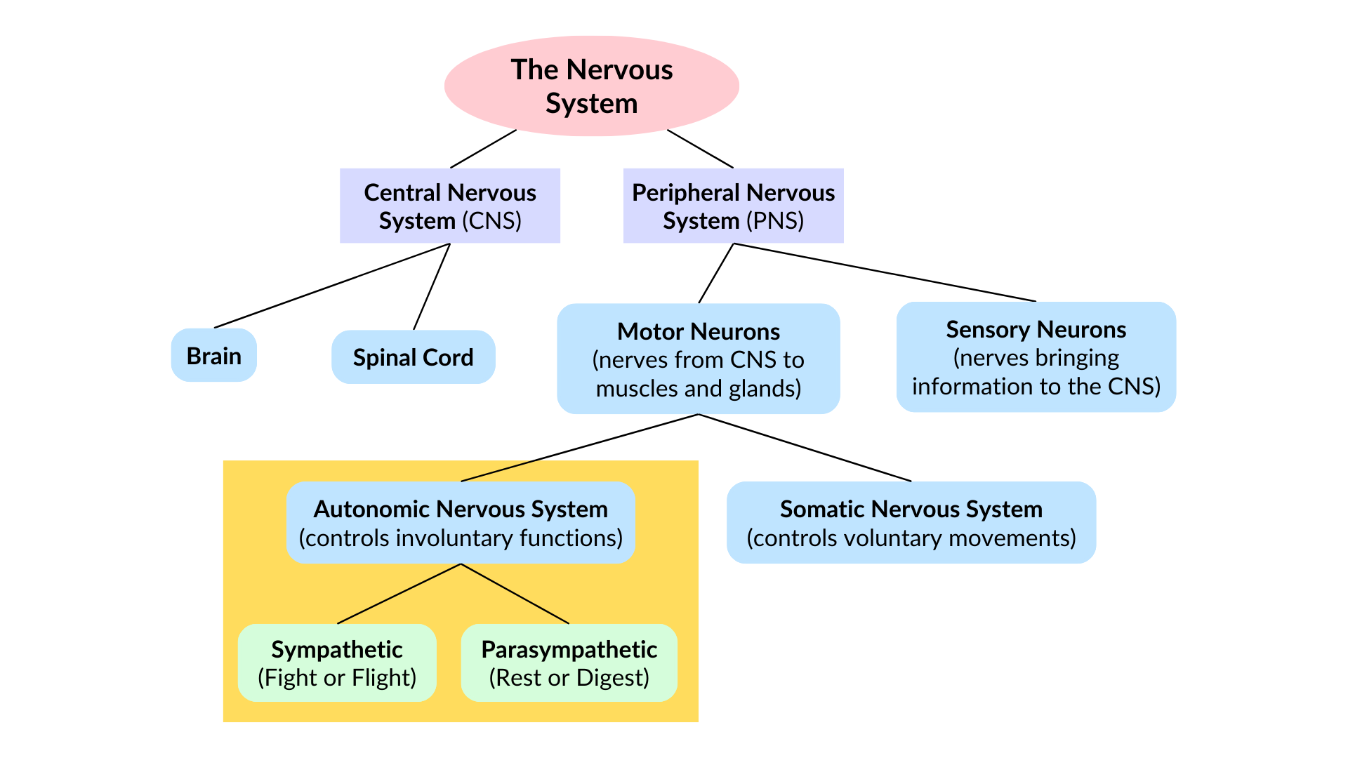
Central Nervous System: Brain and the Spinal Cord, main function is Integration and Control.
Peripheral Nervous System: spinal and Cranial Nerves, as well as the rest of the Nerves and Neuronal Cells outside the CNS. The main function to communicate with the CNS and the other parts of the body.
PNS is divided into 2 Subsystems, these being:
Sensory (Afferent) Division: This contains the Somatic and Visceral Sensory Nerve Fibres, with its main role to conduct impulses from the receptors (in the periphery) to the CNS,
Motor (Efferent) Division: This contains the Motor Nerve Fibres, with its main role to conduct impulses from the CNS to the effectors (muscle or glands).
This can also be split into 2 Further Subdivisions, these being:
Somatic Nervous System: This contains the Somatic Motor (Voluntary) Pathways, with its main function being the conduction of the impulses from the CNS to the skeletal muscles,
Autonomic Nervous System: This contains the Visceral Motor (Involuntary) Pathways, with its main function being to conduct impulses from the CNS to Cardiac Muscles, Smooth Muscles, Glands, etc. This can be further divided into 3 Subdivisions:
Sympathetic Nervous System: The function of this system is to mobilise the body systems during activity (hence the term Fight-or-Flight),
Parasympathetic Nervous System: The function of this system is to promote housekeeping functions within the body (hence the term Rest-and-Digest/ Feed-and-Breed),
Enteric Nervous System: This is sometimes called the Second Brain due to its high level of independence from the CNS, with the function of this system being to regulate the function of the Gastrointestinal Tract. And endocrine system.

What is the autonomic nervous system? What part of the brain regulates it?
Subdivision of the Peripheral Nervous System that is divided into the parasympathetic, sympathetic and enteric nervous system.
The autonomic NS innervates cardiac muscle, smooth muscles and glands, and thus controls the function of the internal organs.
The ANS is regulated by the Hypothalamus and controls vital unconscious functions, such as: Heart Rate, Vasomotor Activity, Digestion, Respiratory Rate, Pupillary Response, Urination, Sexual Arousal Behaviour
What is the enteric nervous system made up pf?
The enteric nervous system consists of a Mesh-Like System of neurons (called Auerbach’s Plexus) that regulate the function of the Gastrointestinal Tract.
The sympathetic and parasympathetic NS are both subdivisions of the ANS and represent antagonistic pathways of short time regulation of the system functions of the body. What are their opposing effects on the cardiac, respiratory, musculoskeletal, pupillary, urinary, genitourinary systems?
The main functions of the Sympathetic Nervous System is to prepare the body for intense physical activity (hence Fight-or-Flight), whereas the Parasympathetic System relaxes the body and inhibits or slows many high energy functions (hence Rest-and-Digest or Feed-and-Breed).
Cardiovascular: The different effects seen are:
SNS: This increases the Contractility and Heart Rate, increasing the Cardiac Output
PNS: This decreases the Contractility and Heart Rate, decreasing the Cardiac Output
Pulmonary: The different effects seen are:
SNS: Bronchial Dilation
PNS: Bronchial Constriction
Musculoskeletal: The different effects seen are:
SNS: Muscular Contraction
PNS: Muscular Relaxation
Pupillary: The different effects seen are:
SNS: Dilation
PNS: Constriction
Urinary: The different effects seen are:
SNS: Decreases Urinary Output and Sphincter Contraction
PNS: Increases Urinary Output and Sphincter Relaxation
Gastrointestinal: The different effects seen are:
SNS: Decreases the Motility of the Stomach and GI Tract, as well as decreasing Secretion
PNS: Increases the Motility of the Stomach and GI Tract, as well as increasing Secretion
What is a reflex arc?
Reflex arc is a neural pathway that goes through Nervous System and control's physiological reaction. The route: stimulus --> action potential travels down afferent nerve into the cell body in the dorsal route ganglion (in the spinal cord). Directly stimulates the efferent neuron or via the interneuron (within the spinal cord). The motor neuron sends the signal to the muscle.

What is the difference between a conditioned vs unconditioned reflex?
Conditioned reflex - an automatic response established by training to an ordinarily neutral stimulus
Unconditioned reflex - an automatic innate instinctive unlearned reaction to a stimulus
What is a synapse?
A synapse is a specialised junction where a neuron passes a signal to another neuron or another type of cell. Signals may be transmitted chemically using neurotransmitters or electrically.
Where are neurotransmitters produced and stored?
Synthesis or Production of Neurotransmitters and their encapsulation into Vesicles within the Soma of the Pre-Synaptic Neuron.
The neurotransmitters are transported to the Pre-Synaptic Terminal (where these vesicles are stored)
What are the steps of chemical synaptic transmission? Including the ion channels involved and including termination of the signal?
When the neuron is stimulated... (1) An action potential is conducted along the axon (via voltage-gated sodium channels) reaches the presynaptic terminal. (2) L-Type Voltage-gated calcium channels open, allowing calcium to enter the terminal cytoplasm. (3) Calcium triggers vesicles to release neurotransmitter into the synaptic cleft (exocytosis) (4) The neurotransmitter binds to receptors on the postsynaptic cell. (5) Receptor activation changes the postsynaptic cell’s activity (inhibitory/excitatory).
(6) The neurotransmitter is cleared from the synapse, ending the signal. By:
Receptor Mediated Endocytosis = the neurotransmitters are reabsorbed back into the pre-synaptic neuron to be recycled/reused.
Empty vesicles are transported back to the cell body (using a complex system of microtubules), where they either merge to the Golgi or ER, or are refilled with neurotransmitters (which then cycles back around to be released).

What is an excitatory postsynaptic potential (EPSP)?
An EPSP is a graded change that makes the postsynaptic neuron more likely to fire an action potential. It often involves depolarisation. E.g. glutamate.
Glutamate binds to receptors on the post-synaptic membrane stimulating a Sodium Ion influx (via the opening of Non-Voltage-Gated Ion Channels) into the post-synaptic cytoplasm (the consequent depolarisation and further propagation of the action potential along the post-synaptic membrane).

What is an inhibitory postsynaptic potential (IPSP)?
An IPSP is a graded change that makes the postsynaptic neuron less likely to fire. It may involve hyperpolarisation. E.g. GABA.
GABAa binds GABAa Ligand-Gated Channel on the post-synaptic membrane causing an influx of Chloride Ion into the Post-Synaptic Cytoplasm. This causes hyperpolarisation – so no further action potential is translated into the post-synaptic membrane.

What is synaptic summation? What are the three types of symmation?
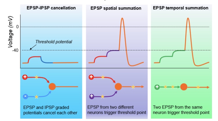
Summation is the combined effect of excitatory and inhibitory graded potentials in a postsynaptic neuron. Their combined effect helps determine whether the neuron reaches the threshold for an action potential.
Either:
EPSP- IPSP cancellation
EPSP spatial summation
EPSP temporal summation

What is EPSP-IPSP Cancellation?
Excitatory postsynaptic potential- inhibitory synaptic potential. This is when the post-synaptic neuron receives one excitatory and one inhibitory signal (of approximately equal intensity), causing them to equally counteract one another and cancel each other out (hence no post-synaptic response),

What is EPSP Spatial Summation?
Excitatory postsynaptic potential summation is when the post-synaptic neuron receives two excitatory signals from different pre-synaptic neurons (both of which are Sub-Threshold signals, hence by themselves they cannot induce EPSP). If these signals both arrive at the post-synaptic neuron simultaneously, then their effects are effectively combined, meaning that the resultant post-synaptic signal reaches the Threshold Level for the Voltage-Gated Sodium Ion Channels, leading to the development of an action potential and therefore the EPSP. This is then propagated to the post-synaptic membrane, allowing the signal to continue being transmitted,

What is EPSP Temporal Summation?
Excitatory postsynaptic potential temporal summation is when the pre-synaptic neuron initially releases excitatory signal (which slightly depolarises the post-synaptic membrane) and then quickly releases another excitatory signal (binding to the post-synaptic neuron when it is in the Relative Refractory Period, causing the depolarisation to increase and reach the Threshold Level). This results in the development of an EPSP and action potential which is then propagated down the neuron to carry on the signal.
Which neurotransmitters are commonly associated with excitation or inhibition?
Excitatory: glutamate, acetylcholine, histamine and ATP.
Inhibitory: GABA and glycine.
Potentially either, depending on receptor and context: dopamine, serotonin, adrenaline and noradrenaline.
What receptor subtype does acetylcholine bind to? And what ion currents are involved?
Acetylcholine is excitatory.
It binds to nicotinic and muscarinic receptors.
Na+ and Ca2+ are involved.
What receptor subtype does GABA bind to? And what ion currents are involved?
GABA is inhibitory.
It binds to GABAa and GABAb.
Cl- and K+ are involved.
What receptor subtype does glutamate bind to? And what ion currents are involved?
Glutamate is excitatory.
It binds to NMDA and AMPA.
Na+ and Ca2+ are involved.
What receptor subtype does Glycine bind to? And what ion currents are involved?
Glycine is inhibitory.
(It binds to GLRa1-4 and GLRb).
Cl- is involved.
What receptor subtype does catecholamines bind to? And what ion currents are involved?
Catecholamines are usually excitatory – adrenaline/noradrenaline.
Binds to alpha and beta receptors.
Na+ and Ca2+ are involved.
What receptor subtype does dopamine bind to? And what ion currents are involved?
Dopamine can be inhibitory or excitatory.
Dopamine binds to D1-5 receptors.
K+ is inhibitory, Ca2+ is excitatory.
What receptor subtype does histamine bind to? And what ion currents are involved?
Histamine is excitatory.
It binds to H1-4 receptors.
Na+ and Ca2+ are involved.
What receptor subtype does serotonin bind to? And what ion currents are involved?
Serotonin is excitatory or inhibitory.
Binds to 5-HT1-7 receptors
Na+ and Ca2+ are involved.
What receptor subtype does ATP bind to? And what ion currents are involved?
ATP is excitatory
Binds to P2X and P2Y
Na+ and Ca2+ are involved.