final exam for lab
 Call Kai
 Learn
 Practice Test
 Spaced Repetition
 Match
 Flashcards
 Knowt Play
1/173

There's no tags or description
Looks like no tags are added yet.
174 Terms
Three Requirements of Science
Natural explanations – Science explains events using natural causes, not supernatural explanations.
Testable explanations – Scientific ideas must be able to be tested through experiments or observations.
Repeatable results – Other scientists must be able to repeat the experiment and get similar results.
Improper Science
Science that uses unreliable methods, biased information, or unsupported claims. Examples include changing data to match a hypothesis, ignoring results, or making claims without evidence.
Scientific Method – Steps
Observation – Notice something happening.
Question – Ask why or how it happens.
Research – Gather background information.
Hypothesis – Make a testable prediction.
Experiment – Test the hypothesis.
Results – Collect and analyze data.
Conclusion – Explain what the results mean and whether they support the hypothesis.
Hypothesis vs. Theory
Hypothesis: A testable prediction about what will happen.
Scientific theory: A well-supported explanation backed by extensive evidence and repeated testing.
Refractometer
A laboratory instrument that measures how much light bends when passing through a solution. It can help estimate the concentration of dissolved substances.
Laboratory Safety Equipment
Eyewash station: Flushes chemicals from eyes.
Safety shower: Rinses chemicals off the body.
Fire extinguisher: Puts out fires.
Fume hood: Removes hazardous fumes.
Fire blanket: Helps extinguish flames on a person or small fire.
First-aid kit: Provides basic first aid.
Laboratory Waste Disposal
Biohazard container: Biological waste.
Sharps container: Needles, blades, and sharp objects.
Broken-glass container: Broken glass.
Chemical waste container: Designated chemical waste.
Regular trash: Ordinary nonhazardous waste.
Basic Laboratory Equipment
Beaker: Holds and mixes liquids.
Graduated cylinder: Measures liquid volume.
Test tube: Holds small samples.
Pipette: Transfers small amounts of liquid.
Erlenmeyer flask: Holds and mixes solutions.
Funnel: Helps transfer liquids.
Microscope: Magnifies small objects.
Digital balance: Measures mass.
Thermometer: Measures temperature.
Experimental Group
The group that receives the treatment or condition being tested in an experiment.
Negative Control Group
A group that does not receive the experimental treatment. It provides a comparison and is generally expected to show no treatment effect.
Positive Control Group
A group that receives a treatment known to produce a positive result. It confirms that the experiment can detect the expected effect.
Independent vs. Dependent Variable
Independent variable: What the researcher changes.
Dependent variable: What the researcher measures in response.
Confounding Variables
Uncontrolled factors that could affect the experiment's results and make it difficult to determine what caused the outcome.
Placebo vs. Placebo Effect
Placebo: A treatment with no active ingredient.
Placebo effect: When someone's expectations cause them to experience a perceived effect from the placebo.
Continuous vs. Categorical Data
Continuous: Measurable numbers that can include decimals (height, mass, temperature).
Categorical: Information separated into groups (colors, species, or blood types).
Line Graph vs. Bar Graph
Line graph: Shows changes and trends, often over time.
Bar graph: Compares different categories or groups.
Graphing Rules
X-axis: Independent variable.
Y-axis: Dependent variable.
-Include a descriptive title.
-Label both axes.
-Include measurement units.
-Use an appropriate, evenly spaced scale.
-Include a legend when multiple data groups are shown.
Metric System Prefixes
Kilo (k) = 1,000
Hecto (h) = 100
Deca (da) = 10
Base unit = 1
Deci (d) = 0.1
Centi (c) = 0.01
Milli (m) = 0.001
Micro (µ) = 0.000001
Metric Units of Measurement
Mass: Grams (g)
Length: Meters (m)
Volume: Liters (L) or milliliters (mL)
Temperature: Degrees Celsius (°C)
Metric Unit Conversions
1 kilogram = 1,000 grams
1 gram = 1,000 milligrams
1 meter = 100 centimeters
1 centimeter = 10 millimeters
1 liter = 1,000 milliliters
-Moving to a smaller unit means multiplying.
-Moving to a larger unit means dividing.
Taring a Digital Balance
Place an empty container or weigh boat on the balance and press the tare/zero button. This resets the reading to zero so only the specimen's mass is measured.
Using a Digital Balance and Weigh Boat
1) Turn on the balance.
2) Place an empty weigh boat on it.
3) Press tare.
4) Add the specimen.
5) Read and record the mass in grams.
Reading a Metric Thermometer
Read the temperature at eye level where the liquid ends. Record the measurement in degrees Celsius (°C).
Solution, Solute, and Solvent
Solution: A uniform mixture.
Solute: The substance being dissolved.
Solvent: The substance doing the dissolving.
-Example: Salt water. Salt is the solute, water is the solvent, and salt water is the solution.
Dissolving vs. Dissociating
Dissolving: A substance spreads throughout a solvent.
Dissociating: An ionic compound separates into individual positive and negative ions.
Volume/Volume Percent (% v/v)
Measures the volume of liquid solute compared with the total solution volume.
Example: 10 mL of alcohol in 100 mL of solution = 10% v/v.

Mass/Volume Percent (% m/v)
Measures the mass of a solute in grams compared with the total solution volume in milliliters.
Example: 5 g of sugar in 100 mL of solution = 5% m/v.

Molar Mass
The total mass of all the atoms in one mole of a molecule. Add the atomic masses of every atom in its chemical formula.
Unit: g/mol.
Example: H₂O = 2(1) + 16 = 18 g/mol.
Molarity (M)
Measures the number of moles of solute dissolved in one liter of solution.
Unit: mol/L.

Calculating Moles
Divide the sample's mass by its molar mass.

Standard Curve
A graph made using solutions with known concentrations. It helps determine the concentration of an unknown sample by comparing its measured result with the graph.
Spectrophotometer
A laboratory instrument that measures how much light a solution absorbs at a selected wavelength. Higher absorbance generally means a higher concentration of the absorbing substance.
Taring a Spectrophotometer
Use a blank containing the appropriate solvent or solution without the substance being measured. Insert it and press blank, zero, or calibrate according to the instrument instructions.
Using a Spectrophotometer
1) Turn on the instrument.
2) Select the correct wavelength.
3) Use a blank to zero the instrument.
4) Insert the experimental sample.
5) Read and record its absorbance.
Acid
A substance that releases hydrogen ions (H⁺) into a solution. Acids have a pH below 7.
Base
A substance that accepts hydrogen ions (H⁺) or releases hydroxide ions (OH⁻). Bases have a pH above 7.
Strong Acid/Base vs. Weak Acid/Base
Strong acid/base: Completely ionizes or dissociates in water.
Weak acid/base: Only partially ionizes or dissociates in water.
pH Scale
pH below 7 = Acidic
pH of 7 = Neutral
pH above 7 = Basic (alkaline)
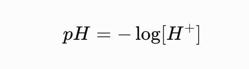
pH Formula
The pH is calculated using the negative logarithm of the hydrogen ion concentration.
Hydrogen Ion Concentration
Use this formula when you know the pH and need to calculate the hydrogen ion concentration.

Hydroxide Ion Concentration
At 25°C, hydrogen and hydroxide ion concentrations multiply to .

How Much Does Each pH Unit Change?
Every change of 1 pH unit represents a 10-fold change in hydrogen ion concentration.
Example:
-pH 3 is 10 times more acidic than pH 4.
-pH 3 is 100 times more acidic than pH 5.
-pH 3 is 1,000 times more acidic than pH 6.
Buffer
A solution that resists sudden changes in pH when small amounts of acid or base are added.
How Does a Buffer Work?
A buffer contains a weak acid and its conjugate base (or a weak base and its conjugate acid). It helps absorb added H⁺ or OH⁻ so the pH stays relatively stable.
Using pH Strips
Dip the pH strip into the solution, remove it, and compare its color to the provided color chart to estimate the pH.
Calibrating a pH Meter
Use known pH buffer solutions to adjust the meter so it gives accurate readings. Rinse the electrode between solutions as instructed.
Using a pH Meter
Rinse the electrode, place it into the experimental solution, wait for the reading to stabilize, and record the pH.
Four Biological Macromolecules
Carbohydrates – Main source of quick energy.
Lipids – Long-term energy storage and cell membranes.
Proteins – Structure, enzymes, transport, and many cell functions.
Nucleic acids – Store and transmit genetic information (DNA and RNA).
Carbohydrates – Structure and Function
Made of carbon, hydrogen, and oxygen, often in a 1:2:1 ratio. They provide energy and can provide structural support.
Examples: Glucose, starch, and glycogen.
Simple Sugars (Monosaccharides)
Single sugar molecules that provide quick energy.
Examples: Glucose and fructose.
Complex Sugars (Polysaccharides)
Long chains of sugar molecules joined together. They are used for energy storage or structural support.
Examples: Starch, glycogen, and cellulose.
Lipids – Structure and Function
Mostly made of carbon, hydrogen, and oxygen. They store long-term energy, provide insulation, and form cell membranes.
Triglyceride Structure
Made of one glycerol molecule attached to three fatty acid chains. Triglycerides are used for long-term energy storage.
Proteins – Structure and Function
Made of amino acids connected by peptide bonds. Proteins help build structures, transport substances, defend the body, and speed up chemical reactions.
Nucleic Acids – Structure and Function
Made of nucleotides. DNA stores genetic instructions, while RNA helps use those instructions to make proteins.
Benedict's Reagent
Tests for reducing sugars, such as glucose.
Positive: Changes from blue to green, yellow, orange, or reddish-brown after heating.
Negative: Remains blue.
Lugol's Iodine (IKI)
Tests for starch, a complex carbohydrate.
Positive: Changes from yellow-brown to dark blue-black.
Negative: Remains yellow-brown.
Biuret Reagent
Tests for proteins by detecting peptide bonds.
Positive: Purple or violet.
Negative: Remains blue.
Sudan III Reagent
Tests for lipids (fats).
Positive: Red/orange staining of the lipid layer or droplets.
Negative: No distinct red-stained lipid layer.
Why Are Chemical Indicators Used?
Indicators react with specific molecules and produce a visible color change. This allows scientists to determine whether a substance is present.
Positive vs. Negative Test Results (chemical indicators)
Positive result: The expected color change occurs, indicating the molecule is present.
Negative result: The expected color change does not occur, indicating the molecule was not detected.
Positive vs. Negative Controls in Macromolecule Tests
Positive control: Contains the molecule being tested for and should produce a positive reaction.
Negative control: Does not contain that molecule and should produce a negative reaction.
Compound Light Microscope
Uses visible light and multiple lenses to magnify small specimens, such as cells and microorganisms. It is commonly used for thin, transparent specimens on slides.
Dissecting Microscope
Used to view larger, solid specimens at lower magnification. It provides a three-dimensional appearance and is useful for observing surface details.
Eyepiece (Ocular Lens)
The lens you look through. Usually magnifies 10x.
Objective Lenses
The lenses closest to the specimen. They provide different magnification levels:
Scanning: 4x
Low power: 10x
High power: 40x
Total Magnification
Multiply the eyepiece magnification by the objective lens magnification.
Total magnification = Eyepiece x Objective
With a 10x eyepiece:
4x objective = 40x
10x objective = 100x
40x objective = 400x
Stage
The platform that holds the microscope slide while you view the specimen.
Stage Clips
Hold the slide in place on the microscope stage.
Diaphragm
Controls the amount of light passing through the specimen.
Illuminator
The light source that shines light through the specimen.
Coarse Adjustment Knob
Moves the stage or body tube significantly to bring the specimen into general focus. Use it on scanning power.
Fine Adjustment Knob
Makes small focusing adjustments to sharpen the image. Use it especially on higher magnifications.
Arm and Base
Arm: Supports the upper microscope and is used when carrying it.
Base: Supports the microscope and keeps it stable.
Parfocal
A microscope is parfocal when the specimen stays mostly in focus when switching between objective lenses.
Field of View
The circular area you can see through the microscope. As magnification increases, the field of view becomes smaller.
Depth of Field
The thickness or depth of the specimen that remains in focus at one time. Higher magnification generally produces a smaller depth of field.
Focusing a Compound Microscope
Start with the scanning objective (4x).
Place and secure the slide.
Use the coarse adjustment to find the specimen.
Use the fine adjustment to sharpen the image.
Center the specimen.
Switch to 10x, then 40x.
Use fine adjustment at higher magnifications.
Prokaryotic vs. Eukaryotic Cells
Prokaryotic: No nucleus or membrane-bound organelles. DNA is in the cytoplasm.
Eukaryotic: Has a nucleus and membrane-bound organelles.
Six Kingdoms of Life
Archaebacteria – Prokaryotic, usually found in distinctive environments.
Eubacteria – Prokaryotic, common bacteria.
Protista – Eukaryotic, mostly single-celled organisms.
Fungi – Eukaryotic, absorb nutrients; includes mushrooms and yeast.
Plantae – Eukaryotic, mostly multicellular and photosynthetic.
Animalia – Eukaryotic, multicellular and obtain food by consuming other organisms.
Microscope Cell Structures
Cell membrane: Controls what enters and leaves.
Cytoplasm: Jelly-like material inside the cell.
Nucleus: Contains DNA and controls cell activities.
Cell wall: Provides support and protection.
Chloroplast: Performs photosynthesis.
Central vacuole: Stores water and helps maintain plant cell pressure.
How Do You Identify Plant vs. Animal Cells?
Plant cells: Usually have a rigid cell wall, rectangular appearance, and large central vacuole. Some have visible chloroplasts.
Animal cells: Have no cell wall or chloroplasts and are often more rounded or irregular.
Storing a Compound Microscope
Remove the slide.
Clean the lenses with lens paper.
Rotate to the scanning objective (4x).
Lower the stage.
Turn off the light.
Wrap the cord properly.
Carry with two hands and store safely.
Plasma Membrane
A thin, flexible barrier surrounding the cell. It controls what enters and leaves the cell and helps maintain a stable internal environment.
Molecules Found in the Plasma Membrane
Phospholipids: Form the main membrane structure.
Proteins: Help transport substances and communicate.
Cholesterol: Helps maintain membrane fluidity.
Carbohydrates: Help with cell recognition and communication.
Phospholipid Structure
A phospholipid has:
Hydrophilic head: Attracted to water.
Hydrophobic tails: Repelled by water.
Hydrophilic vs. Hydrophobic
Hydrophilic: Water-loving; attracted to water.
Hydrophobic: Water-fearing; repelled by water.
Diffusion
The movement of molecules from an area of high concentration to an area of low concentration until they reach equilibrium. It does not require energy.
Osmosis
The diffusion of water across a selectively permeable membrane. Water moves toward the side with the higher solute concentration.
Hypertonic Solution
A solution with a higher solute concentration than the inside of the cell. Water moves out of the cell.
Hypotonic Solution
A solution with a lower solute concentration than the inside of the cell. Water moves into the cell.
Isotonic Solution
A solution with equal solute concentrations inside and outside the cell. Water moves in both directions equally, so there is no net movement.
Shriveled Cell
An animal cell that loses water and shrinks when placed in a hypertonic solution.
Lysed Cell
An animal cell that takes in too much water and bursts when placed in a hypotonic solution.
Plasmolyzed Plant Cell
A plant cell that loses water in a hypertonic solution. The cell membrane pulls away from the cell wall.
Turgid Plant Cell
A plant cell filled with water in a hypotonic solution. The cell becomes firm because water pressure pushes against the cell wall.
Plant Cells in Different Solutions
Hypertonic: Lose water and become plasmolyzed.
Hypotonic: Gain water and become turgid.
Isotonic: No net water movement; the cell is generally flaccid (less firm).
Animal Cells in Different Solutions
Hypertonic: Lose water and become shriveled.
Hypotonic: Gain water and may lyse (burst).
Isotonic: Maintain their normal size and shape.
How Does Molecule Size Affect Diffusion?
Smaller molecules generally diffuse faster than larger molecules because they move more easily through available spaces or pathways.
How Does Concentration Affect Diffusion?
A larger concentration difference creates a steeper concentration gradient, generally increasing the rate of diffusion.