Chapter 7: Axial Skeleton System
 Call Kai
 Learn
Learn Practice Test
 Spaced Repetition
 Match
Match Flashcards
 Knowt Play
1/163

There's no tags or description
Looks like no tags are added yet.
Name | Mastery | Learn | Test | Matching | Spaced | Call with Kai | Chat |
|---|
No analytics yet
Send a link to your students to track their progress
164 Terms
Axial skeleton
Centra supporting axis of the body (includes skull, vertebral column and thoracic cage)
Appendicular skeleton
Upper limbs, pectoral girdle, lower limbs and pelvic girdle
Number of bones at birth
270
Number of bones after fusion
Approx. 206
Sesamoid bones
Form within tendons (ex patella) due to stress
Sutural (wormian) bones
Extra bones of the skull but are not found equally in everyone
Canal
A tubular passage or tunnel in a bone (auditory canal of the skull)
Condyle
A rounded knob (occipital condyles of the skull)
Crest
A narrow ridge (iliac crest of the pelvis)
Epicondyle
A flare superior to a condyle (medial epicondyle of the femur)
Facet
A smooth joint surface that is flat or only slightly concave or convex (Articular factes of the vertebrae)
Fissure
A slit through a bone (orbital fissures behind the eye)
Foramen
A hole through a bone, usually round (foramen magnum of the skull)
Fossa
A shallow, broad or elongated basin (infraspinous fossa of the scapula)
Lines (linea)
A raised, elongated ridge (nuchal lines of the skull)
Meatus
A canal (external acoustic meatus of the tempiral bone)
Process
any bony prominence (mastoid process of the skull)
Protuberance
A bony outgrowth or protruding part (mental protuberance of the chin)
Sinus
A cavity within a bone (frontal sinus of the skull)
Spine
A sharp, slender or narrow process (spine of the scapula)
Sulcus
A groove for a tendon, nerve or blood vessel (intertubercular sulcus of the humerus)
Tubercle
A small, rounded process (greater tubercle of the humerus)
Tuberosity
A rough surface (tibial tuberosity)
Number of bones joined by sutures in the skull
22
Cavities of the brain
- Cranial cavity
- Orbitis (eyes)
- Nasal cavity
- Ear cavities
- Paranasal sinuses
- Oral cavity
Mnemonic for brain cavities
Cranberries Orbiting NASA Eat Paranormals Orally

Foramina
Holes where nerves and blood vessels pass (Foramen-singular)

Major bones of the cranium
Occipital, parietal, frontal, temporal, ethmoid, sphenoid
Mnemonic for bones of the cranium
Old people from texas eat spiders

Major bones of the face
Vomer, Conchae (2), Mandible, Maxilla, Palatine (2), zygomatic (2), and Lacrimal (2)
Mnemonic for major bones of the face
Virgins cannot make my pet zebra laugh

Parts of the frontal bone
Coronal suture and frontal sinus

Parts of the parietal bone
Sagittal suture, coronal suture, lambdoid suture, and squamous suture

Parts of the temporal bone
Squamous suture and 4 regions: squamous, tympanic, mastoid and petrous

Parts of the occipital bone
Lambdoid suture, foramen magnum and occipital condyles

Paranasal sinuses
sphenoidal sinus, frontal sinus, ethmoidal sinus and maxillary sinus
Cranial fossae
anterior, middle, posterior

anterior cranial fossa
formed by the frontal bone and part of the sphenoid bone

middle cranial fossa
holds the temporal lobes of the brain

posterior cranial fossa
supports the cerebellum of the brain

crista galli of ethmoid bone
superior projection in the middle of the cribriform plate of the ethmoid bone; providing a point of attachment for the dura mater, helping to secure the brain within the skull
ethmoidal sinuses of ethmoid bone
Sinuses in the nasal cavity in between the frontal sinus and the sphenoidal sinus

maxillary sinus (maxilla)
sinus on either side of the nasal cavity below the eyes

zygomatic arch
cheek bone

nasal conchae of ethmoid bone
two curled plates of bone; project into the nasal fossa from its lateral wall

coronal suture
the suture between the parietal and frontal bones of the skull

sagittal suture
between the two parietal bones
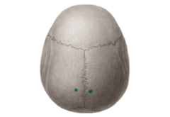
parietal foramen
sometimes occurs near the corner of the lambdoid and sagittal sutures on parietal bones

lambdoid suture
between parietal bones and occipital bone

Temporal bone (lateral surface)
Zygomatic process, external acoustic meatus, mastoid process and styloid process

Temporal bone (medial surface)
Internal acoustic meatus

Sphenoid
Mothlike shape containing: sphenoidal sinuses, greater/lesser wings and sella turcica

Ethmoid
- ethmoidal sinus
- crista galli
- cribriform plate
- superior and middle nasal concha
- nasal septum

Maxillae (2)
Only facial bone in a sinus, intermaxillary suture, alveolar processes and infraorbital foramen

Palatine (2)
hard palate (roof of mouth)

Zygomatic (2)
Angles of cheeks

Lacrimal (2)
- Medial wall of each orbit
- Smallest bone of skull
- Lacrimal fossa

Nasal (2)
Bridge of the nose

inferior nasal conchae (2)
nasal cavity

vomer
means plowshare and has no muscle attached (septum)

Mandible
Only movable bone of the skull and strongest skull bone

Bones of orbital cavity
Ethmoid, lacrimal, palatine, temporal, frontal, maxilla, sphenoid and zygomatic

Key features of mandible
- Coronoid process
- Condylar process
- Mandibular condyles
- Mandibular notch
- Mental foramen
- Mental protuberance
- body
- ramus

coronoid process of mandible
flattened upward projection from the anterior margin of the mandibular ramus

condylar process of mandible
thickened upward projection from posterior margin of mandibular ramus

Mandibular condyles of mandible
two rounded projections on the superior part of the ramus on both sides

mandibular notch of mandible
depression between the condylar process and the coronoid process

mental foramen of mandible
one of two holes located on the anterior surface of the mandible. It permits passage of the mental nerve and vessels.

mental protuberance of mandible
point of chin

body of mandible
horizontal portion

ramus of mandible
vertical portion

Auditory ossicles
bones of the middle ear (malleus, incus and stapes)

Hyoid
Does not articulate with any other bone, located between chin and larynx and fracture here may indicate strangulation

Components of hyoid bone
Body, lesser horn and greater horn

Vertebral Column
Chain of 33 vertebrae and intervertebral discs

Sections of vertebral column
cervical, thoracic, lumbar, sacrum, coccygeal
Cervical spine
The portion of the spinal column consisting of the first seven vertebrae that lie in the neck.
thoracic spine
The 12 vertebrae that lie between the cervical vertebrae and the lumbar vertebrae. One pair of ribs is attached to each of the thoracic vertebrae.
lumbar spine
The lower part of the back, formed by the lowest five nonfused vertebrae; also called the dorsal spine.
Sacral spine
The sacral spine consists of 5 fused sacral vertebrae. It is easily distinguishable as an upside-down triangular shape.
coccygeal spine
4 vertebrae, fused to form the tailbone
Functions of the spine
- Supports skull and trunk
- allows for movement
- protects spinal cord
- absorbs stress from walking, running, etc
- attachment for limbs, thoracic cage and postural muscles
Natural curvature at birth
Column is c-shaped and has primary curvatures
Natural curvatures
occurs at age 3 when column is s-shaped with primary and secondary curvatures
primary curvature
thoracic and pelvic curvatures
secondary curvatures
cervical and lumbar curvatures that develop after lifting head, crawling and walking

Scoliosis
abnormal lateral curvature

Hyperkyphosis (hunchback)
abnormal posterior thoracic curvature
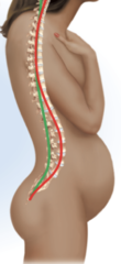
Hyperlordosis (swayback)
abnormal anterior lumbar curvature
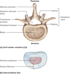
General Structure of a Vertebra
- body
- vertebral foramen
- vertebral arch
- spinous process
- transverse process
- superior and inferior articular processes

intervertebral discs (23)
Pads between vertebrae that absorb stress and protect nerves

Nucelus pulposus
central portion that is a gelatinous consistency

Anulus fibrosus
outer ring of fibrocartilage
Herniated disc ("ruptured" or "slipped" disc)
condition in which the nucleus pulposus protrudes posteriorly through the annulus fibrosus putting pressure on spinal nerves

transverse process
two lateral projections from the vertebral arch

body of vertebrae
the thick, disc-shaped anterior portion which is the weight bearing portion

Inferior articular process
bony process that extends downward from the vertebral arch of a vertebra that articulates with the superior articular process of the next lower vertebra

superior articular process
bony process that extends upward from the vertebral arch of a vertebra that articulates with the inferior articular process of the next higher vertebra

Lamina
part of the vertebral arch

Inferior vertebral notch
indentation of the pedicle for nerve passing