Blood Flow Pathways
 Call Kai
 Learn
 Practice Test
 Spaced Repetition
 Match
 Flashcards
 Knowt Play
1/52

There's no tags or description
Looks like no tags are added yet.
53 Terms
Anterior cerebral artery
Origin:
Internal carotid
Course:
Passes anteriorly along medial aspect of frontal lobe of cerebral cortex
Distribution:
Medial aspect of frontal lobe of cerebral cortex

Anterior cerebral artery and branches
Origin:
Internal carotid
Course:
Passes anteriorly along medial aspect of frontal lobe of cerebral cortex
Comment:
Medial aspect of frontal lobe of cerebral cortex

Basilar artery
Origin:
Junction of paired vertebral arteries
Course:
Unpaired (midline) vessel ascends on anterior (ventral) surface of pons
Distribution:
Pons
Cerebellum

Cerebral arterial circle
Origin:
Internal carotid and basilar arteries
Course:
Circular anastomosis on ventral surface of brain

Internal carotid artery
Origin:
Common carotid
Course:
Ascends in neck to base of cranium
Enters cranial cavity via carotid canal
Distribution:
Eye
Cerebrum
Cerebral arterial circle (of Willis)

Posterior cerebral artery and branches
Origin:
Basilar
Course:
Paired vessels pass laterally around cerebral peduncles
Distribution:
Temporal and occipital lobes of cerebral cortex

Vertebral artery
Origin:
Subclavian
Course:
Paired vessel ascends through neck, via transverse foramina in cervical vertebrae
Enters cranial cavity via foramen magnum
Distribution:
Brain
Spinal cord
Vertebral column

Carotid Pulse
Location:
Neck (lateral)
Between thyroid cartilage and sternocleidomastoid muscle
Description:
Surface projection of pulse in common carotid artery

Common carotid artery
Origin:
Right: brachiocephalic trunk from arch of aorta
Left: arch of aorta
Course:
Exits thorax and ascends in neck
Distribution:
Structures of head and neck

External carotid artery
Origin:
Common carotid (at bifurcation)
Course:
Ascends in neck
Distribution:
Exterior of head (except orbit)
Face
Meninges
Neck structures

External jugular vein
Drainage:
Scalp
Face
Course:
Subcutaneous vessel formed by union of retromandibular and posterior auricular veins near angle of mandible
Descends toward middle of clavicle
Crosses anterior surface of sternocleidomastoid muscle

Internal carotid artery
Origin:
Common carotid
Course:
Ascends in neck to base of cranium
Enters cranial cavity via carotid canal

Internal jugular vein
Drainage:
Cranial cavity, including brain
Face
Neck
Tributaries:
Sigmoid sinus
Inferior petrosal sinus
Facial
Lingual
Pharyngeal
Superior and middle thyroid

Axillary artery
Origin:
Subclavian (at lateral border of rib 1)
Course:
Passes through axilla surrounded by cords and branches of brachial plexus
Divided into 3 parts by pectoralis minor m. First part is proximal to pectoralis minor m., second part is posterior to muscle, and third part is distal to muscle

Axillary vein
Drainage:
Pectoral, shoulder, and scapular regions

Brachial artery
Origin:
Axillary (at inferior border of teres major m.)
Course:
Passes inferiorly along medial aspect of arm

External Jugular vein (2nd view)
Drainage:
Scalp
Face
Course:
Subcutaneous vessel formed by union of retromandibular and posterior auricular veins near angle of mandible
Descends toward middle of clavicle
Crosses anterior surface of sternocleidomastoid muscle

Internal jugular vein (2nd view)
Drainage:
Cranial cavity, including brain
Face
Neck
Tributaries:
Sigmoid sinus
Inferior petrosal sinus
Facial
Lingual
Pharyngeal
Superior and middle thyroid

Left subclavian artery
Origin:
Arch of aorta
Course:
Arches over rib 1

Left subclavian vein
Drainage:
Upper limb
Superficial back and neck

Brachial pulse
Location:
Medial arm
Description:
Surface projection of pulse in brachial artery

Brachial vein
Drainage:
Arm muscles
Humerus
Elbow joint

Brachial pulse
Location:
Medial arm
Description:
Surface projection of pulse in brachial artery
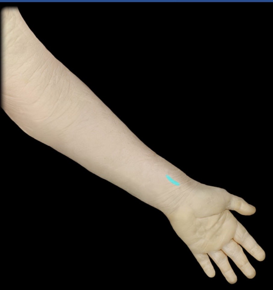
Ulnar pulse
Location:
Anterior medial wrist
Deep to tendon of flexor carpi ulnaris muscle
Description:
Surface projection of pulse in ulnar artery

Ulnar vein
Drainage:
Medial forearm muscles
Hand muscles and joints
Elbow and wrist joints

Brachiocephalic trunk
Origin:
Arch of aorta
Course:
Ascends posterior to right margin of manubrium (sternum)
Ends posterior to right sternoclavicular joint

Left braciocephalic vein
Drainage:
Head and neck (left side)
Upper limb (left)

Left common carotid artery
Origin:
Arch of aorta
Course:
Exits thorax and ascends in neck
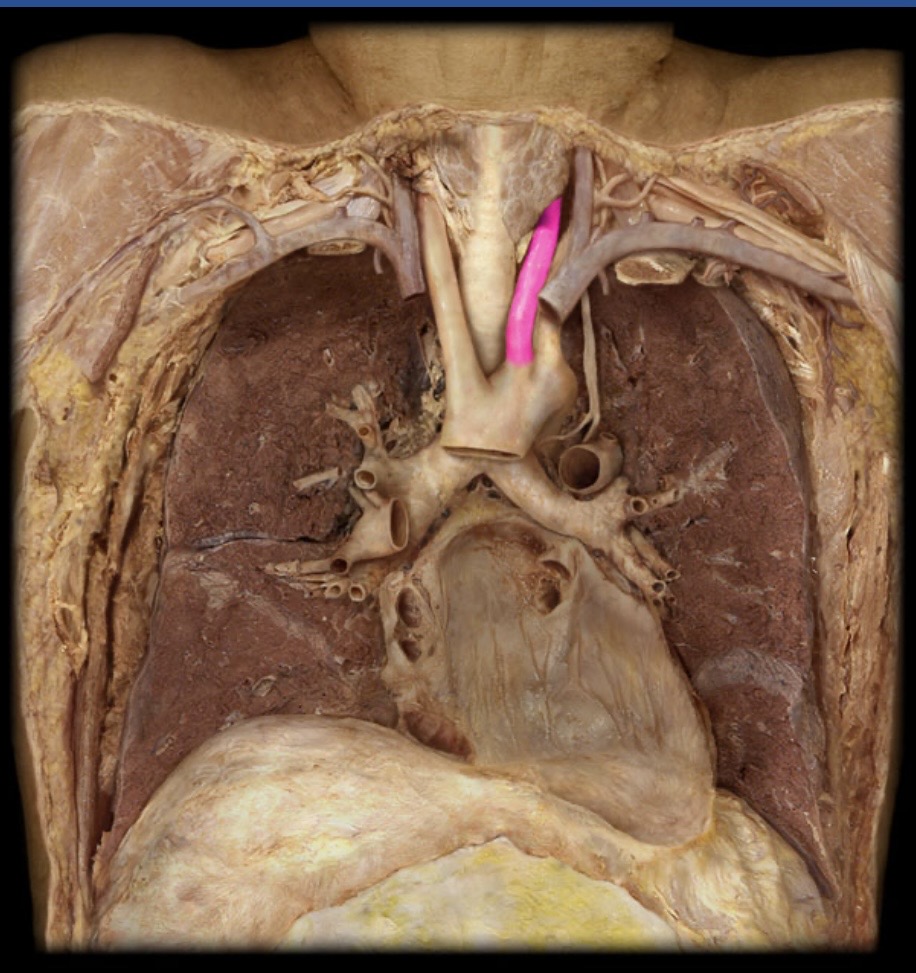
Pulmonary artery
Origin:
Pulmonary trunk
Course:
Left: laterally, inferior to arch of aorta and anterior to left main bronchus
Right: laterally, posterior to ascending aorta and superior vena cava
Through root and hilum of respective lungs

Right brachiocephalic vein
Drainage:
Head and neck (right side)
Upper limb (right)

Right common carotid artery
Origin:
Brachiocephalic trunk from arch of aorta
Course:
Exits thorax and ascends in neck

Right subclavian artery
Origin:
Brachiocephalic trunk from arch of aorta
Course:
Arches over rib 1

subclavian vein
Drainage:
Upper limb
Superficial back and neck

Abdominal Aorta
Origin:
Thoracic aorta
Course:
Parallels vertebral column, slightly left of midline
Divides into common iliac arteries on body of L4 vertebra
Distribution:
Abdominal and pelvic viscera
Walls of abdomen and pelvis
Ovaries and testis

Common illiac artery
Origin:
Abdominal aorta
Course:
Begins anterior to L4 vertebral body
Passes obliquely across L4-5 vertebral bodies
Ends at the level of the sacro-iliac joint (L5-S1)

Inferior vena cava
Drainage:
Everything inferior to diaphragm, except posterior abdominal wall, which drains into azygos system

Left renal artery
Origin:
Abdominal aorta
Course:
Passes laterally to enter kidney

Left renal vein
Drainage:
Kidney
Ureter (upper)
Suprarenal gland
Left testis/ovary
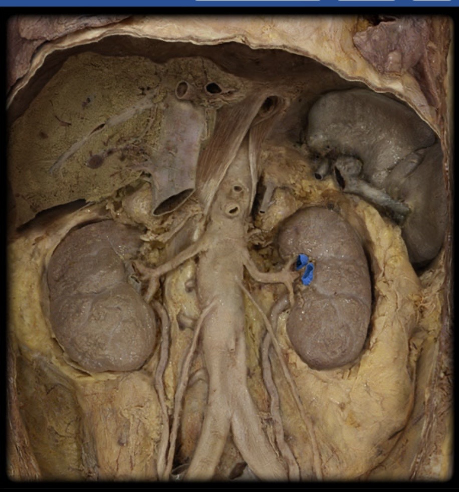
Right renal artery
Origin:
Abdominal aorta
Course:
Passes laterally to enter kidney

Right renal vein
Drainage:
Kidney
Ureter (upper part)

Kidney
Location:
Abdomen (posterior wall)
Retroperitoneal
Left kidney higher than right
Description:
Paired, bean-shaped organ
Concave, medial border is point of entrance/exit of renal vessels and ureter
Contains cortex, medulla, and renal sinus

External iliac artery
Origin:
Common iliac
Course:
Descends laterally along medial border of psoas muscle from level of sacro-iliac joint to inguinal ligament

Femoral artery
Origin:
External iliac
Course:
Descends in anterior and medial thigh
Extends from inguinal ligament to adductor hiatus

Femoral artery pulse
Location:
Proximal anterior thigh
Midway between anterior superior iliac spine and pubic symphysis
Superficial location in femoral triangle
Description:
Surface projection of pulse in femoral artery

Internal iliac artery
Origin:
Common iliac
Course:
Begins at the level of the sacro-iliac joint (L5-S1)
Descends into pelvis and passes posteriorly toward superior margin of greater sciatic notch (ilium)

Common iliac vein
Drainage:
Pelvis
Perineum
Lower limb

External iliac vein
Drainage:
Lower limb
Anterior abdominal wall (inferior)
Tributaries:
Femoral
Inferior epigastric
Deep circumflex iliac

Femoral vein
Drainage:
Abdominal wall (anterior inferior)
External genitalia
Thigh muscles
Femur
Hip and knee joints

Great saphenous vein
Drainage:
Dorsum of foot
Subcutaneous medial leg and thigh

Infreior vena cava
Drainage:
Everything inferior to diaphragm, except posterior abdominal wall, which drains into azygos system

Internal iliac vein
Drainage:
Pelvis
Perineum
Gluteal region
Posterior abdominal wall

Adductor magnus m
Action:
Adduction of thigh
Extension of thigh
Origin:
Pubis (inferior ramus)
Ischium (ramus and tuberosity)

Piriformis m
Action:
Lateral rotation of thigh
Origin:
Sacrum (anterior surface)
