Hole's Essentials of Human Anatomy & Physiology: Chapter 7 - Skeletal System
 Call Kai
 Learn
 Practice Test
 Spaced Repetition
 Match
 Flashcards
 Knowt Play
1/65

Earn XP
Description and Tags
Comprehensive vocabulary flashcards covering the functions, classifications, gross and microscopic anatomy, fracture types, repair, ossification, and hormonal regulation of the skeletal system.
66 Terms
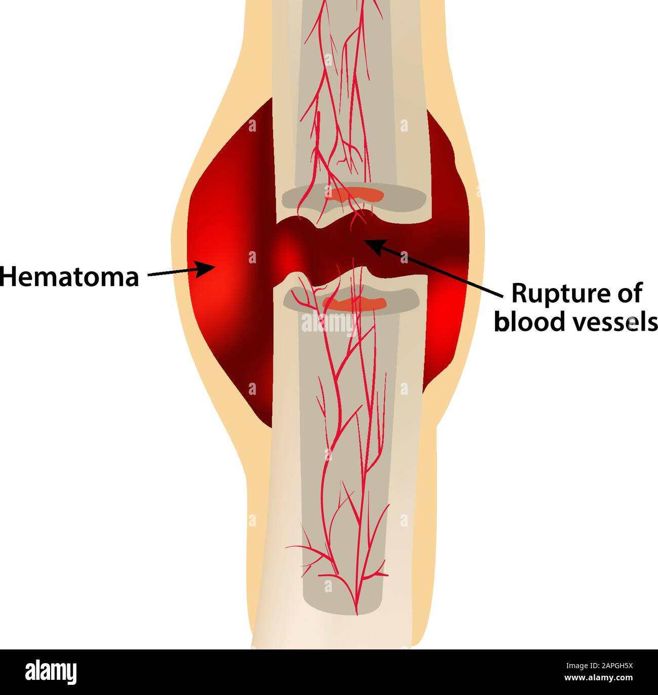
Hematoma (blood clot) is formed
STEP 1
Blood vessels break when the bone fractures → blood escapes and forms a hematoma around break
Periosteum tears
Blood vessels in surrounding tissues dilate→ tissues swell and inflammation occurs
Long Bones
Longer than wide
Have a shaft and heads on both ends
mostly compact bone
EX: Femur, metatarsals

Short Bones
Cube-shaped bones
containing mostly spongy bone
Ex: Carpals and tarsals (not weight bearing→ why tape for gymnastics)

Flat Bones
Thin,
layers of compact bone around a layer of spongy bone
usually curved bones
Ex: sternum, partial bone, scapula, ribs illium (crest)

Irregular Bones
Irregular shape
Do not fit into other bone classification categories
Ex: vertebrae, hip bones, and auditory ossicles.


Sesamoid Bone
rounded shape
Embedded within tendons adjacent to joints
1 bone: Patella
Diaphysis
Shaft
consists of central medullary cavity
surrounded by compact bone

Epiphyses
expanded ends of a long bone
one proximal and one distal
consisting mainly of spongy bone surrounded by a thin layer of compact bone.

Epiphyseal Plate
epiphyses meet the diaphysis
contains hyaline cartilage
Growth (responsible for elongation of long bones)
In kids, Growth plates

Epiphyseal Line
what’s left behind of the Epiphyseal Plate once its done growing
Line
Adults

Periosteum
Peri=surrounding
Osteum=bone
tough, outer dense connective tissue protective covering of diaphysis
contains osteoblasts and osteoclasts (cells that forms and repaid bone tissues)
Insertion for tendons and ligaments.

Endosteum
The thin, inner lining of medullary cavity
contains osteoblasts and osteoclasts.

Medullary Cavity
Hollow space in diaphysis of long bone
yellow bone marrow inside

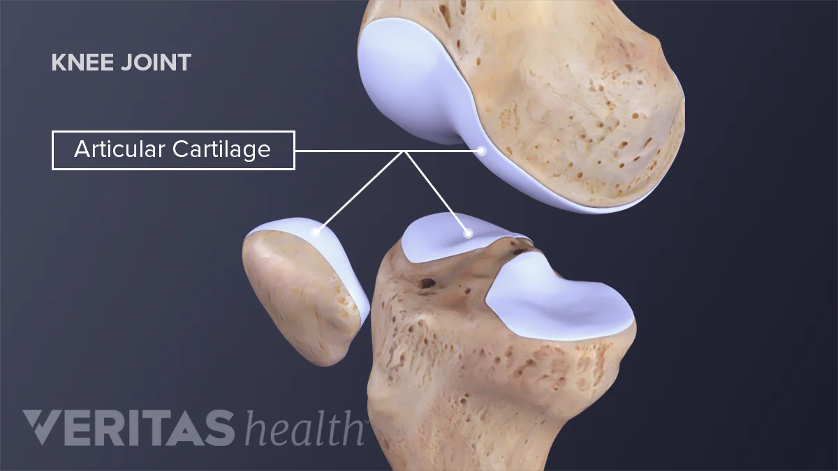
Articular Cartilage
A pad of hyaline cartilage covering the external joint surface of the epiphyses to decrease friction during movement.
osteoarthritis= thinning of Articular Cartilage
Sharpey's Fibers
Collagen fibers that secure periosteum to underlying bone.
Velcro

Osteon (Haversian System)
Whole toilet paper roll
basic structural units of compact bone
bunch of tiny circles/osteon come together to create compact bone
(hemousnous (no gaps))

Osteocytes
Mature, spider-shaped bone cells
located inside lacunae
maintain and monitor bone tissue and cannot undergo mitosis.
alive kids/students in the classroom
(Maintainers)

Lacunae
hallow chambers
containing osteocytes
Classrooms

Lamellae
RINGS of inorganic (dead) minerals (collagen and calcium salts)
Floors

Canaliculi
Connection between each lunate (classroom)
Stairs/Call

Volkmann's Canals (Perforating Canals)
Connecting osteons
Blood vessels go through
Tunnel at LT

Trabeculae
Poorly organized, small needle-like branching pieces or plates of bone that form the structural framework of spongy bone.
Osteoprogenitor Cells
stem cells that undergo mitosis constantly to become osteoblasts
(baby bone cells)
Osteoblasts
BUILDERS
build new bone by secreting collagen
cannot undergo mitosis
(cell that lays new bones based on needs (basketball szn))
Osteoclasts
CLEAR
Cells that break down and remove old bone tissue
Help with bone remodeling
Osteoid
primarily COLLAGE → what gives bone its flexibility (tree branch won’t break off)
lays in-between inorganic and osteoblasts
material that becomes hard bone
Inorganic component of chemical composition of bone
65%
Hydroxyapatite, which is primarily calcium phosphate, gives bone it’s hardness and compression strength.
Closed (Simple) Fracture
The bone breaks but does not pierce the skin
6–8 weeks of healing time.
Open (Compound) Fracture
The broken bone ends break through the skin.
Reduction
realignment of the bone.
afterwards bones are typically immobilized using casts, splints, or braces to allow healing.
Greenstick Fracture
An incomplete break
one side breaks, the other bends
common in children

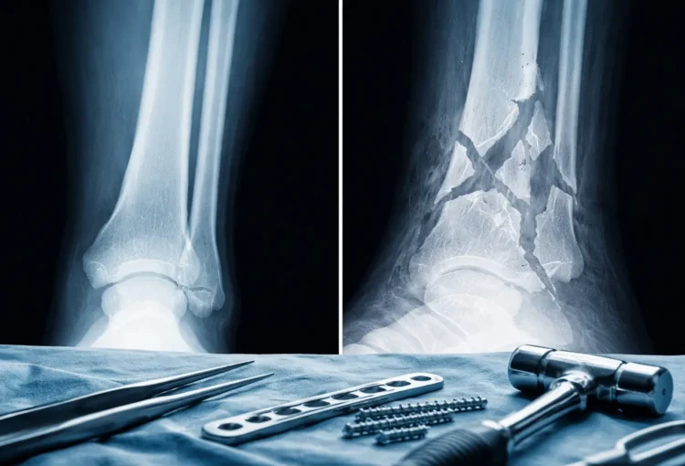
Comminuted Fracture
A complete fracture
that fragments or shatters the bone into multiple pieces.
Intramembranous Ossification
1 st pattern of bone development
where flat bones originate directly between sheetlike layers of connective tissue.
build off of neighboring bone
ex: roofing skull, collar bone (flat bones)
Connective tissue → bone
Endochondral Ossification
2nd pattern of bone development
bone begins as hyaline cartilage masses, the get REPLACED by bone tissue
Most bones, femur, long bones
Hyaline cartilage ex: bridge of nose, parts of ribs, joints, articular cartilage, epiphyseal plates
Cartilage → Bone

Primary Ossification Center
Endochondral ossification
The central region in the DIAPHYSIS of a long bone
where cartilage calcification and bone formation first develop
Primary = First, in the middle
Secondary Ossification Center
Endochondral ossification
develops after primary center
forms in the EPIPHYSES (ends) of long bones
usually develops around birth or childhood.
Secondary= second, at the ends
Appositional Growth
starts with 2 rings
then osteoblasts add new bone (ring) to the outer surface.
Osteoclasts break down the bone (ring) on the inside to make room (now back at 2 rings)
The bone becomes WIDER (medullary cavity gets bigger), but not super heavy.
During infancy and childhood.
Parathyroid Hormone (PTH)
A hormone released by the parathyroid glands when blood calcium is low that stimulates osteoclast activity and kidney calcium reabsorption to elevate blood calcium levels.
Calcitonin
A hormone secreted by the thyroid gland when blood calcium is high that stimulates osteoblast activity and prompts the kidneys to excrete calcium, decreasing blood calcium levels.
Acromegaly
disorder where anterior pituitary gland secretes excess human growth hormone (hGH),
Rly tall
Functions of Bone Tissue
Support: legs and pelvis support the body’s weight, atlas supports head
Protection: encases vital organs like the heart and lungs
Movement: Skeletal muscles attached to bones by tendons serve to move bones
Mineral Homeostasis: Bones store minerals like calcium salts and calcium phosphate, magnesium, sodium, potassium, carbonate ions, releasing them into the bloodstream as needed.
Energy Storage: yellow marrow serves as important chemical energy storage
Hematopoiesis: Blood cell formation
Increase in RBC production→ Lack of Oxygen (Hypoxia) bc rbc get old and don’t have a lot of Oxygen so kidney releases→ hormone ERYTHROPOIETIN and increases production of RBC → new RBC replace old ones in the bone marrow in response to erythropoietin.
Gross Anatomy
typical long bone (ex humerus)
Nutrient arteries
Supplies bone cells with nutrients
Enter bone through nutrient foramen in compact bone.

Compact bone
blue Legos (all the same)
Homogeneous (same), main in long bone (bc bears weight)
continuous extracellular matrix without space
dense concentric circles of osseous tissue
site of red blood cells formation in KIDS

Spongy bone
all different types of lego
Heterogenous (different)
small needle-like pieces of bone
many open spaces
site of red blood cell formation in ADULTS
lightens bones

Central (Haversian) Canals
Hole in middle of each osteon
Blood vessels go through
Toilet paper roll hole
Structure of spongy bone
consists of random trabeculae (small needle-like pieces of bone)
nourished by nearby central canals (Haversian)
Allows for lightness and pockets for blood (Red marrow)

Organic component of chemical composition of bone
35%
Osteoprogenitor
Osteoblasts
Osteocytes
Osteoclasts
Osteoid
Closed reduction
The physician manually moves bone ends back into place WITHOUT surgery
Open reduction
Surgical procedure using pins, plates, screws, or rods to stabilize the bone.
Fractures: Classification by cause
Traumatic: Injury causes the break
Spontaneous: Breaks unexpectedly
Pathologic: Disease weakens the bone
Spiral
Complete fracture
cause by TWISTING of the bone

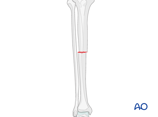
Transverse fracture
Complete fracture
clean break straight across the bone
(at right angle to axis of bone)
Compression Fracture
Complete fracture
Bone is crushed (squished or collapses)
common in vertebrae

Fissured Fracture
Incomplete fracture
incomplete longitudinal break

Oblique Fracture
Complete Fracture
fracture at some other angle not straight (transverse)

Fibrocartilage Callus Formation
STEP 2
Fibrocartilage fills gaps between ends of broken bone from blood vessels
It acts like a splint to hold the broken bone pieces together
A soft bridge connects the broken bone
Osteoblasts from periosteum invade hematoma → blasts form spongy bone near blood vessels
Phagocytes remove blood clot
osteoclasts resorb bone fragments.
Granulation tissue develops

Bony Callus
STEP 3
Fibrocartilage is replaced by boney callus
Cartilage callus breaks down →Bony callus fills space
The soft bridge turns into hard bone.

Bone remodeling
STEP 4
Bony callus is remolded to form a permanent patch
osteoclasts remove excess bony tissues
More bone is produced at site of healing fracture than needed

Ossification
The process of forming new bones tissue
Two types:
Intramembranous: Bone forms directly from connective tissue.
Endochondral: Bone forms by replacing cartilage.
Bone Growth (Epiphyseal plate)
Epiphyseal plate allows for bone lengthening until adulthood.
Cartilage cells are produced by mitosis on the epiphyseal side of the plate
Cartilage cells are destroyed and replaced by bone on the diaphyseal side of the plalte
therefore, the bone on the diaphyseal side increases in length (but thickness stays same)
Factors that impact rate of bone repair, growth and remodel
Exercise, weight, external stress/force
Genes (ethnicity, race)
Hormones
Sex hormones
Testosterone
estrogen
hGH (Human growth hormone)
Parathyroid hormone (PTH) and calcitonin
Age
Diet
Ossification of most bones is completed by age 25 (growth stops with primary and secondary ossification centers meet)
Rate of remodeling
Every 4 months-Femur (bc so weight bearing)
Diaphysis may never be fully replaced during one’s lifetime
Calcium and Phosphorus
Inorganic part of bone
component of hydroxyapatite matrix
Sex hormones
estrogens & testosterone (androgens)
aid osteoblasts activity to promote new bone growth
degenerated cartilage cells in epiphyseal plate (close the epiphyseal plate).
Estrogen effect is greater than androgen (testosterone) effect
Human Growth Hormone (hGH)
Secreted by pituitary gland (brain)
responsible for the general growrh of all tissues
stimulates reproduction of cartilage cells at epiphyseal plate (acro