NRSE360 EXAM#3 postpartum complications
 Call Kai
 Learn
 Practice Test
 Spaced Repetition
 Match
 Flashcards
 Knowt Play
1/70

There's no tags or description
Looks like no tags are added yet.
71 Terms
DVT
Blood clot forms in a deep vein during/after childbirth
The lower extremity can be of superficial or deep veins. PP client @ great risk, can lead to PE
DVT: findings
Leg pain, tenderness, unilateral area of swelling, warmth, redness
Hardened vein over thrombosis, calf tenderness
Diagnostic procedures: Doppler USN, MRI
Nursing assessment: risk factors for DVT
Pregnancy, Operative vaginal birth, C/S (doubles the risk), PE or varicosities
Immobility, Obesity, Smoking, Multiparity, Age > 35 yo, History of thromboembolism, Diabetes, Leg pain
Why are patients at risk for DVT?
hypercoagulability, venous stasis, endothelial injury
Hypercoagulability
Pregnancy and the postpartum period are prothrombotic states—the body increases clotting factors
This adaptation is meant to reduce bleeding risk during delivery, but it also increases the risk of thrombosis.
Coagulation factors remain elevated for several weeks postpartum.
venous stasis
The enlarged uterus compresses the inferior vena cava and pelvic veins during pregnancy, which reduces venous return from the lower extremities.
Immobility during labor, delivery, and the recovery period further contributes to venous stasis.
endothelial injury
Vascular trauma can occur during delivery (especially cesarean section), from instrumentation, or from tissue damage.
This injury to the endothelium activates the coagulation cascade and promotes thrombus formation.
Nursing care: DVT prevention
Compression device before ambulation, ROM with bedrest, Early, frequent ambulation, Elevate legs when sitting, avoid crossing legs, Maintain fluids, No smoking
Nursing care: DVT management
Encourage rest, bedrest, elevate feet above the level of heart, Warm moist compresses, Do not massage legs (Why?), Measure leg circumference, Antiembolism stockings, Treat pain, Anticoagulant Rx
Anticoagulants Medications: Heparin
IV prevents formation of new clots & prevents enlargement of existing clots
IV Heparin for cont. infusion 3-5days with adjusted doses according to labs- (activated partial thromboplastin time- aPTT)
Protamine sulfate antidote
Client should report bleeding, bruising, blood in urine
Anticoagulants Medications: Warfarin (Coumadin)
Used to treat clots PO , 3 months
Phytonadione (Vitamin K) should be available for prolonged clotting time
Monitor PT & INR
Client should watch for bleeding & bruising
Use birth control but no oral contraceptives
Teratogenic effects to fetus
Client education for Anticoagulation Rx
Avoid Aspirin, Motrin/NSAIDS (increases bleeding risk)
Use electric razor for shaving
Avoid alcohol, Use soft toothbrush, Do not rub/massage legs, Elevate legs
Avoid prolonged sitting/leg crossing
Avoid green, leafy veggies Due to Vitamin K, may interact with anticoagulants)
A nurse is caring for a postpartum client diagnosed with a deep vein thrombosis (DVT) in her left leg. Which nursing intervention is most appropriate?
A. Massage the affected leg to promote venous return.
B. Encourage early ambulation to prevent further clot formation.
C. Maintain the affected leg in bed with warm, moist compresses applied.
D. Administer Motrin as prescribed for pain relief.
C
PE
Fragments, clot dislodge into circulation
Complication of DVT, embolism moved into pulmonary artery, lodges into lung, obstructs blood flow to lungs
Expected findings: Apprehension, chest pain, dyspnea, Hemoptysis, Heart murmurs, Peripheral edema, distended neck veins, High temp, hypotension, Hypoxia
PE: treatment
Place pt in semi-fowlers, Give O2, Lung scan, Chest X-Ray, same meds as for DVT & thrombolytics: Alteplase & Streptokinase
Most serious complication of DVT:
Pulmonary Embolism (PE) – Most serious and potentially fatal complication
Cause: A thrombus dislodges and travels to the pulmonary arteries.
Most serious complication of DVT: PE Nursing sings to monitor
Sudden onset dyspnea (shortness of breath)
Chest pain (often sharp or pleuritic)
Tachypnea (rapid breathing), Tachycardia
Hemoptysis (coughing up blood)
Anxiety, restlessness
Low oxygen saturation
Cyanosis or collapse in severe cases
Most serious complication of DVT: PE
if suspected
This is an emergency—call the rapid response team, administer oxygen, monitor vital signs, and prepare for diagnostic imaging (e.g., CT pulmonary angiogram
A nurse is caring for a client who is 3 days postpartum following a cesarean birth. The client suddenly reports shortness of breath and sharp chest pain. The nurse notes tachycardia, anxiety, and a respiratory rate of 32/min. Which of the following is the priority nursing action?
A. Administer oxygen via face mask at 10 L/min
B. Obtain a stat electrocardiogram (ECG)
C. Prepare to administer an anticoagulant
D. Elevate the head of the bed and reassure the client
A
Coagulation Disorder
Disseminated Intravascular Coagulation (DIC)
Complication of underlying pathology (Amniotic fluid embolism)
Coagulation pathways hyperstimulated, breakdown of blood clots (faster than form), quick depletion of clotting factors ➔hemorrhage & death
Coagulation Disorder risk factors
Abruptio placenta, HELLP syndrome, Amniotic fluid embolism
Coagulation Disorder assessment findings
Prolonged uncontrollable uterine bleeding,
Bleeding from IV site, gums, bladder
Purpuric areas at pressure sites (such as BP cuff)
Abnormal clotting study results
Anxiety, S/S of shock (Pale & clammy skin, HR, RR, BP)
DIC: Client-Centered Care .
Unusual bleeding from nose, gums, IV site, incisions, petechiae, bleeding gums, hematuria, tachycardia, hypotension, diaphoresis, oliguria
Volume expanders, fluids, blood, platelets, antibiotics, uterogenic agents etc
Indwelling urinary Catheter
Surgical intervention as needed- Hysterectomy
Critical Care (ICU)
DIC: labs, care, focus
Give Oxygen, Monitor VS
Labs: CBC, Blood type/crossmatch, clotting factor
With DIC: Focus on assessing/correcting underlying cause (Ex preeclampsia, abruption etc)
Amniotic Fluid Embolism:
Anaphylactoid Syndrome of Pregnancy
Amniotic fluid [containing fetal cells, lanugo, vernix] enters maternal vasculature, initiates cascading process➔ cardio-resp. collapse & DIC
Potential areas of entry: cervix, site of placental separation, site of uterine trauma (Lacerations
AFE: Stage 1
Amniotic fluid & fetal cells enter maternal circulation
release of endogenous mediators
pulmonary vasospasm & pulmonary hypotension
Elevated right ventricular pressure & hypoxia
Myocardial & pulmonary capillary damage
Left heart failure and acute respiratory distress
AFE: stage 2
Stage 2: Hemorrhage & death
Amniotic Fluid Embolism (risk factors, assessment findings)
Risk factors: Induction of labor, abruptio placenta, placenta previa
Assessment findings: Dyspnea, Seizures, BP, Cyanosis, CP arrest, Uterine atony (leading to DIC)
Amniotic Fluid Embolism (medical management)
Medical management: No evidence to support any care that improves outcome, Maintain cardiac/respiratory function, stop hemorrhage, correct blood loss, Labs, transfuse blood (Blood, PRBCs, Plts), Chest Xray, ICU, may need heart/lung bypass
Amniotic Fluid Embolism nursing care
Nursing care: Monitor, Report abnormalities, O2, Establish 2 IV sites (for fluid & blood), labs, Blood, Emotional/family support, Transfer to ICU
A 28-year-old woman is 2 hours postpartum after a normal vaginal delivery. She suddenly develops dyspnea, hypotension, and cyanosis, followed by signs of disseminated intravascular coagulation (DIC). Which of the following is the priority nursing action?
A) Administer intravenous oxytocin
B) Prepare for cardiopulmonary resuscitation and oxygen therapy
C) Encourage the patient to cough and deep breathe
D) Monitor vital signs every 15 minutes
B
Pathophysiology of Postpartum Hemorrhage: “5 T’s”
Tone: uterine atony, distended bladder
Tissue: retained placenta and clots
Trauma: vaginal, cervical, or uterine injury
Thrombin: coagulopathy (preexisting or acquired)
Traction: causing uterine inversion
causes if PP hemorrhage
Most common uterine atony (current or past), subinvolution, Overdistended uterus, High parity, Prolonged labor, oxytocin induced or precipitous delivery
Lacerations of the genital tract, Episiotomy, lacerations, hematoma, Retained placental fragments
Uterine inversion, Ruptured uterus, Coagulation disorders, Hematomas of the vulva, vagina, or subperitoneal areas, Pregnancy complications (previa, abruption)
Magnesium Sulfate in labor, Blood loss
Any amount of bleeding that places the mother in hemodynamic jeopardy, May result in hypovolemia and shock
PP hemorrhage blood loss
>500 mL following a vaginal birth
>1,000 mL following a cesarean birth
Postpartum Hemorrhage: Therapeutic Management
• Focus on underlying cause, Uterine massage
• Removal of retained placental fragments
• Antibiotics for infection, Repair of lacerations
• Assess for bladder distention, I&O as needed, Maintain IV as needed
• Elevate legs 20-30o to increase venous return
Medications for Postpartum Hemorrhage
Oxytocin (pitocin), Methylergonovine (Methergine), Misoprostol (Cytotec), Carboprost (Hemabate), Tranexamic Acid (TXA), antibiotics for infection
Oxytocin (Pitocin):
Uterine stimulant, promotes contractions
Methylergonovine (Methergine):
Uterine stimulant, Controls PPH, Do not give to clients with hypertension
Misoprostol (Cytotec):
Uterine stimulant, Controls PPH
Carboprost (Hemabate):
Uterine Stimulant, Controls PPH, Do not give to clients with Asthma
Tranexamic Acid (TXA):
Antifibrinolytic, Improves blood clotting
A nurse is caring for a client who is 2 hours postpartum after a vaginal delivery. The client’s fundus is boggy, and there is bright red bleeding saturating one pad every 5 minutes. Which intervention should the nurse implement first?
A. Administer IV oxytocin as prescribed
B. Massage the fundus until firm
C. Start oxygen via nasal cannula at 2 L/min
D. Insert a urinary catheter to monitor output
B
Hemorrhage: retained placenta (definition and risk factors)
Small portions of the placenta (cotyledons) remain attached to the uterus during the 3rd stage of labor (Inspect placenta!)
Risk factor: Manual removal of placenta
Hemorrhage: retained placenta (assessment findings, medical management, nursing action)
Assessment findings: Bleeding after 1st week, return of lochia rubra after progress to serosa or alba, subinvolution, temp, uterine tenderness (with metritis), pale color, HR, BP
Medical management: D&C, IV antibiotics, Oxytocin (to expel fragments, Terbutaline to relax uterus prior to D&C if Oxytocin was unsuccessful for placental expulsion)
Nursing actions: Patient education on S/S infection, Bleed, Temp, uterine tenderness
Hemorrhage: lacerations
Cervix, vagina, labia, perineum, rectum
Risk for infection
Episiotomy with extension to 3rd, 4th degree laceration
Hemorrhage: lacerations (risk factors, assessment findings, medical management)
Risk factors: Macrosomia, Operative del., Precipitous del.
Assessment findings: Firm uterus with heavy bleed, Tachycardia, hypotension, Oozing of blood with/without clots, excessive lochia rubra, Episiotomy may extend
Medical management: Visual inspection & surgical repair
hemorrhage lacerations: nursing action
Nursing actions: Review documentation, monitor & assess for blood loss, VS,
Notify provider of findings, Pain medication, Prepare woman for pelvic exam, Emotional/family support
vaginal laceration: 1st degree
vaginal mucosa torn
vaginal laceration: 2nd degree
perineal muscles torn
vaginal laceration: 3rd degree
anal sphincter torn
vaginal laceration: 4th degree
rectum torn
Hemorrhage: Hematomas (definition, risk factors)
Collection of blood within the connective tissue of vagina or peri area r/t a vessel that ruptures & continues to bleed
Risk factors: Episiotomy, forceps, prolonged 2nd stage
Hemorrhage: Hematomas (assessment finding)
Assessment findings: Severe pain, HR, BP,
Vaginal: Not visible by RN (Severe pain, heaviness/fullness in vagina, rectal pressure)
Perineal: Swelling, discoloration (blue bulging under skin), tenderness
Hemorrhage: Subinvolution of the uterus (definition, risk factors, labs)
Uterus remains enlarged with continues lochia drainage, can lead to Postpartum hemorrhage
Risk factors: Pelvic infection & endometritis, retained placenta
Prolonged, irregular or excessive bleeding
Enlarged, boggy uterus with fundal height above Umbilicus
Labs: H&H, cultures
May need Dilation & Curettage (D&C)
Hemorrhage: Subinvolution of the uterus
Care: PP Assessment, VS, Breastfeeding, Early ambulation, Frequent voiding, Possible D&C
Medications: uterine stimulants Oxytocin, methylergonovine, antibiotics for infection
Hemorrhage: Hematomas (assessment, interventions, nursing)
Assessment (cont): With 200-500 cc blood, can displace uterus➔atony ➔PPH
Small: Evaluate & monitor
Large: Surgically excised, blood evacuated, open vessel identified & ligated, provides immediate relief of pain
Nursing actions: Review documentation & identify risks, Ice x 24 hrs, Assess/treat pain, Review labs [H&H], notify provider of findings, Emotional support & teaching
The nurse is assessing. a client who had a vaginal delivery 30 minutes ago. The nurse finds the client's perineal pad saturated with blood and blood soaked into the bed linen under the client's buttocks. The nurse's initial action is which of the following?
• A . Call the primary care provider
• B. Administer Pitocin as ordered
• C. Call the primary care provider
• D. Palpate the uterine fundus
D
Metritis: Infection of uterus (risk factors, assessment)
Risk factors: C/S, Prolonged ROM, Prolonged labor, Internal monitoring, mec stained fluid, Cervical exams, Obesity
Assessment findings: temp >100.4 (with or w/o chills), Lower abdominal pain, Uterine tenderness, Tachycardia, Malaise, Heavy/foul-smelling lochia (anaerobic organisms, late sign that entire endometrium involved) or Scant, odorless lochia (Beta-hemolytic, streptococcus present)
Metritis: Infection of uterus (medical management, nursing actions)
Medical management: CBC, Endometrial culture, Blood culture, urinalysis, Antibiotics
Nursing actions: Risk reduction, Hand wash, peri care, change pads, ambulation, fluids, protein, Monitor s/s of infection, Broad Spectrum Antibiotics, pain meds, discharge teaching
Mastitis
Breast infection: Milk stasis from plugged duct, nipple trauma, poor feeding, poor hygiene
Mastitis: risk factors, assessment
Risk factors: History of mastitis (usually S. aureus), cracked/sore nipples, use of antifungal nipple cream (baby with thrush), Diabetes, malnutrition, other infection
Assessment findings: Flu like symptoms (Chills, fever, aches), Pelvic pain, loss of appetite, Hard, tender, palpable mass with surrounding red area, acute pain in unaffected breast, temp, tachycardia, malaise, purulent drainage
Mastitis: medical management, nursing actions
Medical management: Culture of breast milk from affected breast, PO antibiotics 10-14 days
Nursing actions: Risk reduction, Antibiotics, treat pain, Warm compress to affected area, Continue to encourage nursing (empty breasts)
A postpartum client who is breastfeeding reports breast tenderness, warmth, and redness in one breast, along with a fever of 101°F (38.3°C). The healthcare provider diagnoses mastitis. Which nursing instruction is most appropriate for this client?
A. Stop breastfeeding from the affected breast until the infection resolves.
B. Apply ice packs to the affected breast every 2 hours.
C. Continue breastfeeding or pumping frequently from both breasts.
D. Wear a tight-fitting bra to support the breast and decrease pain.
C
UTI (risk factors, expected findings)
Risk factors:
Bladder trauma & catheterizations
Pelvic exams, C/S, epidurals, Genital tract injuries, Hx of UTIs
May progress to kidney damage & failure
Expected findings: Urgency, frequency, dysuria, retention, pelvic pain, elevated temp
UTI: diagnosis and treatment
Urinalysis for WBCs, RBCs, protein, bacteria
Antibiotics (finish course), Tylenol for pain, PO fluids, peri care
Wound Infection: sites, risk factors, assessment findings
Sites: Episiotomy, laceration, c/s
Risk factors: Obesity, Diabetes, Malnutrition, Long labor, Prolonged ROM, Previous infection, Immunodeficiency, Corticosteroid Rx, Poor suturing technique
Assessment findings: Erythema, redness, heat, swelling, tenderness, purulent drainage, low grade fever, pain at site
Wound infections: medical management, nursing actions
Medical management: Culture, For mild to moderate wound infections without purulent drainage (PO antibiotics, heat to area), For infections with purulent drainage (Open & drain wound, IV antibiotics)
Nursing actions: Risk reduction, REEDA assessment, Labs, Antibiotics, Hand washing, Nutrition, Medications, Discharge teaching.
Wound infections: REEDA scale
Clinical tool used by healthcare providers to assess postpartum wound healing
redness, edema, hemosis, drainage, approximation (wound edges)
Know signs of infection
Nursing management: Perineal care
Aseptic technique; hand washing; perineal hygiene
Screening of visitors
The nurse is reinforcing discharge teaching for a postpartum client. Which findings should the client report to the healthcare provider? Select all that apply.
• A. Oral temperature of 100.4
• B. Breast engorgement that only improves with feeding or pumping
• C. Persistent headache, blurred vision, and epigastric pain
• D. Crying, difficulty sleeping and anxiety in the third week PP
• E. Occasional blood clots the size of a golf ball
• F. No bowel movement for 3 days after a vaginal delivery
• G. Pain, redness, warmth and swelling of the breasts
CDEG
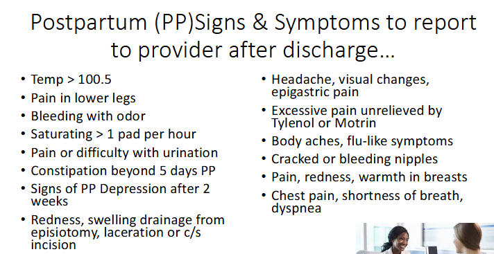
PP signs to report to provider
temp >100.5
saturating >1 pad per hour
constipation >5 days