bsci 202 lab practical 1
 Call Kai
 Learn
 Practice Test
 Spaced Repetition
 Match
 Flashcards
 Knowt Play
1/176

There's no tags or description
Looks like no tags are added yet.
177 Terms
what color is oxygen rich blood?
bright red
What is the average volume of blood in adults?
female – 4-5, male – 5-6L
What type of tissue is blood? Why?
connective tissue, because they are cells within a fluid matrix
What percentage of blood is water?
90%
What is the most numerous blood cell type?
erythrocytes (red blood cells)
What are the formed elements?
erythrocytes and leukocytes
What is in plasma?
other than 95% water is water, electrolytes, and plasma proteins
What are the different types of plasma proteins and their functions
albumin-osmotic balance
fibrinogen-clotting
globulins-antibodies, lipid transport

Name
physical description
# of cells
function
erythrocyte
biconcave,
no nucleus (anucleated)
7-8 micrometer diameter
4-6 million/µL
transport oxygen

name
physical description
# of cells
function
neutrophil multilobed nucleus; pale red and blue cytoplasmic granules, diameter 10-12 3000-7000/ µL phagocytuze pathogens or debris
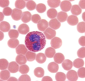
name physical description # of cells and function
eosinophil bilobed nucleus, red cytoplasmic granules 10-14 µm 100-400/ µL kill parasitic worms, kinda phagocytic, involved in allergy/asthma
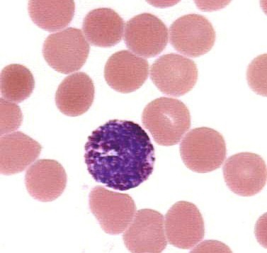
name physical description # of cells and function
basophil
lobed nucleus, large blue cytoplasmic granules
10-14 µm 20-50/ µL release histamine
have heparin (anticoagulant)

name physical description # of cells and function
Lymphocyte: spherical/indented nucleuss, pale blue cytoplasm 5-17 µm 1500-3000/ µL help with immune response by direct cell attack or producing antibodies

name physical description # of cells and function
monocyte U/kidney shaped, gray-blue cytoplasm, 100-700/ µL blood in tissues-develop into macrophages
How many WBC’s/microliter
4800-108000
platelet physical description # of cells and function
cytoplasmic fragments that have granules, deep purple, 150k-400k, blood clotting
leukocytosis
WBC over 11k/mm^3, indicates infection, metabolic disease, hemmorrhage, or poisoning
leukopenia
WBC count below 4000/mm^3, indicate chemical/toxin exposure, leukemia
leukemia
proliferation of abnormal WBCs , comes with reduced # of RBCs and platelets
Polycemia
increased # of RBCs
Anemia
decreased oxygen carrying capacity
Differential WBC count
100 WBCs counted/classified
Hematocrit
amount of RBCs in blood, males-47+/-5 females:42+/- 5
How does oxygen bind to heme
reversibly
What is the normal amount of hemoglobin in blood
12-18g/100 mL, men’s are usually higher than females
Order of Hemostasis
Clotting- inured tissue release tissue factor platelets release phosphatidylserine ; TF and phos interact with other clotting factors and calcium ions to form prothrombin activator ; ->thrombin, thrombin converts fibrinogen into fibrin ; fibrin forms mesh to trap RBCs
Antigens
anything that produces an immune response
Agglutinogens
a type of antigen that causes agglutination
Atherosclerosis
blood vessels blocked by plaque, plaque can serve as start points for blood clots
What is normal value for plasma cholesterol
130-200mg/100ml
What are thrombi
blood clots
What is thrombosis
when blood clot abnormally restricts blood flow
Iron-deficiency anemia
erythrocytes are smaller
Aplastic anemia
bone marrow doesn’t produce enough erythrocytes
Hemolytic anemia
erythrocytes are destroyed too quickly
Sickle cell anemia
sickle-shaped erythrocytes
Hemorrhagic anemia
caused by blood loss, NOT detected by hematocrit
What happens when you are dehydrated?
plasma % decrease (bc plasma is 90% water)
Hematocrit formula
height RBC/height all components X 100
Buffy coat
contains leukocytes and platelets, less than 1% blood
What is the order of conduction of the heart
SA node, AV node, AV bundle, bundle branches, subendocardial conducting network

Sinatrial node
pacemaker, generates impulses


Atrioventricular node
impulses pause for 0.1 second


Atrioventricular bundle
bundle of His, conducts impulse to bundle branches, in interventricular septum


Bundle branches
conduct impulses through interventricular septum


Subendocardial conducting network (purkinge fibers)
depolarizes contractile cells of both ventricles
What happens when AV node-bundle pathway is damaged
stops SA node from interacting with ventricles
Intrinsic conduction system
cardiac pacemaker cells, depolarizes heart sequentially
Segment
region between 2 waves
Interval
segment and one or more waves
What does P hump represent
atrial depolarization
What does R on ecg represent
Ventricular depolarization
What does T represent
ventricular repolarization
Tachycardia
over 100 beats/min
Bradycardia
lower than 60 beats/min
Fibrillation
rapid, uncoordinated heart contractions

what is this
fibrillation

what is this
bradycardia

what is this
tachycardia
what is lub and what is dub
lub: atrioventricular valves close during the beginning of ventricular systolic contraction
dub: semilunar valves close at the end of the ventricular systolic contraction
Why is bradycardia good in athletes
indicates stroke volume has increased
What is stroke volume
amount of blood ejected by ventricle
How many leads are used to record an ECG standard how many are bipolar and how many are unipolar
12, 3 bipolar 9 unipolar
What does enlarged R wave mean
enlarged ventricles
What does prolong P-R interval mean
1st degree heart block, signal from SA node to AV node is delayed longer than normal
What does prolonged Q-T interval mean
increased risk of arrhythmias, interval corresponds to beginning if ventricular depolarization and repolarization
What does elevated S-T segment mean
myocardial infarction
What is the major function of the cardiovascular system
transportation with blood as the transport vehicle
Apex of the heart
pointed tip of the heart that extends slightly to the left
Pericardium
double-walled sac that encloses the heart
Fibrous pericardium
loose-fitting superficial part of pericardium
Serous pericardium
lines internal surface of fibrous pericardium, part of the parietal layer
Visceral layer/epicardium
outer layer of the heart
Myocardium
middle layer, thickest, mainly cardiac muscle, reinforced with dense fibrous connective tissue and has the cardiac skeleton, thicker around the heart valves and base of great vessels leaving the heart
What is the cardiac skeleton?
the thick layer of myocardium that surrounds the heart
What is the thickest layer of the heart?
myocardium
Endocardium
inner lining of the heart, covers heart valves, continuous with inner lining of great vessels
What is the endocardium made of
simple squamous epithelium that rests on areolar connective tissue
What divides the heart longitudinally
interarterial septum
What separates the ventricles
interventricular septum
What forms the bulk of the heart
ventricles, which are discharging chambers
What is the tricuspid valve
right atrioventricular valve, 3 flap/cusps
Mitral valve
left AV valve, two flaps/cusps
What anchors the AV valves and what is it made of
chordae tendons, made of papillary muscle
What happens to the AV valves when ventricles contract
AV valves close to prevent backflow of blood
What happens if AV valves are unanchored
cusps move upward into atria (like turned umbrella)
Pulmonary SL valve location and structure
3 cusps, located between right ventricle and pulmonary trunk
Aortic SL valve location and structure
3 pocketlike cusps, between left ventricle and aorta
During contraction what do SL valves do
SL valves are open against walls of vessel
When ventricles relax what do SL valves do
SL valves close, preventing backflow
What is the order of the blood flow in the pulmonary circuit
Oxygen poor blood from vena cavae go into right atrium 2. Tricuspid valve 3. Right ventricle 4. Pulmonary valve 5. Pulmonary trunk 6. Branch into left and right pulmonary arteries 7. Lungs 8. Pulmonary veins
What is the order of blood flow from the systematic circuit
1.Left atrium (from pulmonary veins) 2. Mitral valve 3. Left ventricle 4. Aortic valve 5. Aorta 6. Arteries 7. Arterioles 8. Capillaries 9. Venules 10. Veins 11. Venae cavae 12. Right atrium
What is the systematic circuit and the pulmonary circuit?
systematic - carries oxygen blood to system pulmonary - carries oxygen poor blood to lungs
What is the largest artery in the body
aorta, leaves from left ventricle of the heart
What supplies the myocardium with blood
coronary circuit
Great cardiac vein
drains anterior part of both ventricles
Coronary sinus
drains the whole heart, middle and small cardiac veins
What are arteries
transport blood away from the heart
Elastic arteries
expand a lot, close to the heart