Test 3- Ch. 12
 Call Kai
 Learn
 Practice Test
 Spaced Repetition
 Match
 Flashcards
 Knowt Play
1/64

There's no tags or description
Looks like no tags are added yet.
65 Terms
second lumbar vertebrae
the spinal cord extends from the foramen magnum to the _____

segments of the spinal cord
cervical (8)
thoracic (12)
lumbar (5)
sacral (5)
spinal nerves
the spinal cord gives rise to 31 pairs of _____
not uniform in diameter through length
cervical enlargement
______ supplies upper limbs
lumbar enlargement
_____ supplies lower limbs
conus medullaris
tampered inferior end of the spinal cord
extends inferiorly to the second lumbar vertebrae

cauda equina
origins of the spinal nerves extending inferiorly from lumbosacral enlargement comes off the conus medullaris
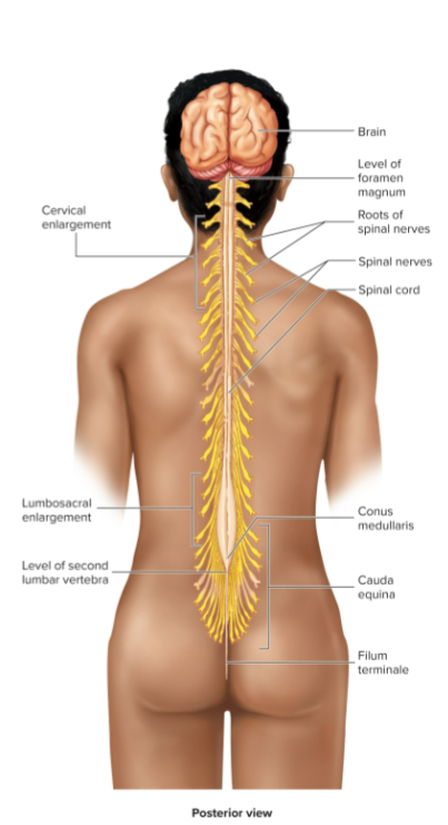
same
a ganglion is a bunch of cell bodies together at axons moving in the _____ direction and boosts signals into the spinal cord. They decide if the signal goes to an interneuron or not.
important info to the brain
survival, food, and reproduction
interneuron
middle man
once activated by a ganglion, it sends the brain a message but tells the arm to move and stimulates it instead of waiting for the brain to decide what to do
Meninges of the Spinal Cord
Connective tissue membranes surrounding the spinal cord and brain
dura mater
arachnoid mater
pia mater
Spaces
epidural
subdural
subarachnoid
Dura mater (Thecal Sac)
continuous with epineurium of the spinal nerves
wrap that goes CNS to PNS
Arachnoid mater
spiny layer
thin and wispy
Pia mater
bound tightly to the surface of the brain and spinal cord
forms the filum terminale, which anchors spinal cord to coccyx and the denticulate ligaments that attach the spinal cord to dura mater
epidural
location for epidural anesthesia
layer of adipose tissue above and around dura mater
contains blood vessels, areolar CT, and fat
subdural
serous fluid
under/below dura mater
subarachnoid
CSF (cerebrospinal fluid) and blood vessels with web-like strands of arachnoid tissue
anterior median fissue and posterior median sulcus
deep clefts partially separating left and right halves of the spinal cord
white mater
myelinated axons forming tracts
three columns (funiculi): ventral, dorsal, lateral
each of these divided into tracts (fasciculi; pathways)
gray matter
neuron cell bodies, dendrites, axons
Horns (multipolar neurons)
posterior (dorsal) horn
anterior (ventral) horn
lateral horn (associated with ANS)
posterior (dorsal) horn
contains neurons that receive information from the dorsal root neurons
anterior (ventral) horn
contains cell bodies of multipolar neurons that innervate skeletal muscle
lateral horn
cell bodies of multipolar neurons of the autonomic nervous system
commissures
connections between left and right halves
gray with central canal in the center
white
roots
spinal nerves that arise as rootlets then combine to form roots
dorsal root
the _____ is sensory
has a ganglion
ventral root
the _____ is motor
laterally
the dorsal and ventral roots merge _____ to form a spinal nerve
ascending and descending nerve tracts
certain spots in the spinal cord consists of ________
ascending is afferent or sensory infromation that goes up to the brain
descending is efferent or motor information being sent to the PNS
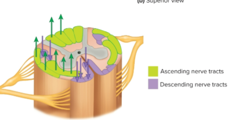
Dorsal Root Ganglion
collections of cell bodies of pseudo-unipolar sensory neurons forming dorsal roots
motor neuron cell bodies are in anterior and lateral horns of spinal cord gray mater
somatic
multipolar ______ mtor neurons are located in the anterior (motor) horn
autonomic
_____ neurons are located in the lateral horn
Axons of motor neurons form ventral roots and pass into spinal nerves
Reflex
self-preservation
autonomic response to a stimulus that occurs without conscious thought; homeostasis
Reflex Arc
neural pathway that controls a reflex; sometimes involves an interneuron
components of a reflex arc
Sensory receptor→ sensory neurpne → interneuron → motor neuron → effector organ (muscle)
reflexes based on number of synapses
monosynaptic reflexes
polysynaptic reflexes
reflexes based on development
innate reflexes
learned reflexes
reflexes based on effector type
somatic reflexes
autonomic (visceral) reflexes
reflexes based on location of integration center
cranial reflexes- brain
spinal reflexes- spinal cord
monosynaptic reflex
no interneurons
polysynaptic reflex
multiple synapses with interneurons
innate reflex
part of normal development
ex: pulling foot back when stepping on lego
learned reflex
develop as activities are repeated
ex: athlete catching a ball coming at its face
somatic reflexes
____ have an effector type of skeletal muscle
autonomic (visceral) reflexes
____ have an effector type of smooth muscle, cardiac muscle, and glands
Reflex Arc Steps
a sensory receptor detects a sstimulus, generating action potentials
a sensory neuron conducts action potentials through a nerve and dorsal root to the spinal cord
in the spinal cord, the action potential is carried to the integra- ion (integration) center, which can involve an interneuron. (Simple reflexes do NOT involve an interneuron)
the interneuron synapses with the motor neuron
the motor neuron axon conducts action potentials through the ventral root and spinal nerve to an effector organ

Example of Reflex Arc
you felt the mosquito bite
action potential happens
sensory receptor sends AP down the dorsal root
into ganglion projecting into the spinal cord
brain says ow, smack it
motor signal goes down spinal cord
out the ventral root it goes
you swing at the mosquito
muscle spindle
specialized muscle cells that respond to a stretch (stretch reflex)
Stretch Reflex Information (monosynaptic reflex)
muscles contract in response to a stretching force applied to them.
(UNIQUE because no interneurons)
muscle spindle: specialized muscle cells that respond to stretch
innervated by specific motor neurons: gamma motor neurons (small diameter neurons) control sensitivity of muscle spindles
sensory neurons innervate the noncontractile centers of the muscle spindle cells
these sensory neurons synapse with motor neurons of the spinal cord called alpha motor neurons which cause the muscle to contract
gamma motor neurons
small diameter neurons that control the sensitivity of muscle spindles
alpha motor neurons
motor neurons of the spinal cord that cause the muscle to contract
Example of Stretch Reflex
when the patellar ligament is tapped, the tendons and muscles of the quadriceps femoris muscle group stretch. Muscle spindle receptors within the muscle stretch, which activates the stretch reflex
axons of these sensory neurons extend to the spinal cord
in the spinal cord, the sensory neurons synapse directly with the alpha motor neurons
alpha motor neurons then stimulate the muscle in which the muscle spindle is embedded
the same muscle that was stretched not contracts in response to the action potentials stimulated by the alpha motor neurons
gamma motor neurons, contract the muscle spindles to allow them to detect further stretching
Teacher Example of a Stretch Reflex
at the doctor’s, they do a patellar test w the hammer. They hit the patellar tendon, reflex causes leg to move to protect the leg muscles. The tendon goes in, pulling the muscle down with it, so response is to pop out the leg so the leg muscles don’t get damaged.
-have spindles wrapped around muscles (muscle spindle fibers) with neurons that can detect movement (sensory neurons)
-tap patellar, sensory neurons alert SC, 1 neuron goes to brain and other connects to Alpha Motor Neuron to cause contraction
Golgi Tendon Reflex
prevents contracting muscles from applying excessive tension to tendons
monosynpatic reflex
contains a golgi tendon organ
Golgi tendon organ
encapsulated nerve endings that have at their ends numerous terminal branches with small swellings associated with bundles of collagen fibers in the tendon. Located in tendon near muscle
prevent damage to tendons that could be caused by excessive tension
produces sudden relaxation of the muscles
Example of a golgi tendon reflex
weight lifter suddenly drops heavy weight. Sudden movement of “clean and jerk” put so musch tension on tendons like achilles, they could break
Teacher example of Golgi Tendon Reflex
a weightlifter is lifting and if the weight becomes too much, the body sends the brain a signal, brain says no and hands open to drop the weight or knee goes down due to inhibitory neuron stopping AP from occuring and Alpha Moton Neuron is affected and stops action potential so muscle relaxes
Components of the Golgi Tendon Reflex
golgi tendon organ → sensory neuron → inhibitory interneurons inhibit alpha motor neuron → muscle relaxes
Explained Steps of Golgi Tendon Reflex
as a muscle contracts, the attached tendons stretch, resulting in increased tension in the tendons
the increased tension stimulates action potentials in the sensory neurons from the Golgi tendon organs. The sensory neurons pass through the dorsal root to the spinal cord and enter the dorsal gray horn
at the integration center in the dorsal horn, the sensory neurons branch and synapse with inhibitory interneurons
the interneurons synapse with alpha motor neurons that innervate the muscle to which the Golgi tendon organ is attached
inhibition of the alpha motor neurons result in relaxation of the muscle and a reduction in tension in the tendon
withdrawl reflex
function is to remove a body limb or other part from a painful stimulus
reciprocal innervation
causes relaxation of extensor muscle when flexor muscle contracts
involved in stretch and withdraw reflexes
where 1 muscle contracts, and 1 reflexes (bicep/tricep)
crossed extensor reflex
when a withdrawal reflex is initiated in one lower limb, the ______ causes extension of opposite lower limb
pain receptors detect a painful stimulus
sensory neurons conduct action potentials to the spinal cord
sensory neurons synapse with excitatory interneurons in the spinal cord that synapse with alpha motor neurons
excitation of the alpha motor neurons results in contraction of the flexor muscles
withdrawal of the limb from the painful stimulus
withdrawal reflex with reciprocal innervation
during the withdarawal reflex, sensory neurons conduct action potentials from pain receptors to the spinal cord
at the integration center sensory neurons synapse with excitatory interneurons, which are part of the withdrawal reflex
collateral axons of sensory neurons that carry action potential from pain receptors synapse with inhibitory interneurons in the dorsal horn of the SC
the inhibitory interneurons synapse with and inhibit alpha motor neurons of extensor (antagonist) muscles. When the withdrawal reflex is initiated, flexor muscles contract and _________ causes the extensor muscles to relax. This reduces the resistance to movement that the extensor muscles would otherwise generate
Withdrawal Reflex with Crossed Extensor Reflex
during the withdrawal reflex, sensory neurons conduct action potentials from pain receptors to spinal cord
at the integration center, sensory neurons synapse with excitatory interneurons, which are part of the withdrawal reflex
collateral axons from the sensory synapse with excitatory interneurons that extend through the white commisure to the opposite side of the spinal cord
these interneurons synapse with alpha motor neurons that innervate extensor muscles in the opposite side of the body. When a withdrawal reflexis initiated in one lower limb, the _______ causes extension of the opposite lower limb
interactions with spinal cord reflexes
sensory information goes to the brain ex: pain
descending tracts from the brain carry info to the reflexes
neurotransmitters produce either EPSP or IPSPs modifying the reflex