Lab 4: Annelida, Bryozoa, Brachiopoda
 Call Kai
 Learn
 Practice Test
 Spaced Repetition
 Match
 Flashcards
 Knowt Play
1/38

There's no tags or description
Looks like no tags are added yet.
39 Terms
Platytrochozoa and onwards + what is lophophore?
Annelida, Brachiopoda and Bryozoa are in the Lophotrochozoa ( a clade of the Platytrochozoa).
Bryozoans and brachiopods both possess a LOPHOPHORE, an extension of the body wall and coelom surrounding the mouth. It usually bears numerous hollow tentacles which are used for gathering food. Recent evidence suggests that the lophophores of the two groups are not homologous.
Annelida basic segmentation and body plan
Annelida have metameric segmentation of the body. the division of an organism's body into a linear series of similar, repeating units called metameres or somites along the head-to-tail axis.
With the exception of the anterior PROSTOMIUM, bearing the "brain", and the posterior PYGIDIUM, bearing the anus, the segments contain repeating units of mesodermally derived tissue.
The second "segment", the PERISTOMIUM surrounding the mouth, is primitively presegmental, but incorporates the first one or two true segments in some taxa. Segments are separated by double transverse SEGMENTAL SEPTA. The youngest segments occur at the posterior end.

Guide for Anterior Region
A. Peristomium - Incorporates segments 1 and 2
B. Protostomium - dorsal surface bearing 4 eyes, pair of forward projecting palps, pair of tentacles
C. Everted Pharynx
D. Jaws
E. Protostomial sensory palps
F. Protostomial Tentacles
G. Peristomial Cirri - 2 pairs on each dorsolateral side of peristomium, adjacent to protostomium, projecting laterally

How is the pharynx everted?
The pharynx is projected/everted when fluid is pushed into the coelomic space of the pharynx by nearby muscular contractions.
This causes the jaws to open (gape). The jaws close on the prey when pharynx is inverted by retractor muscles.
Parapodium Guide
A. Dorsal Notopodium
B. Ventral Neuropodium
C. Bristle like acicula
D. Setae beared on outer margins of acicula
E. Dorsal cirrus
F. Ventral Cirrus

Two types of Musculature of annelids
The musculature of the body wall of annelids is composed of an outer layer of CIRCULAR MUSCLE and inner layer of LONGITUDINAL MUSCLE.

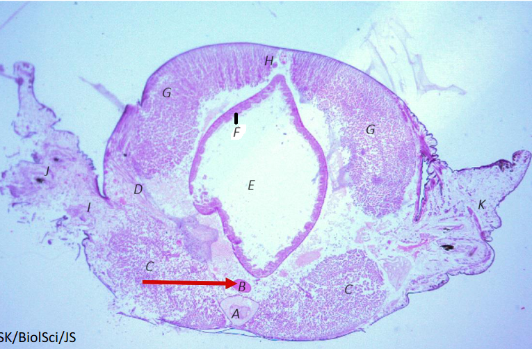
Annelid Nereis Cross Section
A. Ventral Nerve Cord
B. Ventral Blood Vessel
C. Ventral Longitudinal Muscle
D. Oblique Muscle
E. Intestine
F. Gastrodermis, composed of columnar endothelium
G. Dorsal Longitudinal Muscle
H. Dorsal Blood Vessel
I. Acicular Muscle
J. Acicula
K. Parapodium
L. Cuticle
M. Epidermis
N. Circular Muscle
Remember the orientation of the slides. They will be upside-down?
Body wall is composed of L, M, N.
annelid brain plan
The brain is joined to paired ventral nerve cords which run the length of the body. Ganglia are frequently present in each trunk segment.
annelid circulatory system
There is a closed circulatory system composed of vessels, some of which are contractile and function as hearts.
annelid digestive tract
The digestive tract is straight, and complete with mouth and anus, with specialized compartments for storage, mechanical digestion, and chemical digestion/absorption.
Two classes in phylum annelida
Polychaeta and Clitellata
Polychaeta, name 5 traits
Marine, primitive
trochophore larvae, characterisic of trochozoan clade
prostomium often have sensory/feeding appendages
simple eyes usually located on prostomium
each segment has pair of appendages - parapodia as paddles to propel and can be specialized as feeding appendeges
Clitellata, name species and 5 traits, 2 orders
earthworms and leeches
presence of clitellum: a trunk area in which the segments are thickened and glandular. The glands secrete a mucous used in copulation and in the formation of COCOONS.
monoecious
Order Crassiclitellata (earthworms) can be terrestrial or aquatic in marine and freshwater
no parapodia but 1+ pairs of stiff setae w each segment
reduced prostomium, no sensory appendages
Order Hirudinida, leeches, more specialized than crassiclitellates or polychaetes. Marine, freshwater, or terrestrial.
no parapodia and head appendages
monecious
Unlike the earthworms, the trunk surface displays many annular markings which do not coincide with internal segmentation.
Many species have anterior and posterior suckers.
The coelom is reduced and in many leeches forms coelomic channels and sinuses which function as a blood vascular system.
Leeches may be parasitic, feeding on the blood of vertebrates, predacious, capturing small aquatic organisms, or scavengers.
Review classification and comparative annelid characteristics
Two subclasses in polychaeta
Errantia: actively crawl in subsrtate, very diverse, 25% of annelid sp
sedentaria: live in constructed tubes
Nereis sp.
Phylum Annelida → Class Polychaeta → Subclass Errantia → Order Phyllodocida → Nereis sp.
Review structures on pg 5
Called the clam worm
Fast vs slow crawling in polychaete - what body parts are involved?
During slow crawling, only parapodia are involved, effective and recovery strokes alternate on opposite sides of the body.
During fast crawling, the body is thrown into undulating waves. Effective parapodial stroke is on the crest of the wave. Parapodia are recovering on the opposite side, which is also the side on which the longitudinal muscles are contracted

Other polychaete in errantia: sp. Aphrodita and Glycera details
Same classification as nereis
The dorsal surface of the Sea Mouse, Aphrodita sp., is covered by felt-like SETAE. The parapodia, used for crawling over mud or sandy substrate, are short and thickened, operated by complex internal musculature.
The Bloodworm, Glycera sp., is a burrowing, free-living species with four jaws and reduced parapodia. It is a carnivorous species, capturing prey items that move across the mouth of its burrow.

Sedentary Polychaetes - Subclass Sedentaria name 3 species
The Feather-duster Worm, Spirographis sp. has numerous feathery appendages to capture food particles. They live in non-calcareous, parchment-like tubes.
Other sedentary polychaetes burrow into the substrate. The Parchment Worm, Chaetopterus sp., lives in a U-shaped tube buried in mud or sand. Water enters through one opening and leaves through the other. The specialized FAN PARAPODIA are used to move water through the tube. Food particles in the water current are captured in a mucous bag supported by large ALIFORM PARAPODIA and then transferred to the mouth.
The Lugworm, Arenicola sp. is a specialized burrowing sedentary polychaete. There are no head appendages and gills, modified from the notopodia of anterior parapodia, occur on the anterior portion of the body.


Polychaetes all have trochophore larva
The TROCHOPHORE LARVA is roughly top-shaped with an
at: apical tuft of cilia and three bands of cilia round the body.
A thickened epithelium supports the cilia of the pt: prototroch.
st: stomach
mt: mouth
in: intestine
a: anus
teleotroch at the ventral most point

PHYLUM ANNELIDA - CLASS CLITELLATA - ORDER/SUBORDER CRASSICLITELLATA
PG 10 - 16 inclusive

Earthworm Lumbricus External Anatomy
Prostomium: lacking sensory appendages, anterior, bearing the brain
Peristomium: bears only the mouth - second “segment”, but presegmental?
1st segments begin counting after peristomium.
Pygidium: postegmental, bears anus
Setae: four pairs small hairs, on each trunk segment besides first and last
Segments are separated by double transverse segmental septa
Clitellum: swollen segments, 32 to 37 contains glands that produce the egg capsule/cocoon.
Male genetal pore: swellings on ventral surface of segment 15, is the opening to the vas deferens/sperm ducts
Spermatheca/seminal receptacle: pairs of indentations between segments 9 and 10, and between 10 and 11.
Oviducal opening: sometimes referred to as the “female aperture”, segment 14

Earthworm Internal Anatomy
Circulatory System
A. Dorsal Blood Vessel - contractile, moves blood anteriorly, much larger than ventral
B. Pseudohearts - 5 pairs in segments 6 to 10 inclusive. Contractile vessels arch around each side of digestive tract
C. Ventral Blood vessel - pseudohearts connect ventral to dorsal. Blood from ventral vessel distributed throughout each segment via small vessels and eventually returned to dorsal blood vessel
Reproductive System
D. Testes - release immature sperm into coelom
E. Sperm Funnels - receive this sperm
F. Seminal Vesicle - large and obvious, acts as sperm storage sac when sperm comes in from the sperm funnels
G. Sperm ducts(Vas deferens) - mature sperm released from seminal vesicles travel thru sperm ducts to male gonopore which is the hole to the vas deferens. 2 holes from the two sperm duct lines.
H. Spermathecae/Seminal Receptacle: Small white dots on either side of the whole bunch of stuff
Ovary: paired ovaries, close to ventral midline, small and bright white, tiny cluster of grapes
Digestive system - food passes thru in this order
I. Mouth - in ventral peristomium opens to small buccal cavity
J. Pharynx - muscular passes food to K
K. Oesophagus - tubular
L. Crop - thin walled, stores food
M. Gizzard - muscular, grinds up food
N. Calciferous Glands: outgrowths of digestive tract between 4th and 5th pseudohearts - rid the body of excess calcium
O. Intestine

Earthworm nervous system and excretory system
page 15
CRASSICLITELLATA mating
Lumbricus sp. lacks a penis and mutual sperm transfer is accomplished during a form of "copulation."
Worms face in opposite directions such that the male gonopores (= openings to vas deferens) of one align with female gonopores of the other. They are held together with mucous produced by the clitellum. Sperm released by each ( from male gonopores) travel along grooves ( formed by body wall contraction) to the SPERMATHECAL/SEMINAL RECEPTACLES openings of the mate. The worms are then inseminated ( but fertilization has not yet occurred) and separate from each other.
CRASSICLITELLATA fertilization and cocoon formation
A cocoon is formed around the clitellum of each worm. The cocoon moves toward the anterior. As it moves, it receives eggs from the female gonopore and then the sperm from the spermathecal openings (received during insemination). Thus fertilization occurs in the cocoon. Cocoons are deposited into the soil. Earthworms have direct development- i.e. they have no larval stage
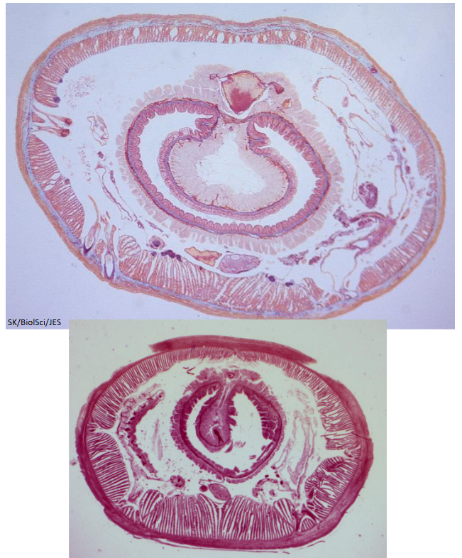
Earthworm cross section
anything within the coelom is probably nephridia related
Look at the staining of the blood vessels. that’s how you can determine the difference between the ventral nerve cord and ventral blood vessel

Earthworm movement
As a worm moves forward a wave of PERISTALSIS passes along the body.
The peristaltic wave is produced by the action of the longitudinal and circular muscles upon the incompressible fluid of the hydraulic skeleton. The body of the earthworm is internally partitioned into segments by septa.
Because the segmental septa limit the movement of the coelomic fluid, body contractions and extensions may be localized; each segment can contract or expand independently of every segment. Coordination is maintained between segments by the ventral nerve cord.
Wave goes from anterior to posterior to move the worm forwards

Order Hirudinida
Phylum Annelida → Class Clitellata → Order Hirudinida
Leeches
review anatomy pg 18

Leech internal anatomy - slides are rhynchobdellid (jawless) leeches
A. Annuli
B. Anterior Sucker - surrounds mouth
C. Mouth
D. Posterior Sucker - larger, surrounds anus
E. Proboscis - exclusive to rychno leeches, everted from proboscis pore in anterior sucker
F. Eye spots
Male and female gonopores open thru clitellum
Numerous paired testes
Rynchobdellid leeches don’t have a penis, monoceoius

How do leeches move
Note that when the leech is at its most elongate and thinnest, the circular muscles are fully contracted and that when the leech is shortest and thickest, the longitudinal muscles are fully contracted. The leech is a fluid-filled bag of unchanging volume, and the longitudinal and circular muscles act antagonistically around this hydrostatic skeleton.
Leeches have an outer layer of circular muscles and inner layer of longitudinal muscles in the body wall, but in addition they possess a double layer of oblique muscles between these two layers. The oblique muscles run as an helical spiral along the length of the body. When the body is long and thin, the spiral fibres reinforce the action of the longitudinal muscles as they contract and the body is shortened.
When the leech is short and thick, they reinforce the contractions of the circular muscles that produce elongation. In an intermediate position, when the spiral muscles are neither lengthening nor shortening, they serve to increase the internal hydrostatic pressure and enable the leech to perch upright on its posterior sucker.

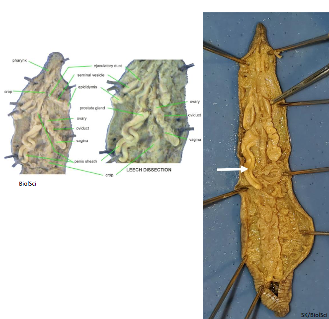
Internal Anatomy of hirudinid leeches
Septa: incomplete! External segments do not line up with internal alignment.

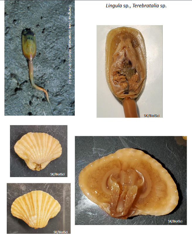
Phylum Brachiopoda basic body plan
The body of brachiopods is contained within a bi-valved shell, with valves covering dorsal and ventral surfaces.
The shell is secreted by a mantle.
Within the shell, the mouth is surrounded by the LOPHOPHORE, a pair of elongate coiled arms bearing hollow tentacles. This arrangement has given the phylum its name, which literally means "arm foot".
Brachiopods are sessile, marine animals, attaching to the substrate by means of an adhesive stalk, the PEDICLE.


Lingula sp. traits
The INARTICULATE brachiopod genus, Lingula sp. is among the longest lived of all animal genera, dating from the Ordovician. The term "inarticulate" refers to the absence of a posterior hinge linking the two valves, which are equal in size. Valves are the top and bottom halves of the “shell”
Although most brachiopods live attached to firm substrates, Lingula sp. lives in vertical burrows in sand or mud bottoms, with the aperture of the shell at the opening of the burrow. Aperture is just opening of the shell
It attaches to the substrate via the elongate pedicle.
SETAE around the edge of the valves channel water currents.

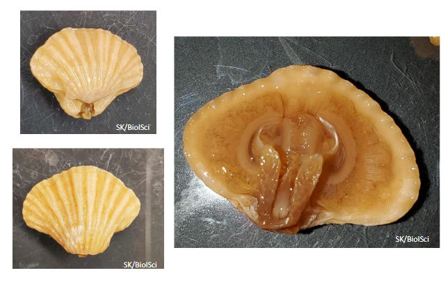
Terebratalia sp.
Terebratalia sp. is an example of the ARTICULATE (hinged) group of brachiopods.
The pedicle passes through an aperture on the dorsal surface of the ventral valve.
The dorsal valve of the shell is smaller than the ventral valve.
Remove the dorsal valve and examine the LOPHOPHORE. The arms are not extended during feeding.
The valve is gaped and water currents, carrying suspended matter, wash over the lophophore and food particles are trapped on the tentacles and then moved by cilia to the base of the arm and thence to the mouth.

Phylum Bryozoa Where do they live?
Bryozoans are colonial and have both marine and freshwater representatives. Brownish colonies are commonly seen attached to wharf pilings, sunken logs, and on stones in lakes and streams.

how do colonies of the bryozoa species Bugula look? What is their body plan?
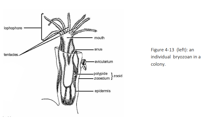
Colonies of Bugula sp. are composed of numerous small vase-shaped ZOOIDS.
The transparent outer non-living covering is composed largely of chitin but is often impregnated with calcium carbonate. This outer covering, together with the epidermis, constitutes the ZOOECIUM/CYSTID.
The visceral mass, lophophore and tentacles together are called the POLYPIDE.
The TENTACLES arise from the LOPHOPHORE, a U-shaped extension of the body wall and coelom surrounding the MOUTH.
The digestive tract is complete, the anus opening just outside the lophophore. You will not be able to see the parts of the digestive system on the whole mounts slides.
The polypide can be retracted within the zooecium by muscles joining the zooecium to the digestive tract. The bryozoans have no circulatory or excretory organs and the nervous system is greatly reduced.
What is a feature of the many individuals in the polymorphic bryozoan colony?
Bryozoan colonies are polymorphic. Many different types of zooids.
In addition to feeding zooids bearing tentacles, there are also special zooids to protect the colony. These are jawed AVICULARIA (sing. AVICULARIUM) attached to the outer surfaces of many feeding zooids. These structures resemble a bird's head with a movable beak. MUSCLES open and close the jaws.
An additional type of zooid, called a VIBRACULUM (not seen in the microscope slide), beats back and forth, setting up water currents which keep the colony free of debris.
How do bryozoans reproduce
Most bryozoans are hermaphroditic. Self-fertilization is common and the eggs develop internally. They also reproduce asexually by budding.