Chapter 13: Skin, Hair, and Nails
 Call Kai
 Learn
Learn Practice Test
 Spaced Repetition
 Match
Match Flashcards
 Knowt Play
1/109

There's no tags or description
Looks like no tags are added yet.
Name | Mastery | Learn | Test | Matching | Spaced | Call with Kai | Chat |
|---|
No analytics yet
Send a link to your students to track their progress
110 Terms
Epidermis
Thinnest, outer layer of skin
Dermis
Middle layer of skin, made of collagen, elastic fibers, blood/lymph vessels, and nerve endings
Subcutaneous
Bottom layer of skin, connects skin to underlying organs, stores energy, absorbs shock, and insulates
High Risk Areas
Between the toes, skin folds, under medical devices, bony prominences, and pressure points
First Level Priorites
Severe dehydration, cyanosis, and extensive burns
Second Level Priorites
Suspicious lesions that have irregular borders, color changes, or rapid growth
Clubbing
Tips of fingers become wide and round, >180 degrees angle at nail base, caused by respiratory or cardiac dysfunction
Schamroth Window Test
Clubbing test in which the patient places dorsal surfaces of opposite fingers together, with clubbing there would be no diamond shaped window
Skin Turgor
The skin’s ability to snap back when pinched up, indicates hydration level, for older adults test the sternum/forehead and for children test the abdomen or medial thigh
Grade 1+
Pitting edema grade with 2 mm depression, mild pitting, and slight indentation
Grade 2+
Pitting edema scale with 4 mm depression, moderate pitting, and indentation subsides rapidly
Grade 3+
Pitting edema scale with 6 mm depression, deep pitting, and indentation remains for short time
Grade 4+
Pitting edema scale with 8 mm depression, very deep pitting, and long lasting indentation
Pressure Injuries
Localized damage to skin and underlying soft tissue, usually over a bony prominence due to constant pressure on the site
Braden Scale
Tool used to assess a patient’s risk for developing pressure injuries, includes sensory perception, moisture, activity, mobility, nutrition, and friction/shear, a score <9 is severe risk and between 15-18 is a baseline
ABCDEs
Melanoma detection tool, includes asymmetry, border irregularity, color, diameter of more than 6 mm, and evolution of lesion over time
Vellus Hair
Fine, faint hair that covers most of the body
Terminal Hair
Darker, thicker hair that grows on the scalp and eyebrows and after puberty the armpits, pubic area, and face/chest in men
Eccrine Glands
Sweat glands that produce a diluted saline solution onto the skin, matures at 2 months old
Apocrine Glands
Sweat glands that produce a thick, milky secretion onto the armpits, genital area, nipples, and navel, become active during puberty
Lanugo
Fine hair on newborns
Keloids
Raised scars that form at a wound site and grow beyond the normal boundaries of the wound
Pseudofolliculitis
Razor bumps/ingrown hairs, occur when shaving too closely
Melasma
Patchy brown discoloration of the face
Mongolian Spot
Blue/black/purple hyperpigmentation area at the sacrum or butt due to deep dermal melanocytes, found in black, asian, native American, and latino newborns

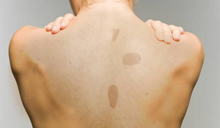
Cafe Au Lait Spot
Large round/oval patch of light brown pigmentation
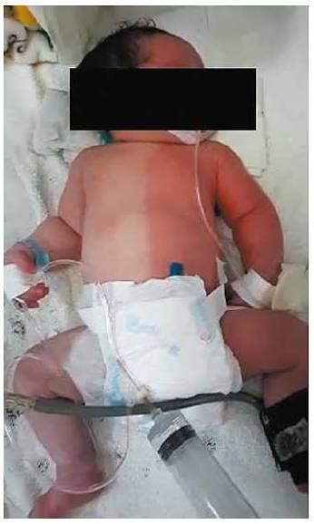
Harlequin Color Change
Distinct line of red/pale color down the midline or horizontal found in newborns, one half of body turns red and blanches
Erythema Toxicum
Common rash in the first 3 to 4 days of life, consists of tiny punctate red macules and papules on the cheeks, trunk, check, back, and butt

Acrocyanosis
Bluish color around the lips, hands, fingernails, toes, and toenails, disappears without warning
Cutis Marmorata
Transient mottling in the trunk and extremities in response to cooler room temps
Physiologic Jaundice
Normal variation of jaundice in about half of all newborns, develops after 3-4 days of life because of increased RBCs
Carotenemia
Yellow/orange color is light skinned people without yellowing of the sclera/mucous membranes, occurs due to ingesting large amounts of foods containing carotene
Milia
Tiny white papules on the face of babies due to sebum occluding opening of follicles
Nevus Simplex
Common vascular birthmark that is flat, irregularly shaped red/pink patch typically on the face or back of neck, usually fades during first year

Striae
Jagged linear stretch marks of pink/silver color that appear during 2nd trimester of pregnancy
Linea Nigra
Brownish/black line down the midline during pregnancy
Solar Lintigines
Common, circumscribed clusters of melanocytes due to chronic sun exposure

Keratoses
Raised, thickened areas of pigmentation that look crusted, scaly, and warty, mostly on the trunk

Actinic Keratosis
Red/tan scaly plaques, premalignant, caused by sun/UV exposure

Acrochordons
Overgrowths of normal skin that form a stalk and are polyp like (skin tags)
Sebaceous Hyperplasia
Raised yellow papules with central depression on the face, more common in men
Annular Lesion
Circular lesion, begins at center and spread (ringworm)

Confluent Lesion
Lesions that run together (hives)

Discrete Lesions
Distinct, individual lesions that remain separate (acne)
Gyrate Lesions
Lesions that are twisted/coiled
Grouped Lesions
Cluster of lesions (contact dermatits)
Linear Lesion
Scratch/streak
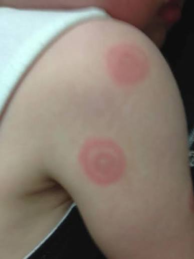
Target Lesions
Iris, concentric rings of color
Zosteriform Lesions
Linear arrangement of lesions along a unilateral nerve route (shingles)

Polycyclic Lesions
Annular lesions that grow together (psoriasis)

Macule
Flat, circumscribed primary lesion, less than 1 cm (freckles/measles)

Papule
Solid, elevated, circumscribed primary lesion, less than 1 cm, caused by superficial thickening of epidermis (mole)

Patch
Macules that are larger than 1 cm (vitiligo)
Plaque
Papules that coalesce to form surface elevation wider than 1 cm (psoriasis)

Nodule
Solid, elevated primary lesion, larger than 1 cm, may extend into dermis (fibroma)

Wheal
Superficial, raised, transient, and erythematous primary lesion, slightly irregular shape (mosquito bite)
Tumor
Primary lesion larger than a few cm, extends into dermis, can be benign or malignant (lipoma)
Urticaria
Wheals that coalesce to form extensive reaction (hives)
Vesicle
Elevated cavity containing free fluid, up to 1 cm (herpes simplex)
Bulla
Primary lesion larger than 1 cm, usually chambered, superficial, and thin walled (friction blister)
Cyst
Encapsulated fluid filled cavity in dermis or subcutaneous layer, causes tensely elevated skin
Pustule
Turbid fluid (pus) in a cavity of a primary lesions, circumscribed and elevated (acne)
Primary Lesion
Lesion that develops on previously unaltered skin
Secondary Lesion
Lesion that develops overtime due to scratching/infection of a previous lesion
Crust
Thickened, dried out exudate left when vesicles/pustules burst or dry up (impetigo)
Scale
Compact, desiccated flakes of skin, silver/white color, caused by shedding of dead excess keratin cells (eczema)
Fissure
Linear crack with abrupt edges, extends into dermis, secondary lesion (cheilosis)
Erosion
Scooped out, but shallow depression, superficial, most, heals without a scar, secondary lesion

Ulcer
Deeper depression extending into dermis, irregular shape, may bleed, will scar, secondary lesion (pressure injury)
Exocoriation
Self induced abrasion, superficial, usually due to scratching
Scar
Connective tissue replaces normal tissue at site of an injury
Atrophic Scar
Scar in which the resulting skin level is depressed with loss of tissue
Lichenification
Tightly packed sets of papules resulting from prolonged, intense scratching, looks like moss

Stage 1
Pressure injury that has non-blanchable erythema and unbroken skin

Stage 2
Pressure injury in which there is epidermal skin loss, exposed dermis, and a red/pink wound bed

Stage 3
Pressure injury that extends into subcutaneous tissue but no muscle/bone/tendon is visible

Stage 4
Pressure injury that exposes muscle, tissue, or bone
Deep Tissue Pressure Injury
Pressure injury with localized, non-blanchable color change to deep red/purple, begins in muscle closest to bone

Hemangioma
Benign proliferation of blood vessels in dermis
Port Wine Stain
Large, flat, hemangioma on the scalp or face, usually along CN V, dark red/purple color, consists of mature capillaries, present at birth (nevus flammeus)
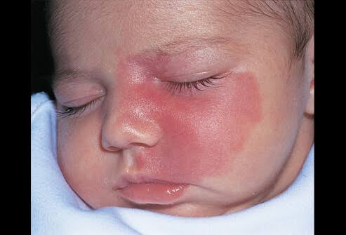
Strawberry Mark
Raised bright red hemangioma with well defined borders, about 2-3 cm, does not blanch, consists of benign growth of endothelial cells (infantile hemangioma)

Deep Hemangioma
Reddish-blue, irregularly shaped, solid, spongy hemangioma
Telangiectasia
Permanently enlarged and dilated blood vessels that are visible on the skin

Spider Telangiectasia
Fiery red, star shaped telangiectasia with solid circular center, develops on face, neck, or chest

Venous Lake
Blue/purple dilation of venules and capillaries in a star-shaped, linear, or flaring pattern, pressure will cause them to empty, common on lips, ears, or face of older adults

Petechiae
Tiny punctuate hemorrhages, 1-3 mm, round, discrete, dark red/purple, caused by bleeding from superficial capillaries, will not blanch, may indicate abnormal clotting factors

Purpura
Confluent and extensive patch of petechiae and ecchymoses, >3 mm, flat, red/purple, occur with old age and thrombocytopenia

Ecchymosis
Purplish patch resulting from excavation of blood into the skin, > 3 mm

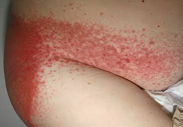
Diaper Dermatitis
Red, moist, maculopapular patch with poorly defined borders in diaper area, caused by skin irritation from ammonia, moisture, or occlusive diapers
Candidiasis
Scalding red, moist patches with sharply demarcated borders, usually in genital area, caused by candida fungus and aggravated by urine, feces, head, and moisture
Impetigo
Moist, thin roofed vesicles with thin red base caused by a highly contagious bacterial infection of skin

Atopic Dermatitis
Chronic inflammatory skin lesion caused by overstimulated immune system, genetics, and environment, causes itchy, localized dry patches and red papules/vesicles (eczema)
Varicella
Small, tight vesicles that are shiny with a red base, extremely itchy, first appears on trunk then face, arms, and legs, not on palms/soles (chickenpox)

Measles
Disease that begins with 2-4 day fever and one or more of a cough, coryza, or conjunctivitis, then rash appears in cephalocaudal direction, does not blanch

German Measles
Pink papular rash first appears on face then spreads, causes neck lymphadenopathy, no Koplik spots
Tinea Corporis
Scales on chest, abdomen, and back of arms forming multiple circular lesions with clear centers (ring worm of the body)

Tinea Pedis
Fungal infection appearing first as small vesicles between toes, on sides of feet, and on soles, grows scaly and hard, found in chronically moist, warm, feet (ring worm of the feet/athletes foot)

Tinea Versicolor
Fine, scaling, round, hypomelanotic patches of pink, tan, or white caused by superficial fungal infection, usually symmetric on neck, trunk, and upper arms

Herpes Zoster
Small, grouped vesicles that emerge along route of sensory nerve, caused by dormant chickenpox, does not cross midline, usually in adults >50
Erythema Migrans
Distinctive bull’s eye, red rash associated with lyme disease, fades in 4 weeks
