Week 6 - Introduction to DNA

0.0(0)
Studied by 0 people
call kaiCall Kai
learnLearn
examPractice Test
spaced repetitionSpaced Repetition
heart puzzleMatch
flashcardsFlashcards
GameKnowt Play
Card Sorting

1/58

encourage image

There's no tags or description

Looks like no tags are added yet.

59 Terms

1
New cards

What are the three essential functions of genetic material?

  • Replication: Genetic material must be copied and passed to the next generation.

  • Growth and development: Genetic material must contain information that controls an organism's growth and development.

  • Adaptation: Genetic material must allow organisms to adapt to environmental changes.


2
New cards

What was Griffith's experiment (1928), and what did it demonstrate?

  • Frederick Griffith studied two strains of Streptococcus pneumoniae:

    • Smooth (S) strain: Virulent (disease-causing).

    • Rough (R) strain: Nonvirulent (not disease-causing).

  • He injected mice with different combinations of living and heat-killed bacteria.

  • His results demonstrated that a substance from dead S bacteria could transform living R bacteria into virulent S bacteria.

  • This process is known as bacterial transformation.

  • Important: Griffith showed that transformation occurred, but he did not identify DNA as the transforming substance.


<ul><li><p>Frederick Griffith studied two strains of <em>Streptococcus pneumoniae</em>:</p><ul><li><p><strong>Smooth (S) strain:</strong> Virulent (disease-causing).</p></li><li><p><strong>Rough (R) strain:</strong> Nonvirulent (not disease-causing).</p></li></ul></li><li><p>He injected mice with different combinations of living and heat-killed bacteria.</p></li><li><p>His results demonstrated that a substance from dead S bacteria could <strong>transform</strong> living R bacteria into virulent S bacteria.</p></li><li><p>This process is known as <strong>bacterial transformation</strong>.</p></li><li><p><strong>Important:</strong> Griffith showed that transformation occurred, but he did <strong>not</strong> identify DNA as the transforming substance.</p></li></ul><p></p>
3
New cards

What was the Avery–MacLeod–McCarty experiment, and how did it demonstrate that DNA is the transforming substance?

  • Researchers investigated which molecule was responsible for bacterial transformation.

  • They obtained material from heat-killed virulent S bacteria.

  • They treated separate samples with enzymes that destroy different molecules:

    • RNase: Destroys RNA.

    • Protease: Destroys proteins.

    • DNase: Destroys DNA.

  • Each treated sample was combined with living, nonvirulent R bacteria.

Results:

  • RNase treatment → Transformation still occurred.

  • Protease treatment → Transformation still occurred.

  • DNase treatment → No transformation occurred.

Conclusion: DNA was the transforming substance, providing evidence that DNA carries genetic information.

4
New cards

Useful Diagram

knowt flashcard image
5
New cards

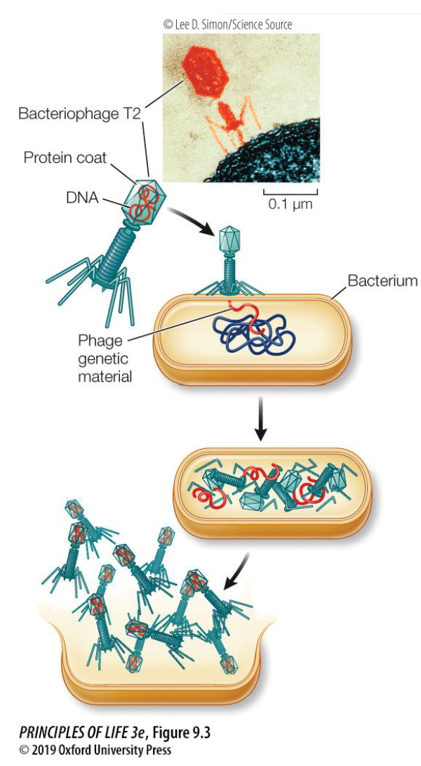
What was the purpose of the Hershey–Chase experiment (1952)?

  • Alfred Hershey and Martha Chase used T2 bacteriophages (viruses that infect bacteria).

  • They wanted to determine whether DNA or protein was the hereditary material.

  • They used radioactive isotopes to track DNA and proteins during bacterial infection.


<ul><li><p>Alfred Hershey and Martha Chase used <strong>T2 bacteriophages</strong> (viruses that infect bacteria).</p></li><li><p>They wanted to determine whether <strong>DNA or protein</strong> was the hereditary material.</p></li><li><p>They used radioactive isotopes to track DNA and proteins during bacterial infection.</p></li></ul><p></p>
6
New cards

Why did Hershey and Chase use radioactive phosphorus (³²P) and sulfur (³⁵S)?

  • Radioactive phosphorus (³²P): Used to label DNA, because DNA contains phosphorus (PO4) in its phosphate backbone.

  • Radioactive sulfur (³⁵S): Used to label proteins, because some amino acids contain sulfur (SO4), whereas DNA does not.

  • These radioactive markers allowed researchers to determine which molecule entered the bacterial cells during infection.

  • Important: Remember ³²P = DNA and ³⁵S = protein!

<ul><li><p><strong>Radioactive phosphorus (³²P):</strong> Used to label <strong>DNA</strong>, because DNA contains phosphorus (PO4) in its phosphate backbone.</p></li><li><p><strong>Radioactive sulfur (³⁵S):</strong> Used to label <strong>proteins</strong>, because some amino acids contain sulfur (SO4), whereas DNA does not.</p></li><li><p>These radioactive markers allowed researchers to determine which molecule entered the bacterial cells during infection.</p></li><li><p><strong>Important:</strong> Remember <strong>³²P = DNA</strong> and <strong>³⁵S = protein</strong>!</p></li></ul><p></p>
7
New cards

How was the Hershey–Chase experiment performed, and what were the results?

  • Researchers produced two groups of bacteriophages:

    • ³²P-labelled phages: Contained radioactive DNA.

    • ³⁵S-labelled phages: Contained radioactive proteins.

  • Each group was allowed to infect bacteria.

  • Researchers examined where the radioactive material ended up.

Results:

  • ³²P (DNA): Radioactivity was detected inside the bacterial cells.

  • ³⁵S (protein): Most radioactivity remained outside the bacterial cells, associated with the phage coats.

Conclusion: DNA, rather than protein, enters bacteria and contains the genetic information required to produce new bacteriophages.

<ul><li><p>Researchers produced two groups of bacteriophages:</p><ul><li><p><strong>³²P-labelled phages:</strong> Contained radioactive DNA.</p></li><li><p><strong>³⁵S-labelled phages:</strong> Contained radioactive proteins.</p></li></ul></li><li><p>Each group was allowed to infect bacteria.</p></li><li><p>Researchers examined where the radioactive material ended up.</p></li></ul><p><strong>Results:</strong></p><ul><li><p><strong>³²P (DNA):</strong> Radioactivity was detected inside the bacterial cells.</p></li><li><p><strong>³⁵S (protein):</strong> Most radioactivity remained outside the bacterial cells, associated with the phage coats.</p></li></ul><p><strong>Conclusion:</strong> DNA, rather than protein, enters bacteria and contains the genetic information required to produce new bacteriophages.</p>
8
New cards

What are the key conclusions about genetic material from these experiments?

  • Griffith: Demonstrated that bacterial transformation occurs.

  • Avery–MacLeod–McCarty: Identified DNA as the transforming substance.

  • Hershey–Chase: Demonstrated that DNA, rather than protein, carries the genetic information of T2 bacteriophages.

  • DNA stores genetic information in most living organisms.

  • Some viruses use RNA as their genetic material instead.


9
New cards

Who discovered the double-helical structure of DNA, and when?

  • James Watson and Francis Crick developed their model of DNA's double-helical structure in 1953.

  • Their model combined existing experimental evidence about DNA.

  • They published their findings in the journal Nature in April 1953.

  • Watson, Crick, and Maurice Wilkins received the 1962 Nobel Prize in Physiology or Medicine for their contributions to determining DNA's structure.


<ul><li><p><strong>James Watson and Francis Crick</strong> developed their model of DNA's double-helical structure in <strong>1953</strong>.</p></li><li><p>Their model combined existing experimental evidence about DNA.</p></li><li><p>They published their findings in the journal <em>Nature</em> in April 1953.</p></li><li><p>Watson, Crick, and Maurice Wilkins received the <strong>1962 Nobel Prize in Physiology or Medicine</strong> for their contributions to determining DNA's structure.</p></li></ul><p></p>
10
New cards

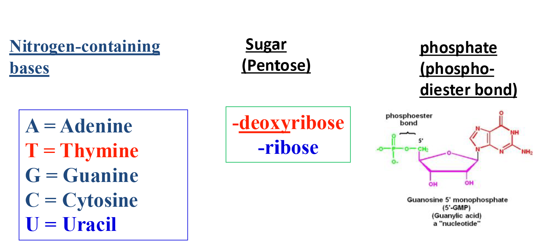
What are nucleotides, and what are their three components?

  • Nucleotides are the building blocks (monomers) of nucleic acids, including DNA and RNA.

  • Each nucleotide contains three components:

    • Nitrogenous base: Adenine (A), Thymine (T), Guanine (G), Cytosine (C), or Uracil (U).

    • Pentose sugar: A five-carbon sugar, either deoxyribose (DNA) or ribose (RNA).

    • Phosphate group: Participates in forming the sugar-phosphate backbone of DNA and RNA.

<ul><li><p><strong>Nucleotides</strong> are the building blocks (monomers) of nucleic acids, including DNA and RNA.</p></li><li><p>Each nucleotide contains three components:</p><ul><li><p><strong>Nitrogenous base:</strong> Adenine (A), Thymine (T), Guanine (G), Cytosine (C), or Uracil (U).</p></li><li><p><strong>Pentose sugar:</strong> A five-carbon sugar, either deoxyribose (DNA) or ribose (RNA).</p></li><li><p><strong>Phosphate group:</strong> Participates in forming the sugar-phosphate backbone of DNA and RNA.</p></li></ul></li></ul><p></p>
11
New cards

What is the difference between ribose and deoxyribose sugars?

  • Ribose: The five-carbon sugar found in RNA.

    • Contains a hydroxyl group (–OH) on its 2′ carbon.

  • Deoxyribose: The five-carbon sugar found in DNA.

    • Contains a hydrogen atom (–H) instead of a hydroxyl group on its 2′ carbon.

  • This difference distinguishes the sugar components of DNA and RNA.

  • Both are considered pentose sugars


<ul><li><p><strong>Ribose:</strong> The five-carbon sugar found in RNA.</p><ul><li><p>Contains a <strong>hydroxyl group (–OH)</strong> on its 2′ carbon.</p></li></ul></li><li><p><strong>Deoxyribose:</strong> The five-carbon sugar found in DNA.</p><ul><li><p>Contains a <strong>hydrogen atom (–H)</strong> instead of a hydroxyl group on its 2′ carbon.</p></li></ul></li><li><p>This difference distinguishes the sugar components of DNA and RNA.</p></li><li><p>Both are considered <strong>pentose</strong> sugars</p></li></ul><p></p>
12
New cards

What are purines and pyrimidines, and which nitrogenous bases belong to each group?

  • Nitrogenous bases are divided into two categories according to their structure.

Purines — two-ring structures:

  • Adenine (A)

  • Guanine (G)

Pyrimidines — one-ring structures:

  • Cytosine (C)

  • Thymine (T) — found in DNA.

  • Uracil (U) — found in RNA.


<ul><li><p>Nitrogenous bases are divided into two categories according to their structure.</p></li></ul><p><strong>Purines — two-ring structures:</strong></p><ul><li><p><strong>Adenine (A)</strong></p></li><li><p><strong>Guanine (G)</strong></p></li></ul><p><strong>Pyrimidines — one-ring structures:</strong></p><ul><li><p><strong>Cytosine (C)</strong></p></li><li><p><strong>Thymine (T)</strong> — found in DNA.</p></li><li><p><strong>Uracil (U)</strong> — found in RNA.</p></li></ul><p></p>
13
New cards

What is a phosphodiester bond, and what is its role in DNA and RNA?

  • A phosphodiester bond is a covalent bond that joins adjacent nucleotides together.

  • It connects the 3′ carbon of one sugar to the 5′ carbon of another sugar through a phosphate group.

  • These bonds form the sugar-phosphate backbone of nucleic acids.

  • Phosphodiester bonds are present in both DNA and RNA.

  • Their arrangement gives nucleic acid strands a 5′ to 3′ directionality.


<ul><li><p>A <strong>phosphodiester bond</strong> is a covalent bond that joins adjacent nucleotides together.</p></li><li><p>It connects the <strong>3′ carbon of one sugar</strong> to the <strong>5′ carbon of another sugar</strong> through a phosphate group.</p></li><li><p>These bonds form the <strong>sugar-phosphate backbone</strong> of nucleic acids.</p></li><li><p>Phosphodiester bonds are present in <strong>both DNA and RNA</strong>.</p></li><li><p>Their arrangement gives nucleic acid strands a <strong>5′ to 3′ directionality</strong>.</p></li></ul><p></p>
14
New cards

What is the difference between the 5′ and 3′ ends of a nucleic acid strand?

  • 5′ end: The end associated with the 5′ carbon of the sugar, typically bearing a free phosphate group.

  • 3′ end: The end with a free hydroxyl (–OH) group attached to the 3′ carbon.

  • Because the ends are chemically different, DNA and RNA strands have a specific direction or polarity.

  • Nucleic acid sequences are conventionally written in the 5′ → 3′ direction.


15
New cards

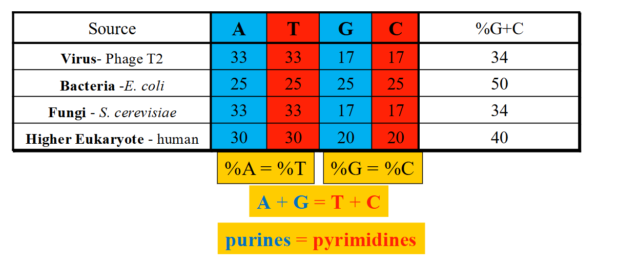
What is Chargaff's Rule, and what did it reveal about DNA?

  • Erwin Chargaff analyzed the nitrogenous base composition of DNA from different organisms.

  • He discovered that, in double-stranded DNA:

    • The amount of adenine (A) equals thymine (T).

    • The amount of guanine (G) equals cytosine (C).

  • Therefore, A = T and G = C.

  • The total amount of purines (A + G) equals the total amount of pyrimidines (T + C).

  • Chargaff's findings provided important evidence for complementary base pairing in DNA.
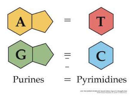

<ul><li><p><strong>Erwin Chargaff</strong> analyzed the nitrogenous base composition of DNA from different organisms.</p></li><li><p>He discovered that, in double-stranded DNA:</p><ul><li><p>The amount of <strong>adenine (A) equals thymine (T)</strong>.</p></li><li><p>The amount of <strong>guanine (G) equals cytosine (C)</strong>.</p></li></ul></li><li><p>Therefore, <strong>A = T</strong> and <strong>G = C</strong>.</p></li><li><p>The total amount of <strong>purines (A + G)</strong> equals the total amount of <strong>pyrimidines (T + C)</strong>.</p></li><li><p>Chargaff's findings provided important evidence for <strong>complementary base pairing</strong> in DNA.</p></li></ul><p></p>
16
New cards

How can Chargaff's Rule be used to calculate the percentage of DNA bases?

  • In double-stranded DNA, %A = %T and %G = %C.

  • All four base percentages must add up to 100%.

  • If one base percentage is known, the remaining percentages can be calculated.

Example: If DNA contains 30% adenine:

  • Thymine = 30%

  • Remaining percentage = 100% − 60% = 40%

  • Guanine = 20%

  • Cytosine = 20%


<ul><li><p>In double-stranded DNA, <strong>%A = %T</strong> and <strong>%G = %C</strong>.</p></li><li><p>All four base percentages must add up to <strong>100%</strong>.</p></li><li><p>If one base percentage is known, the remaining percentages can be calculated.</p></li></ul><p><strong>Example:</strong> If DNA contains 30% adenine:</p><ul><li><p>Thymine = <strong>30%</strong></p></li><li><p>Remaining percentage = 100% − 60% = 40%</p></li><li><p>Guanine = <strong>20%</strong></p></li><li><p>Cytosine = <strong>20%</strong></p></li></ul><p></p>
17
New cards

How did Rosalind Franklin contribute to discovering the structure of DNA?

  • Rosalind Franklin used X-ray diffraction to investigate DNA's structure in the early 1950s.

  • X-rays passing through DNA produced a diffraction pattern that revealed information about its molecular organization.

  • Her work provided evidence that:

    • DNA has a helical (spiral) structure.

    • DNA has a uniform diameter.

    • The nitrogenous bases are located inside the helix.

  • Her findings were essential to developing the double-helical model of DNA.


<ul><li><p><strong>Rosalind Franklin</strong> used X-ray diffraction to investigate DNA's structure in the early 1950s.</p></li><li><p>X-rays passing through DNA produced a <strong>diffraction pattern</strong> that revealed information about its molecular organization.</p></li><li><p>Her work provided evidence that:</p><ul><li><p>DNA has a <strong>helical (spiral) structure</strong>.</p></li><li><p>DNA has a <strong>uniform diameter</strong>.</p></li><li><p>The <strong>nitrogenous bases are located inside</strong> the helix.</p></li></ul></li><li><p>Her findings were essential to developing the double-helical model of DNA.</p></li></ul><p></p>
18
New cards

What did William Astbury, Rosalind Franklin, and Maurice Wilkins reveal about DNA's structure?

  • William Astbury (1947): Used X-ray diffraction analysis to show that DNA contains stacked bases.

  • Rosalind Franklin and Maurice Wilkins (1950–1953): Produced X-ray diffraction evidence supporting the conclusion that DNA is a helix.

  • Together, these discoveries helped establish the structural features used in the Watson–Crick model.
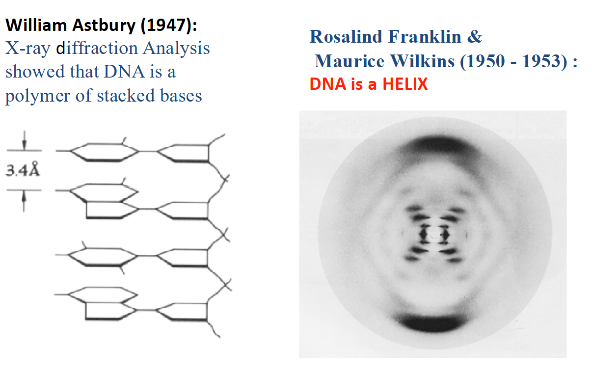

<ul><li><p><strong>William Astbury (1947):</strong> Used X-ray diffraction analysis to show that DNA contains <strong>stacked bases</strong>.</p></li><li><p><strong>Rosalind Franklin and Maurice Wilkins (1950–1953):</strong> Produced X-ray diffraction evidence supporting the conclusion that <strong>DNA is a helix</strong>.</p></li><li><p>Together, these discoveries helped establish the structural features used in the Watson–Crick model.</p></li></ul><p></p>
19
New cards

What evidence contributed to the Watson–Crick model of DNA, and what structural features did it propose?

Existing evidence:

  • Chargaff's Rule: A = T and G = C.

  • X-ray diffraction: DNA has a helical structure.

  • Base stacking: Nitrogenous bases are stacked on top of one another.

Features of the Watson–Crick model:

  • DNA forms a double helix consisting of two strands.

  • The two strands are antiparallel, running in opposite directions.

  • The sugar-phosphate backbone is located on the outside of the helix.

  • The nitrogenous bases are located on the inside.

  • Complementary bases are held together by hydrogen bonds.


20
New cards

How do complementary nitrogenous bases pair in DNA?

  • DNA contains two types of complementary base pairs:

    • Adenine (A) pairs with thymine (T) through 2 hydrogen bonds.

    • Guanine (G) pairs with cytosine (C) through 3 hydrogen bonds.

  • Each pair contains one purine and one pyrimidine.

  • This arrangement helps maintain DNA's uniform diameter of 2 nm.

  • Hydrogen bonds help hold the two DNA strands together.


<ul><li><p>DNA contains two types of complementary base pairs:</p><ul><li><p><strong>Adenine (A) pairs with thymine (T)</strong> through <strong>2 hydrogen bonds</strong>.</p></li><li><p><strong>Guanine (G) pairs with cytosine (C)</strong> through <strong>3 hydrogen bonds</strong>.</p></li></ul></li><li><p>Each pair contains <strong>one purine and one pyrimidine</strong>.</p></li><li><p>This arrangement helps maintain DNA's <strong>uniform diameter of 2 nm</strong>.</p></li><li><p>Hydrogen bonds help hold the two DNA strands together.</p></li></ul><p></p>
21
New cards

What are the main structural dimensions of the DNA double helix?

  • Diameter: Approximately 2 nm.

  • One complete turn: Approximately 3.4 nm.

  • Base pairs per turn: Approximately 10 base pairs.

  • Distance between adjacent base pairs: Approximately 0.34 nm.

  • DNA contains alternating major and minor grooves along its double helix.


<ul><li><p><strong>Diameter:</strong> Approximately <strong>2 nm</strong>.</p></li><li><p><strong>One complete turn:</strong> Approximately <strong>3.4 nm</strong>.</p></li><li><p><strong>Base pairs per turn:</strong> Approximately <strong>10 base pairs</strong>.</p></li><li><p><strong>Distance between adjacent base pairs:</strong> Approximately <strong>0.34 nm</strong>.</p></li><li><p>DNA contains alternating <strong>major and minor grooves</strong> along its double helix.</p></li></ul><p></p>
22
New cards

What determines the polarity (directionality) of a DNA strand?

  • DNA strand polarity is determined by the arrangement of sugar-phosphate bonds.

  • Adjacent nucleotides are connected by phosphodiester bonds between the 3′ carbon of one sugar and the 5′ carbon of the next.

  • The two ends of a DNA strand are chemically different:

    • 5′ end: Typically contains a free phosphate group attached to the 5′ carbon.

    • 3′ end: Contains a free hydroxyl (–OH) group attached to the 3′ carbon.

  • DNA sequences are conventionally written in the 5′ → 3′ direction.

  • Example: 5′-CTAACG-3′.


<ul><li><p>DNA strand polarity is determined by the arrangement of <strong>sugar-phosphate bonds</strong>.</p></li><li><p>Adjacent nucleotides are connected by <strong>phosphodiester bonds</strong> between the 3′ carbon of one sugar and the 5′ carbon of the next.</p></li><li><p>The two ends of a DNA strand are chemically different:</p><ul><li><p><strong>5′ end:</strong> Typically contains a free phosphate group attached to the 5′ carbon.</p></li><li><p><strong>3′ end:</strong> Contains a free hydroxyl (–OH) group attached to the 3′ carbon.</p></li></ul></li><li><p>DNA sequences are conventionally written in the <strong>5′ → 3′ direction</strong>.</p></li><li><p>Example: <strong>5′-CTAACG-3′</strong>.</p></li></ul><p></p>
23
New cards

What does it mean for DNA strands to be antiparallel?

  • Antiparallel means that the two DNA strands run in opposite directions.

  • One strand runs 5′ → 3′, while the complementary strand runs 3′ → 5′.

  • The strands have opposite chemical polarity, determined by the orientation of their deoxyribose sugars and sugar-phosphate backbones.

  • This antiparallel arrangement is a fundamental feature of DNA's double-helical structure.


<ul><li><p><strong>Antiparallel</strong> means that the two DNA strands run in <strong>opposite directions</strong>.</p></li><li><p>One strand runs <strong>5′ → 3′</strong>, while the complementary strand runs <strong>3′ → 5′</strong>.</p></li><li><p>The strands have opposite <strong>chemical polarity</strong>, determined by the orientation of their deoxyribose sugars and sugar-phosphate backbones.</p></li><li><p>This antiparallel arrangement is a fundamental feature of DNA's <strong>double-helical structure</strong>.</p></li></ul><p></p>
24
New cards

What is complementary base pairing, and how can the sequence of one DNA strand be used to determine the other?

  • DNA strands exhibit complementary base pairing, meaning each nitrogenous base pairs with a specific partner.

    • A pairs with T through 2 hydrogen bonds.

    • G pairs with C through 3 hydrogen bonds.

  • The two strands run antiparallel, meaning they have opposite 5′ and 3′ orientations.

  • Knowing the sequence of one strand allows the complementary strand to be determined.


<ul><li><p>DNA strands exhibit <strong>complementary base pairing</strong>, meaning each nitrogenous base pairs with a specific partner.</p><ul><li><p><strong>A pairs with T</strong> through 2 hydrogen bonds.</p></li><li><p><strong>G pairs with C</strong> through 3 hydrogen bonds.</p></li></ul></li><li><p>The two strands run <strong>antiparallel</strong>, meaning they have opposite 5′ and 3′ orientations.</p></li><li><p>Knowing the sequence of one strand allows the complementary strand to be determined.</p></li></ul><p></p>
25
New cards

What is Polymerase Chain Reaction (PCR), and what is its purpose?

  • Polymerase Chain Reaction (PCR) is a molecular technique used to amplify (make many copies of) a specific DNA sequence.

  • It builds on the natural process of DNA replication and complementary DNA base pairing (hybridization).

  • PCR can generate large quantities of a target DNA sequence from a very small amount of starting material.

  • Advantages: It is relatively fast, inexpensive, highly versatile, and requires little starting DNA.

  • PCR was developed by Kary Mullis, who shared the 1993 Nobel Prize in Chemistry.


<ul><li><p><strong>Polymerase Chain Reaction (PCR)</strong> is a molecular technique used to amplify (make many copies of) a specific DNA sequence.</p></li><li><p>It builds on the natural process of <strong>DNA replication</strong> and <strong>complementary DNA base pairing (hybridization)</strong>.</p></li><li><p>PCR can generate large quantities of a target DNA sequence from a very small amount of starting material.</p></li><li><p><strong>Advantages:</strong> It is relatively fast, inexpensive, highly versatile, and requires little starting DNA.</p></li><li><p>PCR was developed by <strong>Kary Mullis</strong>, who shared the 1993 Nobel Prize in Chemistry.</p></li></ul><p></p>
26
New cards

What are the three steps of PCR, and what happens during each step?

  • 1. Denaturation (~95°C):

    • Double-stranded DNA is heated to separate it into two single strands.

    • Hydrogen bonds between complementary bases are disrupted.

  • 2. Annealing (~52°C):

    • The temperature is lowered to allow primers to bind to complementary sequences on the template DNA.

    • Primers identify the DNA region that will be amplified.

  • 3. Elongation (~72°C):

    • DNA polymerase extends the primers by adding complementary nucleotides.

    • New DNA strands are synthesized.

  • These three steps are repeated over multiple cycles, resulting in exponential amplification of the target DNA.


<ul><li><p><strong>1. Denaturation (~95°C):</strong></p><ul><li><p>Double-stranded DNA is heated to separate it into two single strands.</p></li><li><p>Hydrogen bonds between complementary bases are disrupted.</p></li></ul></li><li><p><strong>2. Annealing (~52°C):</strong></p><ul><li><p>The temperature is lowered to allow <strong>primers</strong> to bind to complementary sequences on the template DNA.</p></li><li><p>Primers identify the DNA region that will be amplified.</p></li></ul></li><li><p><strong>3. Elongation (~72°C):</strong></p><ul><li><p><strong>DNA polymerase</strong> extends the primers by adding complementary nucleotides.</p></li><li><p>New DNA strands are synthesized.</p></li></ul></li><li><p>These three steps are repeated over multiple cycles, resulting in <strong>exponential amplification</strong> of the target DNA.</p></li></ul><p></p>
27
New cards

How does PCR amplify DNA exponentially, and how can the number of copies be calculated?


<p></p>
28
New cards

What five components are required for PCR, and what is the function of each?

  • 1. DNA template:

    • Contains the target DNA sequence that will be amplified.

  • 2. Primers:

    • Short DNA sequences that bind to complementary regions of the template DNA.

    • Provide a starting point for DNA polymerase.

  • 3. dNTPs (dATP, dTTP, dCTP, dGTP):

    • Deoxyribonucleotide triphosphates that serve as the building blocks for newly synthesized DNA.

  • 4. DNA polymerase:

    • Enzyme that synthesizes new DNA strands by adding complementary nucleotides.

  • 5. Salts and buffer:

    • Maintain suitable chemical conditions for DNA polymerase activity.


<ul><li><p><strong>1. DNA template:</strong></p><ul><li><p>Contains the target DNA sequence that will be amplified.</p></li></ul></li><li><p><strong>2. Primers:</strong></p><ul><li><p>Short DNA sequences that bind to complementary regions of the template DNA.</p></li><li><p>Provide a starting point for DNA polymerase.</p></li></ul></li><li><p><strong>3. dNTPs (dATP, dTTP, dCTP, dGTP):</strong></p><ul><li><p>Deoxyribonucleotide triphosphates that serve as the <strong>building blocks</strong> for newly synthesized DNA.</p></li></ul></li><li><p><strong>4. DNA polymerase:</strong></p><ul><li><p>Enzyme that synthesizes new DNA strands by adding complementary nucleotides.</p></li></ul></li><li><p><strong>5. Salts and buffer:</strong></p><ul><li><p>Maintain suitable chemical conditions for DNA polymerase activity.</p></li></ul></li></ul><p></p>
29
New cards

Why does PCR produce exponential rather than linear DNA amplification?

  • After each PCR cycle, the newly synthesized DNA molecules can serve as templates for the next cycle.

  • Both the original DNA and newly produced DNA are copied in subsequent cycles.

  • Therefore, the amount of DNA ideally doubles with every cycle.

  • This produces exponential amplification, allowing millions of copies to be generated from a small amount of starting DNA.


<ul><li><p>After each PCR cycle, the newly synthesized DNA molecules can serve as <strong>templates</strong> for the next cycle.</p></li><li><p>Both the original DNA and newly produced DNA are copied in subsequent cycles.</p></li><li><p>Therefore, the amount of DNA ideally <strong>doubles with every cycle</strong>.</p></li><li><p>This produces <strong>exponential amplification</strong>, allowing millions of copies to be generated from a small amount of starting DNA.</p></li></ul><p></p>
30
New cards

What are PCR primers, and why are two primers required?

  • Primers are short, single-stranded DNA sequences that bind to complementary regions of a DNA template.

  • They provide a starting point for DNA polymerase to synthesize new DNA strands.

  • PCR requires two primers:

    • Forward primer: Anneals to the bottom template strand and has the same sequence as the corresponding region of the top strand.

    • Reverse primer: Anneals to the top template strand and has the reverse-complement sequence of the corresponding region of the top strand.

  • The two primers flank the target DNA region that will be amplified.

  • DNA polymerase extends both primers in the 5′ → 3′ direction.


31
New cards

How do forward and reverse primers determine which DNA region is amplified during PCR?

  • The forward primer binds to a complementary sequence on the bottom DNA strand.

  • The reverse primer binds to a complementary sequence on the top DNA strand.

  • Both primers have their 3′ ends facing toward the target region.

  • DNA polymerase adds nucleotides to the 3′ ends of the primers, extending the new strands in the 5′ → 3′ direction.

  • Repeated PCR cycles preferentially amplify the DNA region between the two primer-binding sites.
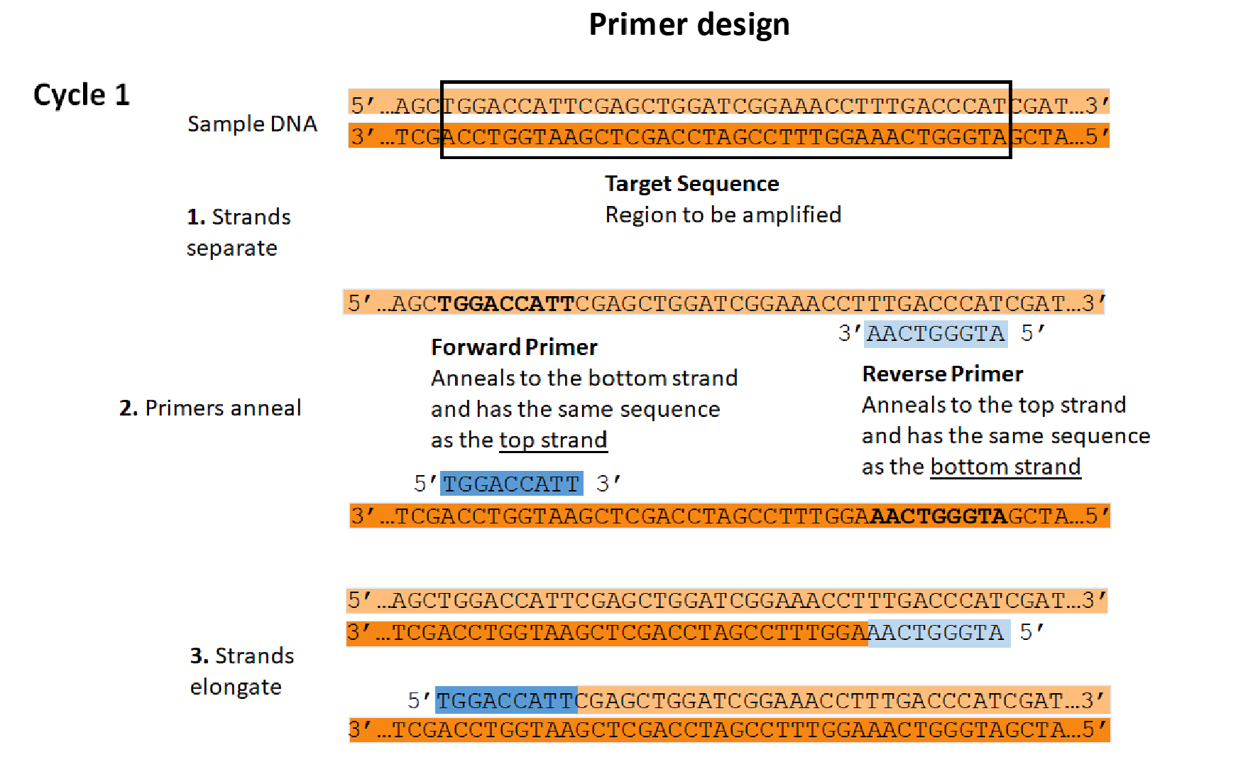

<ul><li><p>The <strong>forward primer</strong> binds to a complementary sequence on the bottom DNA strand.</p></li><li><p>The <strong>reverse primer</strong> binds to a complementary sequence on the top DNA strand.</p></li><li><p>Both primers have their <strong>3′ ends facing toward the target region</strong>.</p></li><li><p>DNA polymerase adds nucleotides to the <strong>3′ ends</strong> of the primers, extending the new strands in the 5′ → 3′ direction.</p></li><li><p>Repeated PCR cycles preferentially amplify the DNA region <strong>between the two primer-binding sites</strong>.</p></li></ul><p></p>
32
New cards

What is gel electrophoresis, and how does it separate DNA fragments?

  • Gel electrophoresis is a molecular technique used to separate DNA fragments according to their size.

  • DNA samples are loaded into wells in a gel, and an electric field is applied.

  • DNA has a negative charge due to the phosphate groups in its backbone.

  • Therefore, DNA fragments migrate toward the positive electrode (anode).

  • The gel acts as a molecular sieve:

    • Smaller DNA fragments migrate faster and travel farther.

    • Larger DNA fragments migrate more slowly and remain closer to the wells.

  • The separated fragments form bands that can be visualized and analyzed.


33
New cards

How can you determine which DNA fragment is largest or smallest on an electrophoresis gel?

  • Largest DNA fragments:

    • Migrate the shortest distance through the gel.

    • Appear closest to the wells.

  • Smallest DNA fragments:

    • Migrate farther through the gel.

    • Appear farthest from the wells, toward the positive electrode.

  • Fragments are separated by their movement through the gel, allowing researchers to compare their sizes.


<ul><li><p><strong>Largest DNA fragments:</strong></p><ul><li><p>Migrate the shortest distance through the gel.</p></li><li><p>Appear <strong>closest to the wells</strong>.</p></li></ul></li><li><p><strong>Smallest DNA fragments:</strong></p><ul><li><p>Migrate farther through the gel.</p></li><li><p>Appear <strong>farthest from the wells</strong>, toward the positive electrode.</p></li></ul></li><li><p>Fragments are separated by their movement through the gel, allowing researchers to compare their sizes.</p></li></ul><p></p>
34
New cards

What are restriction enzymes, and what is their natural function in bacteria?

  • Restriction enzymes are bacterial endonucleases that recognize and cut DNA at specific sequences.

  • Their natural function is to help protect bacteria against foreign DNA, such as DNA introduced by viruses.

  • They recognize specific double-stranded DNA sequences called restriction sites.

  • Restriction sites are typically 4–8 base pairs long and are often palindromic.

  • Restriction enzymes can cut DNA at particular locations wherever their recognition sequences occur, making them valuable tools in molecular biology.


<ul><li><p><strong>Restriction enzymes</strong> are bacterial <strong>endonucleases</strong> that recognize and cut DNA at specific sequences.</p></li><li><p>Their natural function is to help protect bacteria against <strong>foreign DNA</strong>, such as DNA introduced by viruses.</p></li><li><p>They recognize specific double-stranded DNA sequences called <strong>restriction sites</strong>.</p></li><li><p>Restriction sites are typically <strong>4–8 base pairs</strong> long and are often <strong>palindromic</strong>.</p></li><li><p>Restriction enzymes can cut DNA at particular locations wherever their recognition sequences occur, making them valuable tools in molecular biology.</p></li></ul><p></p>
35
New cards

What are restriction sites, and what does it mean for a restriction site to be palindromic?

  • Restriction sites are specific double-stranded DNA sequences recognized and cleaved by restriction enzymes.

  • These recognition sequences are usually 4–8 base pairs long.

  • Many restriction sites are palindromic, meaning the sequences read identically in the 5′ → 3′ direction on both complementary strands.

  • For example, EcoRI recognizes the sequence:

    • 5′-GAATTC-3′

    • 3′-CTTAAG-5′

  • Restriction enzymes cut DNA wherever their specific restriction sites occur.


<ul><li><p><strong>Restriction sites</strong> are specific double-stranded DNA sequences recognized and cleaved by restriction enzymes.</p></li><li><p>These recognition sequences are usually <strong>4–8 base pairs long</strong>.</p></li><li><p>Many restriction sites are <strong>palindromic</strong>, meaning the sequences read identically in the 5′ → 3′ direction on both complementary strands.</p></li><li><p>For example, <strong>EcoRI</strong> recognizes the sequence:</p><ul><li><p>5′-GAATTC-3′</p></li><li><p>3′-CTTAAG-5′</p></li></ul></li><li><p>Restriction enzymes cut DNA wherever their specific restriction sites occur.</p></li></ul><p></p>
36
New cards

What is the difference between sticky ends and blunt ends produced by restriction enzymes?

  • Sticky ends:

    • Produced when a restriction enzyme makes staggered cuts in the two DNA strands.

    • Leave short, single-stranded overhangs.

    • These overhangs can form complementary base pairs with compatible DNA ends.

    • Example: EcoRI.

  • Blunt ends:

    • Produced when a restriction enzyme cuts both DNA strands at the same position.

    • Do not contain single-stranded overhangs.

    • Example: SmaI.

  • Both types of ends can be used in DNA cloning.


<ul><li><p><strong>Sticky ends:</strong></p><ul><li><p>Produced when a restriction enzyme makes <strong>staggered cuts</strong> in the two DNA strands.</p></li><li><p>Leave short, single-stranded overhangs.</p></li><li><p>These overhangs can form complementary base pairs with compatible DNA ends.</p></li><li><p>Example: <strong>EcoRI</strong>.</p></li></ul></li><li><p><strong>Blunt ends:</strong></p><ul><li><p>Produced when a restriction enzyme cuts both DNA strands at the same position.</p></li><li><p>Do not contain single-stranded overhangs.</p></li><li><p>Example: <strong>SmaI</strong>.</p></li></ul></li><li><p>Both types of ends can be used in DNA cloning.</p></li></ul><p></p>
37
New cards

What are the major applications of restriction enzymes in biotechnology?

  • Gene cloning: Inserting foreign DNA fragments into plasmids.

  • Genetic engineering: Creating recombinant DNA by combining DNA from different sources.

  • Diagnostics and forensic science: Analyzing DNA patterns for identification.

  • Molecular research: Studying genes through gene mapping and genome editing.
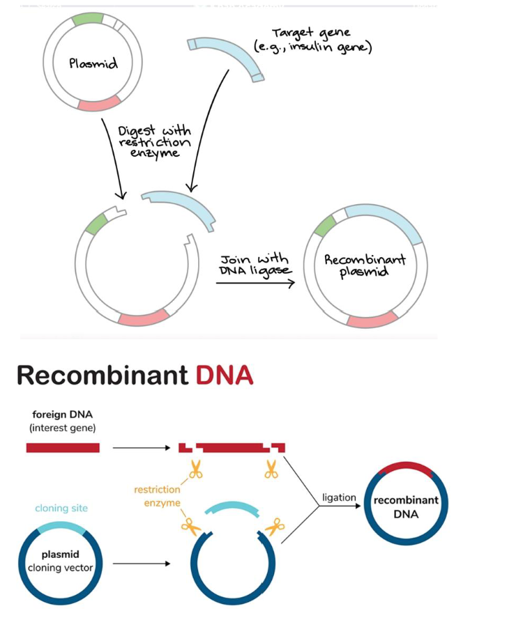

<ul><li><p><strong>Gene cloning:</strong> Inserting foreign DNA fragments into plasmids.</p></li><li><p><strong>Genetic engineering:</strong> Creating <strong>recombinant DNA</strong> by combining DNA from different sources.</p></li><li><p><strong>Diagnostics and forensic science:</strong> Analyzing DNA patterns for identification.</p></li><li><p><strong>Molecular research:</strong> Studying genes through gene mapping and genome editing.</p></li></ul><p></p>
38
New cards

What is a cloning plasmid, and what three essential components must it contain?

  • A cloning plasmid is a DNA molecule engineered to carry and replicate foreign DNA within a host cell.

  • Plasmids occur naturally in organisms such as bacteria and yeast but can be modified for laboratory use.

  • A cloning plasmid requires three main components:

    • 1. Multiple Cloning Site (MCS): Contains numerous restriction sites where foreign DNA can be inserted.

    • 2. Origin of Replication (ORI): Allows the plasmid to replicate within the host cell.

    • 3. Selectable Marker: Allows researchers to identify or select cells containing the plasmid, often using an antibiotic resistance gene (Ex. will survive in the presence of ampicillin, while others would not).


<ul><li><p>A <strong>cloning plasmid</strong> is a DNA molecule engineered to carry and replicate foreign DNA within a host cell.</p></li><li><p>Plasmids occur naturally in organisms such as bacteria and yeast but can be modified for laboratory use.</p></li><li><p>A cloning plasmid requires three main components:</p><ul><li><p><strong>1. Multiple Cloning Site (MCS):</strong> Contains numerous restriction sites where foreign DNA can be inserted.</p></li><li><p><strong>2. Origin of Replication (ORI):</strong> Allows the plasmid to replicate within the host cell.</p></li><li><p><strong>3. Selectable Marker:</strong> Allows researchers to identify or select cells containing the plasmid, often using an <strong>antibiotic resistance gene </strong>(Ex. will survive in the presence of ampicillin, while others would not).</p></li></ul></li></ul><p></p>
39
New cards

What are the four main steps involved in DNA cloning using a plasmid?

  • 1. Digestion:

    • Restriction enzymes cut the plasmid vector and foreign DNA at specific restriction sites.

    • Compatible DNA ends are produced.

  • 2. Ligation:

    • The foreign DNA fragment is inserted into the opened plasmid.

    • DNA ligase joins the DNA fragments by forming phosphodiester bonds, producing a recombinant plasmid.

  • 3. Transformation:

    • The recombinant plasmid is introduced into bacterial host cells.

  • 4. Selection:

    • Bacteria are grown on a medium containing an antibiotic, such as ampicillin.

    • Cells containing the resistance plasmid survive and form colonies.

    • Cells without the plasmid cannot grow under these conditions.


40
New cards

Useful Diagram

knowt flashcard image
41
New cards

How do antibiotic resistance genes function as selectable markers during plasmid cloning?

  • Cloning plasmids often contain an antibiotic resistance gene, such as ampᴿ.

  • After transformation, bacteria are grown on medium containing the corresponding antibiotic.

  • Transformed bacteria carrying the plasmid can survive and form colonies.

  • Non-transformed bacteria lacking the plasmid are unable to grow.

  • This allows researchers to select bacteria that have taken up a plasmid.


42
New cards

How can gel electrophoresis be used to analyze recombinant plasmids?

  • Gel electrophoresis separates DNA fragments according to their sizes.

  • Researchers can compare DNA from plasmids containing an insert with DNA from plasmids lacking the insert.

  • A recombinant plasmid contains both the original plasmid DNA and the inserted DNA fragment.

    • Original plasmid = 3,200 bp.

    • Foreign DNA insert = 1,200 bp.

    • Recombinant plasmid = 4,400 bp.

  • A DNA ladder provides reference fragment sizes for comparison.

  • Appropriate gel analysis can help determine whether the expected DNA insert is present.
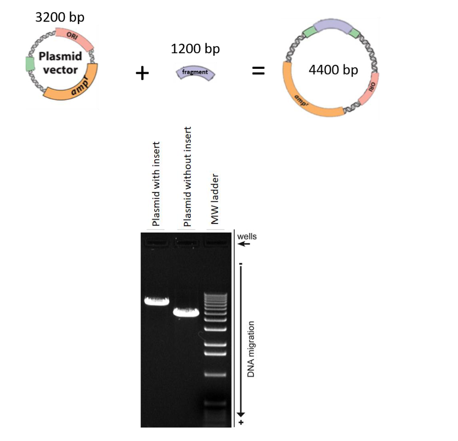

<ul><li><p><strong>Gel electrophoresis</strong> separates DNA fragments according to their sizes.</p></li><li><p>Researchers can compare DNA from plasmids containing an insert with DNA from plasmids lacking the insert.</p></li><li><p>A recombinant plasmid contains both the original plasmid DNA and the inserted DNA fragment.</p><ul><li><p>Original plasmid = <strong>3,200 bp</strong>.</p></li><li><p>Foreign DNA insert = <strong>1,200 bp</strong>.</p></li><li><p>Recombinant plasmid = <strong>4,400 bp</strong>.</p></li></ul></li><li><p>A <strong>DNA ladder</strong> provides reference fragment sizes for comparison.</p></li><li><p>Appropriate gel analysis can help determine whether the expected DNA insert is present.</p></li></ul><p></p>
43
New cards

What is DNA supercoiling, and why is it important?

  • DNA supercoiling occurs when the DNA double helix undergoes additional twisting and coils upon itself.

  • Supercoiling helps compact long DNA molecules, allowing them to fit inside cells.

  • DNA in living cells (in vivo) is often maintained in a supercoiled state.

  • In prokaryotes and viruses, DNA can be organized into supercoiled domains.
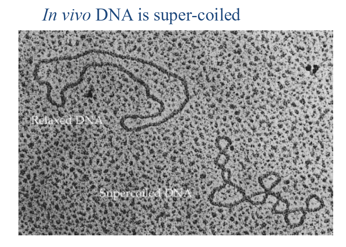

<ul><li><p><strong>DNA supercoiling</strong> occurs when the DNA double helix undergoes additional twisting and coils upon itself.</p></li><li><p>Supercoiling helps <strong>compact long DNA molecules</strong>, allowing them to fit inside cells.</p></li><li><p>DNA in living cells (<em>in vivo</em>) is often maintained in a <strong>supercoiled state</strong>.</p></li><li><p>In prokaryotes and viruses, DNA can be organized into <strong>supercoiled domains</strong>.</p></li></ul><p></p>
44
New cards

How does circular DNA become supercoiled?

  • Circular DNA can undergo additional twisting beyond its normal double-helical structure.

  • In the diagram, one DNA strand is temporarily nicked (cut).

  • The DNA is rotated to introduce additional twisting:

    • Overwinding introduces positive supercoiling.

    • Underwinding introduces negative supercoiling.

  • The nick is then sealed, maintaining the altered twisting.

  • The DNA responds by coiling upon itself, forming a supercoiled structure.


<ul><li><p>Circular DNA can undergo additional twisting beyond its normal double-helical structure.</p></li><li><p>In the diagram, one DNA strand is temporarily <strong>nicked (cut)</strong>.</p></li><li><p>The DNA is rotated to introduce additional twisting:</p><ul><li><p><strong>Overwinding</strong> introduces positive supercoiling.</p></li><li><p><strong>Underwinding</strong> introduces negative supercoiling.</p></li></ul></li><li><p>The nick is then sealed, maintaining the altered twisting.</p></li><li><p>The DNA responds by <strong>coiling upon itself</strong>, forming a supercoiled structure.</p></li></ul><p></p>
45
New cards

Why must prokaryotic DNA be highly condensed, and how is it organized in E. coli?

  • Escherichia coli is described as monoploid, meaning it contains one genome.

  • Its chromosome consists of a circular DNA molecule approximately 1.5 mm long.

  • However, an E. coli cell is only about 2 µm long and 1 µm wide.

  • Therefore, the DNA must be condensed more than 1,000-fold to fit inside the cell.

  • This condensation occurs through DNA folding and supercoiling, producing a compact bacterial chromosome.


<ul><li><p><em>Escherichia coli</em> is described as <strong>monoploid</strong>, meaning it contains one genome.</p></li><li><p>Its chromosome consists of a <strong>circular DNA molecule</strong> approximately <strong>1.5 mm long</strong>.</p></li><li><p>However, an <em>E. coli</em> cell is only about <strong>2 µm long and 1 µm wide</strong>.</p></li><li><p>Therefore, the DNA must be condensed <strong>more than 1,000-fold</strong> to fit inside the cell.</p></li><li><p>This condensation occurs through <strong>DNA folding and supercoiling</strong>, producing a compact bacterial chromosome.</p></li></ul><p></p>
46
New cards

How is the bacterial chromosome compacted through folding and supercoiling?

  • Bacterial DNA is compacted through a combination of loop formation and supercoiling, assisted by RNA and proteins.

  • The circular DNA molecule is first organized into approximately 40–50 loops (domains).

  • The DNA within these loops undergoes further supercoiling and folding.

  • This reduces the physical space occupied by the bacterial chromosome.


<ul><li><p>Bacterial DNA is compacted through a combination of <strong>loop formation and supercoiling</strong>, assisted by RNA and proteins.</p></li><li><p>The circular DNA molecule is first organized into approximately <strong>40–50 loops (domains)</strong>.</p></li><li><p>The DNA within these loops undergoes further <strong>supercoiling and folding</strong>.</p></li><li><p>This reduces the physical space occupied by the bacterial chromosome.</p></li></ul><p></p>
47
New cards

What are the key characteristics of prokaryotic and viral chromosome organization?

  • DNA molecules in prokaryotic and viral chromosomes can be organized into supercoiled domains.

  • Bacterial chromosomes commonly contain circular DNA molecules.

  • In E. coli, the chromosome is organized into approximately 50 domains.

  • Folding and supercoiling allow long DNA molecules to be packaged into small cells.


48
New cards

How is human DNA organized, and why must it be tightly packaged?

  • Humans are diploid, meaning they typically possess two sets of chromosomes (46 chromosomes total).

  • The human haploid genome contains 23 chromosomes.

  • The total length of DNA in a diploid human cell is approximately 2 metres.

  • However, the human nucleus is only approximately 5–10 µm in diameter.

  • Therefore, DNA must be extensively folded and packaged into multiple chromosomes to fit inside the nucleus.


<ul><li><p>Humans are <strong>diploid</strong>, meaning they typically possess <strong>two sets of chromosomes</strong> (46 chromosomes total).</p></li><li><p>The human <strong>haploid genome</strong> contains <strong>23 chromosomes</strong>.</p></li><li><p>The total length of DNA in a diploid human cell is approximately <strong>2 metres</strong>.</p></li><li><p>However, the human nucleus is only approximately <strong>5–10 µm in diameter</strong>.</p></li><li><p>Therefore, DNA must be <strong>extensively folded and packaged into multiple chromosomes</strong> to fit inside the nucleus.</p></li></ul><p></p>
49
New cards

What is chromatin, and what are the main components of a eukaryotic chromosome?

  • Each eukaryotic chromosome consists of one large, linear DNA molecule associated with proteins.

  • Chromatin is the complex of DNA and proteins that forms eukaryotic chromosomes.

  • Chromatin contains two major categories of proteins:

    • Histones: Highly positively charged proteins that help package DNA.

    • Non-histone proteins: A diverse group of proteins involved in chromosome organization and other functions.

  • DNA packaging in chromatin occurs through three major levels of compaction.


<ul><li><p>Each eukaryotic chromosome consists of <strong>one large, linear DNA molecule</strong> associated with proteins.</p></li><li><p><strong>Chromatin</strong> is the complex of DNA and proteins that forms eukaryotic chromosomes.</p></li><li><p>Chromatin contains two major categories of proteins:</p><ul><li><p><strong>Histones:</strong> Highly positively charged proteins that help package DNA.</p></li><li><p><strong>Non-histone proteins:</strong> A diverse group of proteins involved in chromosome organization and other functions.</p></li></ul></li><li><p>DNA packaging in chromatin occurs through <strong>three major levels of compaction</strong>.</p></li></ul><p></p>
50
New cards

Why are histones positively charged?

  • DNA has a negatively charged phosphate backbone.

  • The positive charges on histones help them associate with negatively charged DNA, allowing DNA to wrap around these proteins.


51
New cards

What are histones, and what roles do they play in eukaryotic cells?

  • Histones are positively charged proteins that help package DNA into chromatin.

  • They organize DNA into compact structures called nucleosomes.

  • There are two major categories of histones:

    • Core histones: H2A, H2B, H3, and H4.

    • Linker histone: H1.

  • Histones help organize DNA and are involved in gene expression, DNA replication, and DNA repair.


<ul><li><p><strong>Histones</strong> are positively charged proteins that help package DNA into <strong>chromatin</strong>.</p></li><li><p>They organize DNA into compact structures called <strong>nucleosomes</strong>.</p></li><li><p>There are two major categories of histones:</p><ul><li><p><strong>Core histones:</strong> H2A, H2B, H3, and H4.</p></li><li><p><strong>Linker histone:</strong> H1.</p></li></ul></li><li><p>Histones help organize DNA and are involved in <strong>gene expression, DNA replication, and DNA repair</strong>.</p></li></ul><p></p>
52
New cards

What is a histone octamer, and which histones does it contain?

  • A histone octamer is a group of eight histone proteins that forms the core of a nucleosome.

  • It contains:

    • 2 H2A histones

    • 2 H2B histones

    • 2 H3 histones

    • 2 H4 histones

  • Altogether, these form 8 histone proteins.

  • Approximately 147 base pairs of DNA wrap around this histone octamer to form a nucleosome core.


<ul><li><p>A <strong>histone octamer</strong> is a group of eight histone proteins that forms the core of a nucleosome.</p></li><li><p>It contains:</p><ul><li><p><strong>2 H2A histones</strong></p></li><li><p><strong>2 H2B histones</strong></p></li><li><p><strong>2 H3 histones</strong></p></li><li><p><strong>2 H4 histones</strong></p></li></ul></li><li><p>Altogether, these form <strong>8 histone proteins</strong>.</p></li><li><p>Approximately <strong>147 base pairs of DNA</strong> wrap around this histone octamer to form a nucleosome core.</p></li></ul><p></p>
53
New cards

What is a nucleosome, and how does it form the first level of DNA packaging?

  • A nucleosome is the basic structural unit of chromatin.

  • It forms when DNA wraps around a histone octamer containing eight core histone proteins.

  • Approximately 147 base pairs of DNA wrap around the octamer about 1¾ turns.

  • Adjacent nucleosomes are connected by linker DNA.

  • This arrangement forms an approximately 11-nm-wide structure, often described as “beads on a string.”

  • Nucleosome formation represents the first level of DNA packaging in eukaryotic chromosomes.
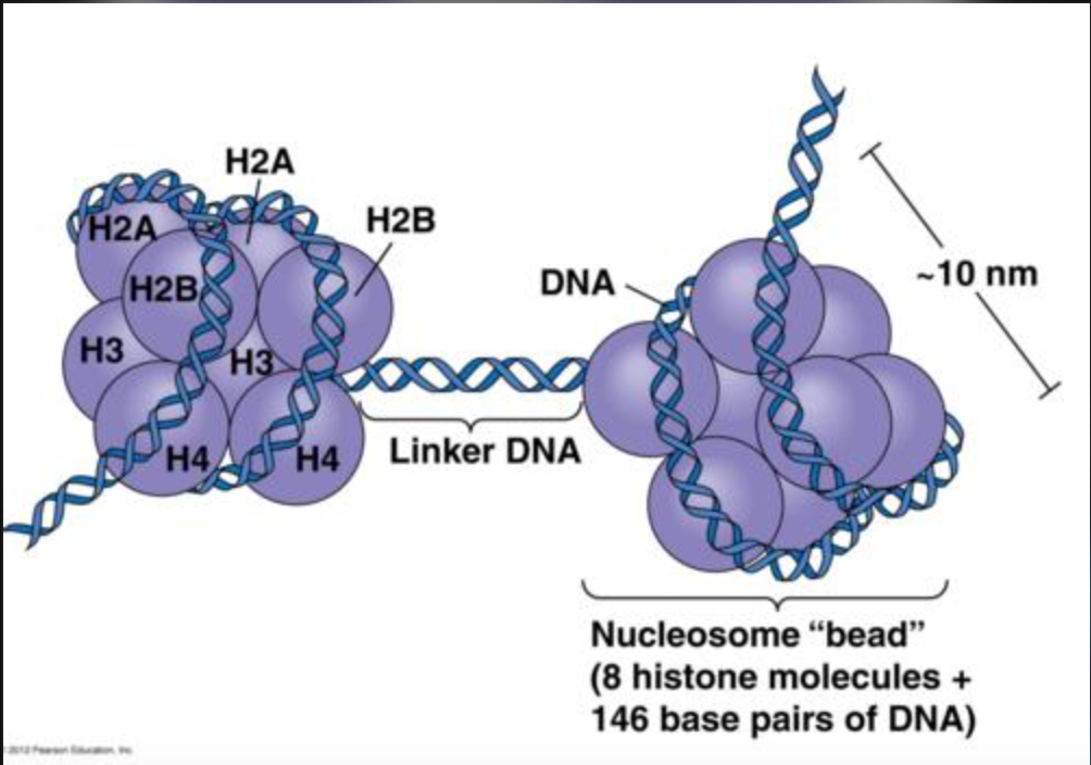

<ul><li><p>A <strong>nucleosome</strong> is the basic structural unit of chromatin.</p></li><li><p>It forms when DNA wraps around a <strong>histone octamer</strong> containing eight core histone proteins.</p></li><li><p>Approximately <strong>147 base pairs of DNA</strong> wrap around the octamer about <strong>1¾ turns</strong>.</p></li><li><p>Adjacent nucleosomes are connected by <strong>linker DNA</strong>.</p></li><li><p>This arrangement forms an approximately <strong>11-nm-wide structure</strong>, often described as <strong>“beads on a string.”</strong></p></li><li><p>Nucleosome formation represents the <strong>first level of DNA packaging</strong> in eukaryotic chromosomes.</p></li></ul><p></p>
54
New cards

What is the difference between the nucleosome core and the complete nucleosome, and what is the role of linker DNA?

  • Nucleosome core: Contains a histone octamer (2 copies each of H2A, H2B, H3, and H4) wrapped by approximately 147 bp of DNA.

  • Linker DNA: Connects adjacent nucleosomes and varies in length from 8–114 bp.

  • Complete nucleosome: Includes the nucleosome core and histone H1, which associates with linker DNA and helps promote further chromatin compaction.


<ul><li><p><strong>Nucleosome core:</strong> Contains a histone octamer (2 copies each of H2A, H2B, H3, and H4) wrapped by approximately <strong>147 bp of DNA</strong>.</p></li><li><p><strong>Linker DNA:</strong> Connects adjacent nucleosomes and varies in length from <strong>8–114 bp</strong>.</p></li><li><p><strong>Complete nucleosome:</strong> Includes the nucleosome core and <strong>histone H1</strong>, which associates with linker DNA and helps promote further <strong>chromatin compaction</strong>.</p></li></ul><p></p>
55
New cards

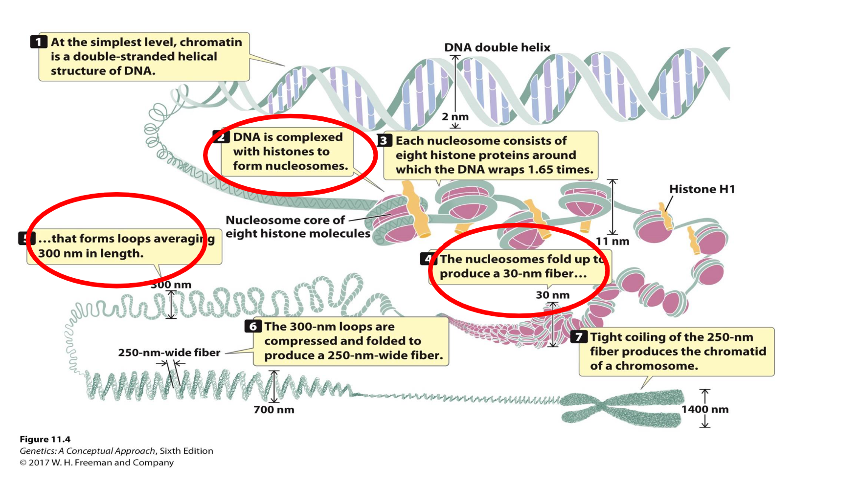
What is the second level of DNA packaging in eukaryotes?

  • The second level of DNA packaging involves the further coiling and compaction of nucleosomes into a 30-nm chromatin fiber.

  • Nucleosomes are arranged into a solenoid or zigzag structure, producing a thicker fiber.

  • Histone H1 helps stabilize nucleosome organization and promotes higher-order chromatin compaction.

  • This structure is approximately 30 nm in diameter, compared with the approximately 11-nm nucleosome fiber.
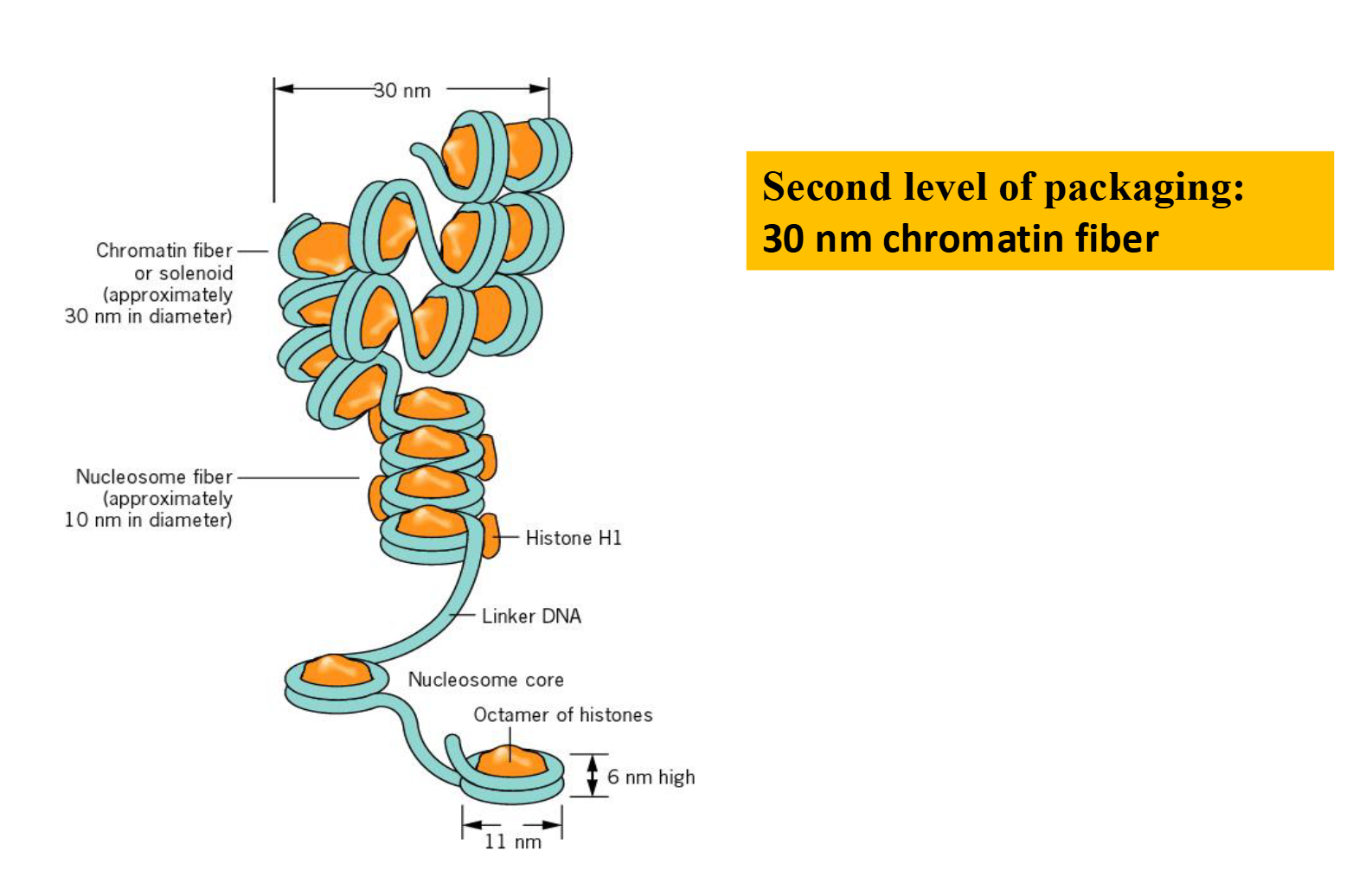

<ul><li><p>The second level of DNA packaging involves the further coiling and compaction of <strong>nucleosomes</strong> into a <strong>30-nm chromatin fiber</strong>.</p></li><li><p>Nucleosomes are arranged into a <strong>solenoid or zigzag structure</strong>, producing a thicker fiber.</p></li><li><p><strong>Histone H1</strong> helps stabilize nucleosome organization and promotes higher-order chromatin compaction.</p></li><li><p>This structure is approximately <strong>30 nm in diameter</strong>, compared with the approximately 11-nm nucleosome fiber.</p></li></ul><p></p>
56
New cards

What is the third level of DNA packaging in eukaryotes?

  • The third level of DNA packaging involves the formation of chromatin loops from the 30-nm chromatin fiber.

  • These loops attach to a chromosome scaffold composed of non-histone proteins.

  • The resulting looped domains are approximately 300 nm in diameter.

  • These structures undergo additional folding and condensation to form interphase chromosomes.

  • Interphase chromosomes are less condensed than mitotic chromosomes but remain organized within the nucleus.


<ul><li><p>The third level of DNA packaging involves the formation of <strong>chromatin loops</strong> from the 30-nm chromatin fiber.</p></li><li><p>These loops attach to a <strong>chromosome scaffold</strong> composed of <strong>non-histone proteins</strong>.</p></li><li><p>The resulting looped domains are approximately <strong>300 nm in diameter</strong>.</p></li><li><p>These structures undergo additional folding and condensation to form <strong>interphase chromosomes</strong>.</p></li><li><p>Interphase chromosomes are less condensed than mitotic chromosomes but remain organized within the nucleus.</p></li></ul><p></p>
57
New cards

How does chromatin become further condensed during mitosis?

  • During mitosis, chromatin undergoes additional folding and condensation.

  • The looped chromatin domains become more tightly packed, producing highly condensed metaphase chromosomes.

  • According to the diagram on slide 20:

    • DNA double helix: 2 nm.

    • Nucleosome fiber: Approximately 11 nm.

    • Chromatin fiber: 30 nm.

    • Chromatin loops: Approximately 300 nm.

    • Condensed chromatid: Approximately 700 nm.

    • Metaphase chromosome: Approximately 1,400 nm wide.

  • This extensive packaging allows chromosomes to be organized and separated during cell division.


58
New cards

Useful Diagram

knowt flashcard image
59
New cards

What are the three main levels of DNA packaging in eukaryotic chromosomes?

  • First level — Nucleosome formation:

    • DNA wraps around histone octamers to form approximately 11-nm nucleosomes.

  • Second level — 30-nm chromatin fiber:

    • Nucleosomes coil and compact into a 30-nm chromatin fiber.

  • Third level — Chromatin loops:

    • The 30-nm fiber forms approximately 300-nm looped domains attached to a scaffold of non-histone proteins.

  • During mitosis, further condensation produces highly compact metaphase chromosomes.