32. surgery for ear disease
 Call Kai
 Learn
 Practice Test
 Spaced Repetition
 Match
 Flashcards
 Knowt Play
1/18

There's no tags or description
Looks like no tags are added yet.
19 Terms
what causes aural hematomas?
secondary to trauma from ear disease
shear forces → dead space, fills with blood
violent head shaking, ear scratching
ALWAYS do a thorough ear exam → must treat underlying disease
outcomes of doing nothing to treat aural hematoma
clot granulates
cavity fibroses, cartilage ossifies
disfigurement ± chronic irritation
aural hematoma treatment — drain ± steroids
drain only
clip & prep in sterile fashion!
least effective treatment; typically recurs, multiple treatments
can introduce infection to the site
+ steroids
conflicting results; will likely require multiple treatments
what type of surgical drain is preferred for aural hematomas? which are not?
preferred → closed suction drain
penrose drain (not recommended)
teat cannula (messy everywhere)
aural hematoma treatment — incision drainage
can use 1st line; definitely for chronic, severe, or recurrent cases
S-shaped incision through skin
remove clot and fibrin, flush
place large, full-thickness, non-resorbable, interrupted mattress sutures over entire convex surface of pinna
MUST be parallel to vasculature
ear ends up thickened
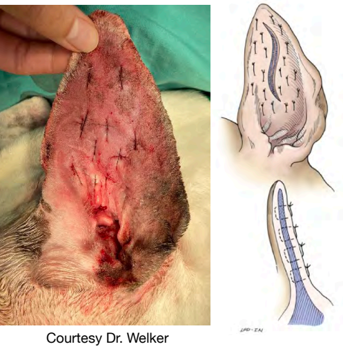
aural hematoma — post-op care & complications
need to bandage ear (compression)
fold ear onto dorsum of head
change every 2-3 days until skin adhering down
treat underlying cause
complications
infection
recurrence
what is the most common neoplasia of the pinna?
squamous cell carcinoma in cats (sunny climates)
white cats have increased risk
tend not to spread if only on pinna
other tumors are possible
hemangioma/hemangiosarcoma
basal cell tumors, mast cell tumors, histiocytomas, etc.
pinna neoplasia — diagnosis
FNA or incisional biopsy
staging dependent on tumor type
pinna neoplasia — treatment
subtotal or total pinnectomy
amputate pinna at least 1-2 cm from tumor
cauterize vessels
pull skin on concave surface over cut edge and suture
don’t suture cartilage
diagnostic work up for diseases of the ear canal / bulla
otoscopy: should be done on every animal presenting for ear disease (+ routine exam)
palpation: is ear canal ossified? (indicates end stage disease)
otic cytology and biopsy (try to obtain from horizontal canal)
FNA, biopsy, impression smear if mass seen in canal
diagnostic imaging: CT ideal
how is the bulla anatomically different in dogs vs. cats?
septum of the tympanic bulla is present in dogs & cats, but separates bulla into 2 compartments in cats (communicate medially through a slit-like opening)
what is the most common ear canal neoplasia?
ceruminous gland adenocarcinoma
indications for TECA / TECABO surgery
severe, irreversible chronic obstructive ear disease (end stage disease)
calcification of ear canals
patency of ear canals minimal or absent
neoplasia of the ear canals that extend into the horizontal canal or beyond
post-op complications of TECA surgery
bleeding (retroglenoid/retroarticular vein)
can be life-threatening
facial nerve paralysis or paresis
horner’s syndrome
cats > dogs due to anatomic location of post-ganglionic sympathetic fibers
post-op fistula
can occur if epithelium left in bulla, ear canal not completely removed, or debris left behind
draining sinuses may not appear for months to years
warn owners that dogs will technically be deaf after surgery
where do feline polyps originate?
within auditory tube → usually grow into nasopharynx
from rostral lateral (smaller) compartment of the tympanic bulla → usually grow out the ear canal
can grow in both directions
feline polyps — diagnosis
CBC, chem, UA, FeLV/FIV testing
under general anesthesia
examine ears
evaluation of pharynx via rostral reaction of the soft palate or with a scope
CT scan
feline polyps — removal via traction + medical treatment
steady traction can be applied to polyps for removal
can occur via oral cavity or ear, depending on location
medical treatment
antibiotics: base on culture if associated with ear infection, otherwise broad spectrum
prednisolone (typically 2 wks)
analgesics for 4+ days (e.g. buprenorphine)
indications for VBO surgery
most commonly done in cats for polyps originating in the middle ear
usually try traction 1st
gives access only to middle ear
also undertaken in cases of severe middle ear infections where ear canal is still patent (dogs & cats)
rarely done in dogs; also much more challenging due to location of bulla
feline polyps — post-op complications / prognosis
horner’s syndrome
often resolves within weeks; if still around at 6 wks, likely permanent
vestibular signs (can occur with TECAs as well)
many pre-date surgery due to pressure on labyrinthine structures from material in ear
iatrogenic: can occur from either osteotomy or curettage during polyp removal
unlike TECA, facial nerve paralysis is uncommon