Lecture 2 - Introduction to Microscopy
 Call Kai
 Learn
 Practice Test
 Spaced Repetition
 Match
 Flashcards
 Knowt Play
1/73

There's no tags or description
Looks like no tags are added yet.
74 Terms
Microbe of the Day: Lacticaseibacillus casei
Type & Metabolism: Genomically diverse, facultative heterofermenter (produces lactic, acetic, formic, and propionic acids).
Food & Preservation: Used in cheeses, dairy, kimchi, and pickles; its lactic acid acts as a natural preservative.
Probiotic Use: Used to treat IBS and other digestive disorders.
Spoilage: Major spoilage organism in beer and wine.

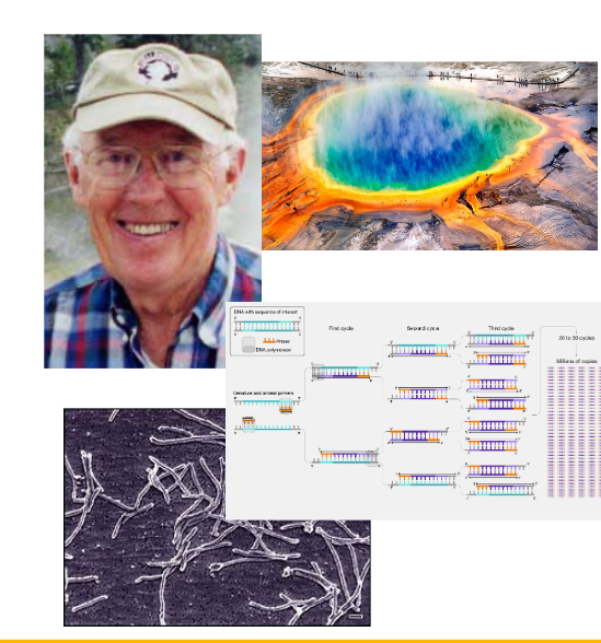
Microbe of the Day: Thermis aquaticus
Type & Growth: First recorded thermophilic bacterium (optimal growth temperature: 65–70°C).
Biotech Significance: Source of Taq DNA polymerase, an essential enzyme used in PCR (polymerase chain reaction).
Discovery: Discovered in ~1969 by Thomas Brock.
What is microscopy?
Scientific technique to study small objects to small to be seen by the naked eye.
What is magnification in microscopy?
The apparent visual increase in the size of an object (no theoretical limit).
What is resolution (resolving power) in microscopy?
The ability to distinguish between two separate objects.

What is the resolution limit?
The minimum distance between two separate objects so you can tell them apart.
What is detection in microscopy?
The ability to observe a signal. (e.g light emitted from a single fluorescent molecule).
What happens when light enters a new medium?
It bends (refracts), the refraction of index measured the relative of light into that medium.

How do lenses create different magnifications?
Lenses refract light into a single focal point.
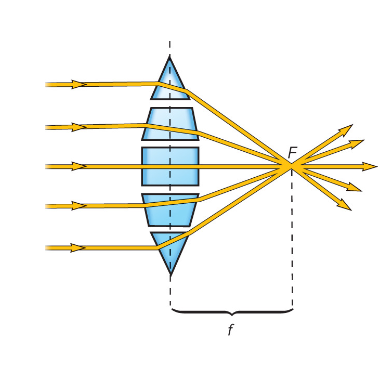
How does focal lenght effect magnification?
The shorter the focal length (lens to focal point), the higher the magnification.

What type of microscope was first invented and still widely used?
Light microscopes.
What is the resolution limit for light microscopes? Why?
Approximately 0.2 μm.
What is the function of the Bright-Field Microscope?
- Examines both stained and unstained specimens by producing a dark image against a brighter background.
- Multiple objective lenses.
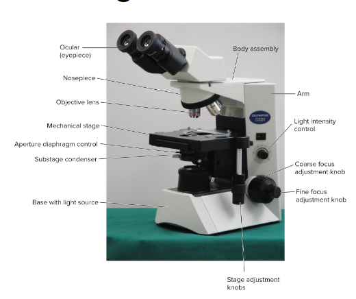
How can we calculate the total magnification of a Bright-Field Microscope?
Multiply the magnification of the ocular lens by the magnification of the objective lens.
How does diffraction lead to a resolution limit?
As light passes from a specimen through lenses, optical diffraction causes the waves to interfere and blur of two close objects, creating a resolution limit that makes it difficult to visually separate two closely spaced objects.

How do we measure the resolution limit?
Microscopic objects emit as a circle of blurred light, if the distance between 2 objects > radius, you see distinct objects. If D < R, you see a merged blob (resolution limit).

How do scientists reduce the resolution limit?
Shorter wavelengths of light lead to smaller diffraction patterns and thus greater resolution (e.g. blue filters)
What happens to brightness when light waves interfere?
Constructive (in phase): Brightness increases
Destructive (out of phase): Brightness decreases

What is working distance?
The distance between lens and cover glass when the specimen is in sharp focus.

What is the relationship between a microscopes numerical apertures/light-gathering ability (NA) and resolution. {Niche}
Proportional.

What is the purpose of adding immersion oil between the slide and objective lenses?
Immersion oil has a similar refraction of index to glass. Replacing air with immersion oil, reduces light refraction and reflection at the surfaces. This allows more light rays to enter the objective lens, increasing the numerical aperture and improving resolution.

Why are unpigmented microbes hard to see under a regular microscope?
Transparent and not colored; there is not enough contrast to distinguish them from the background.
What 3 Microscopes can observe unstained specimens?
1 Dark-field microscope
2 Phase-contrast microscope
3 Differential interference contract (D I C) microscope
How does a Dark-field microscope work? What do you see?
- It blocks direct light from reaching the objective lens and only captures light that bounces off the specimen.
- Produces a bright image of the specimen against a dark background.

When is a Dark-Field Microscope most useful?
Useful for observing living, unstained specimens and identifying bacteria with unique morphologies (e.g Treponema pallidum).

How does a Phase-contrast microscope work?
Light slows down slightly as it passes through denser parts of a cell. This microscope converts those small light-speed changes into differences in brightness, making unstained cells appear darker against a lighter background.

When is a Phase-Contrast Microscope most useful?
Observing microbial movement and internal structures.

How does a Differential Interference Contrast (DIC) Microscope work?
Detects differences in refractive indices and thickness within a specimen, creating a bright, 3D-like image where the specimen appears to "pop out" from the background.

When is a Differential Interference Contrast (DIC) Microscope most useful?
Viewing 3D surface features and fine internal organelles in living cells. It provides sharper edges and is useful for observing cell walls, endospores, and vacuoles.

Who created the Phase-Contrast Microscope and hid Jewish people from the Nazi’s?
Lili Bleeker actually made it, but inventor Frits Zernike took credibility and won the Nobel Prize in 1953.

What is the main purpose of fluorescence microscopy?
A technique that uses fluorescent tags to label specific cellular components.

How does fluorescence microscopy generate a visual image?
You shine light (lamp/lazer) of a specific wavelength (excitation light) at the sample. This excites the fluorescent tag, causing it to glow and emit light of a different wavelength (emission light).

What types of fluorescent tags/probes can be used?
1. Dyes: Chemical stains (e.g., fluorescein, rhodamine) that color specific cell parts.
2. DNA Probes: Fluorescent tags that attach to specific genetic sequences.
3. Antibodies: Fluorescently tagged immune proteins that lock onto specific targets.
4. GFP (Green Fluorescent Protein): A protein originally found in jellyfish (Aequorea victoria) that can be genetically fused directly to a cell's own proteins to track them live.
Where does GFP come from and how is it used in living cells?
GFP was originally discovered in the jellyfish Aequorea victoria. Scientists attach its genetic code directly to a target protein's gene, allowing them to track that protein's location and movement inside a live cell.
Who discovered fluorescent proteins?
Discovery was awarded the Nobel Prize in 2008 (Osamu Shimomura, Martin Chalfie and Roger Y. Tsien)

What is the biggest advantage of using fluorescent tags over general cell stains?
Specificity. Instead of staining the entire cell, you can light up only the single protein, gene, or organelle you want to study.
What makes confocal microscopy different from standard fluorescence microscopy?
It uses a laser beam for precise illumination and a pinhole aperture to block out-of-focus light, producing much sharper images.

Does confocal fluorescent only produce a single image?
Algorithms can then “reassemble” these images to produce high-resolution 3D images. Useful for bioflims.

Why is staining important in microscopic examination of bacteria and archea?
- Increase visibility
- Emphasis features
- Preserves specimen for future
What is cell fixation and why is it used?
Fixation preserves a cell's structure and shape by inactivating enzymes and toughening cellular components, so they can be viewed under a microscope.
How does heat fixation work, and what is its main drawback?
It uses heat to quickly lock the cell's overall shape onto a glass slide.
Drawback: Preserves overall morphology but destroys fine subcellular structures.
How does chemical fixation work, and why is it used instead of heat?
It uses chemical solutions to preserve cells gently. Protects fine cellular substructures and morphology (e.g., using formalin).
What is simple staining, and what does it tell you?
Simple staining uses only a single dye. It is used to easily see the size, shape, and arrangement of bacterial cells under a microscope.

What is the main difference between basic dyes and acidic dyes?
1. Basic dyes: Have a positive charge (+) and bind to negatively charged (-) cell components.
2. Acidic dyes: Have a negative charge (-) and bind to positively charged (+) cell components.

Why do basic dyes stain bacterial cells so well?
Most bacterial cell surfaces, proteins, and nucleic acids are negatively charged (-). Because opposites attract, positively charged (+) basic dyes stick directly to them.
What is differential staining?
Unlike simple staining (which uses one dye), differential staining uses multiple steps/dyes to divide bacteria into different groups based on their physical or structural properties.
What feature does the Gram stain divide bacteria by?
Divides bacteria into Gram-positive (stain purple) and Gram-negative (stain pink/red) based on cell wall structure.
What does Gram-positive mean and show?
Cell Structure: It means the bacterium has a very thick layer of peptidoglycan in its cell wall (and no outer membrane).
Under the Microscope: It shows up Purple.
What does Gram-negative mean and show?
It means the bacterium has a very thin layer of peptidoglycan plus an extra outer membrane.
Under the Microscope: It shows up Pink or Red.
What are the 4 steps in Gram Staining?
Crystal Violet (Primary Stain): All cells stain purple.
Iodine (Mordant): Enhances dye binding; cells remain purple.
Alcohol (Decolorizer): Gram-positive cells remain purple; Gram-negative cells become colorless.
Safranin (Counterstain): Gram-positive cells remain purple (repel counterstain); Gram-negative cells appear red (abosorb dye).

Why do bacteria like Mycobacterium tuberculosis require Acid-Fast staining?
They have a waxy cell wall (rich in lipids) that acts as a waterproof barrier, preventing standard stains from entering.

How does the Ziehl-Neelsen Acid-Fast method force stain into waxy cells?
Heat is used to "melt" the waxy layer so the dye (carbol fuchsin) can penetrate. Once cooled, the waxy layer traps the dye inside so acid cannot wash it out.
What are 3 specific stains methods to observe specific microbial structures?
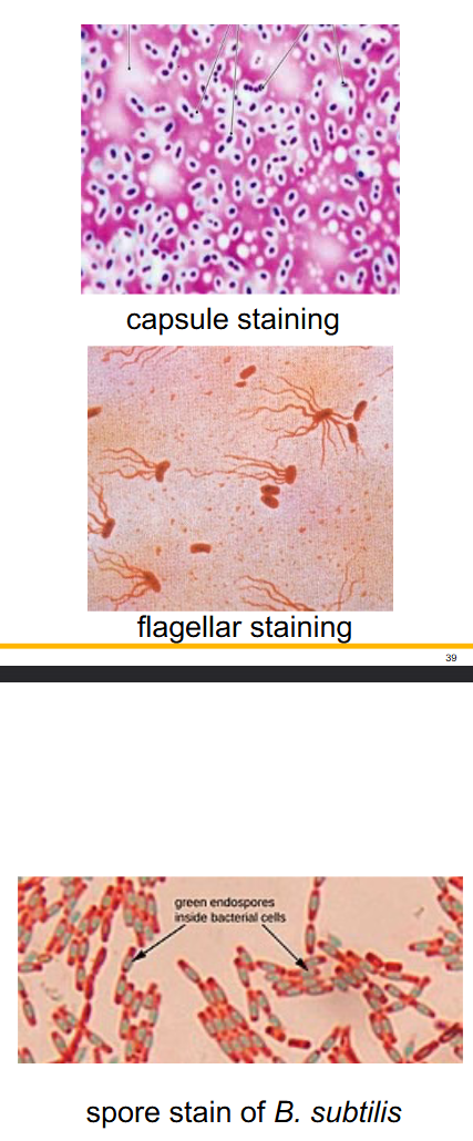
Capsule staining: Negative stained background, to show colourless capsules.
Flagella staining: Use of mordants (to increase thickness).
Endospores staining: Uses heat to drive stains into resistant structures (like endospores).

Electron microscopy: Why does an electron microscope have much higher resolution than a light microscope?
It uses electrons instead of light. The wavelength of an electron beam is 100,000 times shorter than visible light, allowing you to see much smaller details.

How does a Transmission Electron Microscope (TEM) form an image?
Uses a focused beam of electrons that pass through a very thin specimen.
Denser regions scatter electrons more, appearing darker.
Why must TEM operate inside a high vacuum?
Reduce scatter, producing clearer image.
Why must TEM specimens be cut extremely thin?
Electrons cannot penetrate thick, solid objects. If the specimen is too thick, the electron beam gets completely absorbed or scattered.
Why are heavy metals used to stain TEM specimens?
Contrast: Heavy metals scatter electrons well, creating clear dark regions.
Protection: They protect the delicate biological sample from being destroyed by the high-energy electron beam.
How does negative staining work to prepare for TEM, and what is it used for?
Specimen is spread in a thin film with heavy metal salts, producing a bright image against a dark background. Useful for viruses and surface structures.

What is shadowing to prepare for TEM, and what is it useful for?
Specimen is coated with a heavy metal on one side, creating a 3D effect. Useful for small parts like flagella.

How does freeze-etching work to prepare for TEM?
Cells are rapidly frozen in liquid nitrogen, then fractured to reveal internal structures, particularly membranes.

What is the main purpose of a Scanning Electron Microscope (SEM)?
To create realistic, detailed 3D images of a specimen's surface features.
How does an SEM create an image?
A narrow electron beam is scanned back and forth across the surface, and exites secondary electrons. They are detected, and converted into a 3d digital picture, instead of 2D like TEM.

What is the main difference between TEM and SEM?
TEM (Transmission): Electrons pass through the specimen to show internal structure (like an X-ray).
SEM (Scanning): Electrons bounce off the surface to show 3D surface shape (like a photo).

What is Cryo-Electron Microscopy (Cryo-EM) mainly used for?
Creating 3D models of biomolecules (like proteins, lipids, and nucleic acids).

How is the final 3D image constructed?
Multiple 2D TEM images of the frozen sample, combined into a 3D structure.

How are samples prepared for Cryo-EM, and do they require staining?
Plunge Freezing (Vitrification): Hydrated (wet) samples are rapidly frozen in liquid nitrogen, protected from high energy electrons. No staining is needed, similar, but not freeze-etching.
How does Scanning Probe Microscopy (SPM) work?
Moves an extremely sharp tip (probe) across the surface of a sample to map out its features. It passes an electrical current (tunneling current) between the probe tip and the sample. Can map single atoms.

How does an Atomic Force Microscope (AFM) detect surface shapes?
The probe scans the surface, and the up-and-down movement of the cantilever due to surface topography is detected by a reflected laser beam.
Can image protein-protein and protein-DNA interactions, though imaging is slow.

What is the main idea behind Expansion Microscopy?
Instead of building a bigger or more complex microscope, it physically swells/expands the sample itself (embedded in polyelectrolyte hydrogel and water) so that tiny structures become large.

Which of the following types of microscopy is best suited for visualizing swimming behavior in live bacteria?
Correct Answer: Phase contrast microscopy (D)
Phase contrast microscopy (D) enhances contrast by converting differences in refractive index between unstained live cells and water into visible light/dark variations, making it the best choice for tracking live bacterial motility.
Gram-stained light microscopy (A) requires heat-fixing, which kills the bacteria.
Live bright-field microscopy (B) offers very poor contrast for small, transparent, unstained bacteria.
Electron microscopy (C) requires sample fixation and a high vacuum, so organisms cannot be alive.
Which of the following types of microscopy is best suited for visualizing the location of protein complexes in live bacteria?
Correct Answer: B) fluorescent microscopy
Fluorescent microscopy (B) uses specific fluorescent tags (like GFP fusions) to target and reveal the precise spatial location of specific protein complexes within live cells.
Dark-field microscopy (A) and Phase contrast microscopy (C) provide contrast for overall cell morphology and motility, but cannot selectively pinpoint specific protein complexes.
Electron microscopy (D) requires cells to be fixed, dehydrated, and placed under a vacuum, making live observation impossible.
Which of the following types of microscopy is best-suited for observing molecular-level structures of macromolecular protein complexes?
Correct Answer: C) cryo-electron microscopy
Cryo-electron microscopy (C) preserves macromolecular protein complexes in a frozen-hydrated native state and provides near-atomic (molecular-level) structural resolution.
Dark-field microscopy (A) is an optical light microscopy technique used to observe small live organisms, with resolution limited to the wavelength of light.
Fluorescent microscopy (B) is ideal for tracking specific tagged proteins inside whole cells, but it cannot resolve individual molecular-level protein structures.
Electron microscopy (D) is a broad category that includes TEM and SEM, but cryo-EM is specifically tailored for macromolecular protein complexes without damaging them during sample preparation.
Which of the following types of microscopy is best-suited for observing intracellular movement of live organelles/processes in unstained organisms?
Correct Answer: A) differential interference microscopy or C) Phase contrast microcopy
Differential interference contrast (DIC) microscopy (A) provides high-resolution, pseudo-3D images using polarized light. Its fine "optical sectioning" ability makes it ideal for viewing internal 3D structures and live intracellular movement without producing distracting halo artifacts.
Phase contrast microscopy (B) generates bright "halos" around thick structures, which obscure delicate internal organelles and details.
Bright field microscopy (C) lacks contrast for transparent, unstained internal structures.
Transmission electron microscopy (D) kills and fixes cells in a vacuum, preventing the observation of live processes.