audiology quiz 1
 Call Kai
 Learn
Learn Practice Test
 Spaced Repetition
 Match
Match Flashcards
 Knowt Play
1/82

There's no tags or description
Looks like no tags are added yet.
Name | Mastery | Learn | Test | Matching | Spaced | Call with Kai | Chat |
|---|
No analytics yet
Send a link to your students to track their progress
83 Terms
scala vestibuli
upper most layer of cochlea, filled with perilymph
scala media
middle layer of cochlea, filled with endolymph
scala tympani
bottom layer of cochlea, filled with perilymph
outer hair cells
12,000 in three rows, primarily efferent, make contact with tectorial membrane
inner hair cells
3,500 in one single row, afferent
high frequency waves of basilar membrane
concentrated near the base
medium frequency waves in basilar membrane
cause maximum vibration near middle of membrane
low frequency waves of basilar membrane
concentrated near the apex of basilar membrane
conductive hearing loss
issue in outer or middle ear (ear canal)
sensorineural hearing loss
issue with the nerves
mixed hearing loss
both conductive and sensorinueral
factors that affect classroom acoustics
noise, directionality, distance, reverberation
x-axis of audiogram
pitch/frequency
y-axis of audiogram
hearing threshold level (decibels)
db SPL (sound pressure level)
used in measuring hearing aid performance
db IL (intensity level)
used in calibration of audiometers
db HL(hearing level)
used on hearing tests/audiograms
db SL (sensation level)
used for speech perception testing
human hearing range
20-20,000Hz
normal decibel hearing range
-10-20dB
mild hearing loss range dB
21-40
moderate hearing loss range (dB)
41-70
severe hearing loss range dB
71-90
frequency
number of complete cycles per unit of time
period
amount of time needed to complete one cycle
what type of wave is sound
longitudinal
transverse wave
particle movement perpendicular to energy
longitudinal wave
particle movement parallel to energy
cerumen
ear wax
stenosis
narrow ear canal, easy for wax or water to get trapped
acute external otitis
swimmer’s ear
atresia
little or no ear formation, no ear canal
microtia
very small ear, may or may not have open ear canal
Perichondritis
infection and inflammation of outer ear cartilage (red, swollen, hot, tender)
otitis media
middle ear infection
serous otitis media
effusion is thin and clear
mucoid otitis media
thicker, yellowish effusion
suppurative otitis media
thick, yellow pus
tympanic membrane perforation
hole/tear in the eardrum, can be caused by a foreign object, blow to the head, scuba diving or flying, or otitis media
cholesteatoma
benign growth behind eardrum filled with skin cells and debris
mastoiditis
infectious invasion of the mastoid bone
ostosclerosis
fixation of the ossicular chain
ankylosis
fixation of only the stapes
vestibular schwannoma
unilateral slow growing tumor of facial nerve
meniere’s disease
inner ear disorder usually seen as hearing loss, vertigo, and tinnitus
noise induced hearing loss
inner ear disorder, repeated exposure leads to progressive loss
presbycusis
inner ear disorder, aging of the ear
otalgia
ear pain
Otorrhea
foul odor emanating from the ear
Occluding/non-occluding
Blocked or not blocked
acoustic reflex test results in patients with auditory neuropathy spectrum disorder
elevated or absent
otoacoustic admissions test results in patients with auditory neuropathy spectrum disorder
normal
Auditory Brainstem Response results in patients with auditory neuropathy spectrum disorder
absent
waardenberg syndrome
10 most common syndromes, pigment issues
a rare genetic condition that causes hearing loss and changes in the coloring of the hair, skin, and eyes
treacher-collins syndrome
10 most common syndromes, craniofacial
affects the development of the bones and tissues in the face
pendred syndrome
10 most common syndromes, thyroid
causes early hearing loss and an enlarged thyroid gland
usher syndrome
10 most common syndromes, causes deaf-blindness
branchio-oto-renal syndrome
10 most common syndromes, affects the development of tissues in the neck and causes malformations of the ears and kidneys
stickler syndrome
10 most common syndromes: genetic connective tissue disorder caused by mutations in genes responsible for making collagen - mild to severe hearing loss
alport syndrome
10 most common syndromes, renal
leading to progressive kidney disease, hearing loss, and eye abnormalities
NFII
10 most common syndromes, tumors on facial nerve
Jervell-Lange-Neilson syndrome
10 most common syndrome, cardiac
causes deafness
CHARGE syndrome
10 most common syndromes, affects multiple organ systems
otoscopy
technically not a test but is the visual inspection of the ear via otoscope, verifies the ear canal is not occluded and checks for signs of trauma or infection
AuD becomes familiar with shape and size of patient ear canal
tympanometry
measures the flexibility of the eardrum
“A” tympanogram
normal pressure, normal compliance, seen in normal middle ear
“As” tympanogram
normal pressure, decreased compliance, seen in otosclerosis
“Ad” tympanogram
normal pressure, increased compliance, seen in ossicular discontinuity
“B” tympanogram
flat curve, seen when there is fluid in ME, or Tympanic membrane perforation
“C” tympanogram
negative pressure, normal compliance, seen in eustachian tube obstruction
otoacoustic emissions
assess outer hair functions
transient evoked otoacoustic emissions (TEOAE)
typically used as a screening for children or a crosscheck for adults
uses a broadband click stimulus (not specific to any one frequency)
Distortion Product otoacoustic emissions (DPOAE)
used for diagnostic information, uses two frequencies at 2 different intensities
inverse square law
signal decreases by 6dB for every doubling of distance
air conduction left/right symbols
x, O
air conduction masked left and right symbols
square, triangle
bone conduction symbols left/right
>, <
bone conduction masked symbols left/right
], [
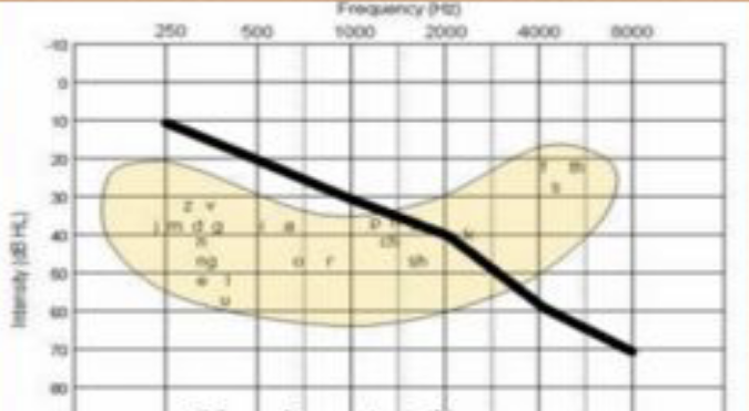
what type of audiogram
sloping

what type of audiogram is this
reverse slope

what type of audiogram is this
cookie bite

what type of audiogram is this
tent shaped

what type of audiogram is this
flat