Applied Immunodiagnostics - Dr. Zimm 10/7
 Call Kai
 Learn
Learn Practice Test
 Spaced Repetition
 Match
Match Flashcards
 Knowt Play
1/38

There's no tags or description
Looks like no tags are added yet.
Name | Mastery | Learn | Test | Matching | Spaced | Call with Kai | Chat |
|---|
No analytics yet
Send a link to your students to track their progress
39 Terms
⭐ 1. Q: How do plasma and serum differ?
Plasma is the fluid portion of unclotted blood and contains clotting proteins, whereas serum is obtained after blood clots and lacks fibrinogen and most coagulation proteins.
🔎 What this means:
Plasma
Collected in an anticoagulant tube
Contains albumin, globulins, clotting factors, and fibrinogen
Serum
Collected in a no-additive or serum-separator tube
Contains albumin and globulins
Does not contain fibrinogen
💡 Think: Plasma still has the clotting stuff; serum has already clotted.
2. Q: How are total protein, albumin, and globulin evaluated on a routine chemistry profile?
Total protein and albumin are measured, whereas globulin is calculated by subtracting albumin from total protein.
🔎 What this means:
TP = total protein
ALB = albumin
GLOB = globulin
Globulin is not directly measured:
GLOB = TP − ALB
TP and albumin can be evaluated using either serum or plasma.
3. Q: How does the refractometric method estimate total protein, and what can falsely increase the result?
A refractometer estimates total protein from how much light is refracted by plasma, but lipemia or other increased solids can falsely increase the result.
🔎 What this means:
Plasma comes from a microhematocrit tube.
The lecture associates this with an EDTA lavender/purple-top tube.
Light bending reflects the amount of total solids.
The instrument converts the refractive index into an estimated TP.
💡 Because it measures solids rather than protein alone, extra nonprotein solids can interfere.
4. Q: How is heat-precipitated fibrinogen measured, and what is the major limitation of this method?
Fibrinogen is estimated by subtracting total protein after heating from total protein before heating, but this method can detect increases rather than decreases reliably.
🔎 What this means:
Uses plasma from an EDTA microhematocrit tube
Heat at 56°C for 3 minutes
Heating precipitates fibrinogen.
Compare refractometric TP before vs after heating.
The lecture emphasizes that increased fibrinogen is clinically significant, while decreases are not detectable by this method
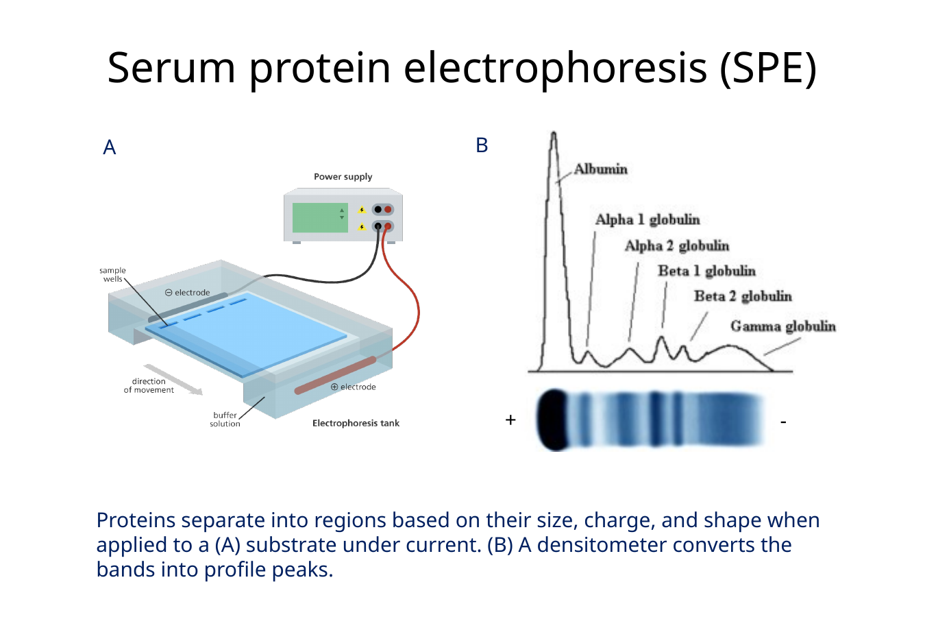
⭐ 5. Q: How does serum protein electrophoresis work, and what information does it provide?
Serum protein electrophoresis separates serum proteins into regions according to their size, charge, and shape, and a densitometer converts the separated bands into profile peaks.
🔎 What this means:
The sample is placed on a substrate under an electrical current.
Different proteins migrate differently, producing distinct protein regions that can then be evaluated for abnormal patterns
Focus on: the separated bands becoming the peaks on the right.
⭐ 6. Q: What major protein regions are seen on serum protein electrophoresis?
The major SPE regions are albumin, α1, α2, β1, β2, and γ globulins.
🔎 What this means:
Important proteins emphasized:
Albumin: migrates the most
α2: haptoglobin and α2-macroglobulin
β1: complement C3 and C4
β2: complement, transferrin, IgM, IgA
γ: primarily IgG
VM 523 Applied Immunodiagnostic…

⭐ 7. Q: What clinically important distinction can serum protein electrophoresis make when evaluating gammopathies?
Serum protein electrophoresis can help differentiate polyclonal from monoclonal gammopathies.
🔎 What this means:
A gammopathy is an abnormality involving immunoglobulins in the globulin region.
The lecture specifically emphasizes recognizing whether the SPE pattern is:
Polyclonal
Monoclonal
⭐ 8. Q: What information is provided by the erythrogram, leukogram, thrombogram, and blood-smear review?
The erythrogram quantifies RBC data, the leukogram quantifies WBC data, the thrombogram quantifies platelet data, and blood-smear review provides qualitative cell-morphology information.
In list form:
Erythrogram
RBC mass indicators
RBC indices
RDW
Reticulocytes
Leukogram
Total WBC concentration
Differential counts
Abnormal cells
Thrombogram
Platelet concentration
MPV
Plateletcrit
Blood smear
Cell morphology
⭐ 9. Q: When is buccal mucosal bleeding time used, and what does it evaluate?
Buccal mucosal bleeding time evaluates platelet-dependent primary hemostasis when a primary hemostatic disorder is suspected despite a normal platelet concentration.
🔎 What this means:
BMBT = buccal mucosal bleeding time
A standardized incision is made and the time until bleeding stops is measured.
It may be prolonged with significant:
Platelet-function defects
von Willebrand factor defects
The lecture emphasizes that BMBT:
Lacks sensitivity
Is user dependent
Is independent of the coagulation cascade
⭐ 10. Q: What do secondary-hemostasis diagnostic tests evaluate?
Secondary-hemostasis tests evaluate coagulation-factor function using in-vitro coagulation testing.
🔎 What this means:
Secondary hemostasis concerns the coagulation factors, rather than platelet function.
The lecture also emphasizes the vitamin K-dependent factors:
II, VII, IX, and X
2, 7, 9, 10
Trick to memorize: 2+7=9 and one more is 10
and notes that specialized immunodiagnostic testing may be needed to detect individual factor deficiencies

⭐ 11. Q: What does tertiary hemostasis refer to?
Tertiary hemostasis refers to fibrinolysis, the processes involved in breaking down fibrin clots.
⭐ 12. Q: How do bone-marrow aspirate cytology and bone-marrow core histology differ in processing?
Bone-marrow aspirates are made into air-dried squash smears for cytology, whereas core samples are fixed, decalcified, paraffin embedded, sectioned, and stained for histology.
Aspirate → cytology
Stain like blood smears
Faster turnaround
Core → histology
Place in formalin or B5 fixative
Decalcify
Paraffin embed
Section
H&E stain
Longer turnaround

13. Q: How are aspirates, biopsies, and necropsy samples associated with cytology, histopathology, and anatomy?
An aspirate is evaluated by cytology
A biopsy is evaluated by histopathology
A necropsy involves gross anatomy.
The lecture also reminds you that hematopoietic evaluation is not limited to bone marrow; organs such as the thymus and spleen may also be important.
14. Q: What two major immunologic principles are exploited by immunodiagnostic tests?
Immunodiagnostic tests can measure an animal’s specific adaptive immune response or use manipulated immune reagents to detect antigens or components of that immune response.
🔎 Essentially you can look for:
The patient's response to something, such as antibodies against a pathogen.
The thing itself, such as pathogen antigen, using antibodies as diagnostic reagents.
🟡 LOWER PRIORITY 15. Q: What is screening, and when are screening programs useful?
Screening is presumptive testing of apparently healthy individuals to separate those likely to have disease from those likely not to have disease, and it is useful when early detection improves treatment or reduces transmission.
🔎 What this means:
Veterinary examples include screening for:
Brucellosis
Tuberculosis
Equine infectious anemia
Heartworm disease
16. Q: What is serology, and what can it be used for?
Serology basically diagnostic testing that using antibodies/antigens in a body-fluid sample to look for evidence of disease or immune response, usually involving serum.
serology is basically immune testing from serum/body fluids.
🔎 What this means:
Uses emphasized include:
Infectious-disease diagnosis
Epidemiology and disease control
Monitoring vaccine responses
Characterizing some cancer cells
Autoimmune and endocrine disease diagnosis
Blood typing/crossmatching
⭐ 17. Q: Why does finding pathogen-specific antibody not necessarily prove that an animal currently has an infection?
Antibodies can persist in serum after they are produced, so detecting antibody may reflect previous exposure rather than current infection.
18. Q: What patient samples and antibody reagents are commonly used in serologic testing?
Serologic tests commonly use serum or other body fluids as the patient sample and may use monoclonal antibodies or antiglobulins as antibody reagents.
🔎 What this means:
Serum or other body fluids contain the material being tested and can be frozen for later testing.
Monoclonal antibody: an antibody produced from a single clone of cells, so the antibodies are chemically uniform.
Antiglobulin: an antibody that binds to another immunoglobulin (antibody).
VM 523 Applied Immunodiagnostic…
⭐ 19. Q: How does an immunodiagnostic test differ when detecting antibody versus detecting antigen?
Antibody tests use known pathogen antigen to capture the patient’s antibodies, whereas antigen tests use known antibodies to capture antigen in the patient sample.
⭐ 20. Q: How does a radioimmunoassay work, and how is the result interpreted?
A radioimmunoassay is a highly sensitive competitive assay in which patient antigen competes with radioactive antigen for antibody binding
🔎 What this means:
For antigen testing:
Patient sample + specific antibody
Add radiolabeled antigen
Both compete for antibody binding
Measure radioactivity
High radioactivity → low patient antigen
Low radioactivity → high patient antigen
Common uses include hormone measurement and trypsin-like immunoreactivity (TLI) for evaluating exocrine pancreatic insufficiency.
⭐ 21. Q: How does a direct fluorescent-antibody test work, and what does it detect?
A direct fluorescent-antibody test uses a fluorescently labeled antibody that binds directly to antigen in a tissue or cell sample.
🔎 What this means:
The lecture specifically says FA tests for antigen.
Example:
Rabies diagnosis
Sample: brain tissue
Reagent: FITC-labeled anti-rabies antibody
Other examples include Listeria, clostridial organisms, and FeLV in infected WBCs.

⭐ 22. Q: How does indirect fluorescent-antibody testing differ from direct fluorescent-antibody testing?
IFA uses a fluorescently labeled antiglobulin to detect a primary antibody, making it more sensitive and allowing it to detect antibody in serum or antigen in tissues.
🔎 What this means:
Direct FA
Fluorescent label is on the primary antibody
Primary antibody binds directly to antigen
Fewer layers
Usually detects antigen
Indirect FA / IFA
Primary antibody binds first
A fluorescent antiglobulin binds the primary antibody
Multiple fluorescent secondaries can bind → stronger signal
Can detect antibody in serum or antigen in tissue
Can identify antibody class
If you’re still having trouble understanding it like me:
Think of direct FA vs indirect FA like putting a glow stick on the target (direct) vs. putting glow sticks on a person who is already holding the target (indirect)
Direct fluorescent antibody = the antibody itself is glowing
You make an antibody that specifically recognizes that suspect and attach a fluorescent glow tag directly to that antibody.
Indirect fluorescent antibody = a second glowing antibody detects the first antibody
The first antibody binds its target. Then you add a second fluorescent antibody called an antiglobulin, which recognizes and binds to the first antibody.
⭐ 23. Q: How does an indirect ELISA detect antibody in a patient sample?
ELISA = enzyme-linked immunosorbent assay. It is an immunologic test that uses antigen-antibody binding, then an enzyme reaction that produces a visible color change so you can tell whether the target was present.
An indirect ELISA uses known antigen to capture patient antibodies, then an enzyme-labeled antiglobulin binds those antibodies and produces a color change after substrate is added.
In list form:
Known antigen is attached to the plate.
Matching patient antibody binds the antigen.
An enzyme-labeled antiglobulin binds the patient antibody.
Substrate is added.
Color change indicates that the patient had antibody against that antigen.
The reason it is called indirect is that the detectable label is not on the patient antibody itself. A second antibody binds to the patient antibody and carries the enzyme label.

24. Q: Can ELISA detect both antigen and antibody, and what veterinary examples are given?
ELISA can be configured to detect either antigen or antibody depending on the assay design.
🔎 What this means:
Antigen examples
Canine heartworm
FeLV
Canine parvovirus
Antibody examples
FIV
Bovine respiratory syncytial virus
Hog cholera
Streptococcus equi SeM protein
⭐ 25. Q: What is immunohistochemistry used for?
Immunohistochemistry uses antibodies to visualize specific antigens within histologic tissue sections and can identify pathogens or characterize neoplasia.
🔎 What this means:
The lecture example shows a puppy with diarrhea.
A pathologist saw bacteria in colonic crypts and used an antibody specific for Campylobacter.
Campylobacter antigen staining in the crypts confirmed the diagnosis.
⭐ 26. Q: How does PCR for Antigen Receptor Rearrangement help diagnose lymphoma?
PARR = PCR for Antigen Receptor Rearrangement, it is a special PCR test used to look for clonal populations of lymphocytes, which can support a diagnosis of lymphoma
Different normal lymphocytes generally have different antigen-receptor DNA arrangements.
Now imagine lymphoma 🫠
Lymphoma is a neoplasm of lymphocytes.
A neoplasm starts when one abnormal cell proliferates repeatedly.
So now instead of having:
🟢 3481
🔵 9572
🟣 1026
🟠 7734
you might have:
🔴 3481
🔴 3481
🔴 3481
🔴 3481
🔴 3481
🔴 3481
They're copies descended from the same original lymphocyte, so they share the same antigen-receptor DNA arrangement.
That's what clonal population means:
A bunch of cells derived from the same original cell.
So what does PARR actually do?
PARR uses PCR to examine the DNA encoding the lymphocytes' antigen receptors.
Lots of different arrangements
→ more consistent with a mixed population of lymphocytes
One dominant identical arrangement
→ suggests a clonal lymphocyte population
→ supports lymphoma
Again. ⭐ 26. Q: What is PARR, and how can it help diagnose lymphoma?
PARR is a PCR test that looks for a large population of lymphocytes with the same antigen-receptor DNA pattern, which supports lymphoma.
🔎 What this means:
PARR = PCR for Antigen Receptor Rearrangement.
Normal B and T lymphocytes have different antigen-receptor DNA arrangements.
In lymphoma, many lymphocytes are copies of the same original abnormal cell, so they share the same receptor DNA pattern. This is called a clonal population.
Many different DNA patterns? → mixed lymphocyte population
One dominant identical pattern? → clonal population → supports lymphoma
It can be performed on fluid, cytology slides, biopsy samples, or necropsy samples; the slide gives canine performance as 80% sensitivity and 94% specificity
27. Q: What are disposable immunoassays, and how do immunofiltration and immunochromatography differ?
An immunoassay is a test that uses antigen–antibody binding to detect something.
Disposable just means it is a single-use test device.
The lecture says these tests work by the sample wicking through a porous material and lists two types:
Immunofiltration
Immunochromatography
Immunofiltration captures the target before labeling it, whereas immunochromatography labels the target first and captures it later at a test line.
Immunofiltration: ex. snap tests
Imagine the thing you are trying to detect, such as heartworm antigen, is a package.
You have special antibodies that recognize that package.
Immunofiltration = catch it first, label it afterward
Imagine the package immediately reaches a checkpoint containing a capture antibody.
A capture antibody is basically an antibody fixed in place whose job is:
“If my target comes through here, I’m grabbing it.”
So:
1. Sample is added.
2. Target antigen gets caught by the capture antibody.
3. Then a labeled antibody is added and binds the captured antigen.
4. A visible color develops if the target is present.
Catch first → label second.
Immunochromatography: ex. rapid bacc urine test, covid rapid test, pregnancy tests
This is the style that looks more like a rapid test strip.
Now imagine the package first encounters a colored antibody while it is traveling.
So:
1. Sample containing antigen is added.
2. Antigen binds a colored/labeled antibody.
3. The antigen-antibody complex travels along the strip.
4. A second antibody at the test line catches that complex.
5. The accumulated colored complexes create a visible line.

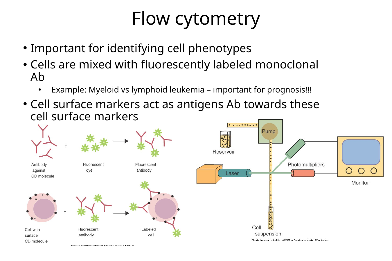
⭐ 28. Q: How does flow cytometry identify cell phenotypes, and what veterinary condition can it help characterize?
Flow cytometry figures out what kind of cells you have by looking at the “ID tags” on their surfaces.
It identifies cell phenotypes using fluorescently labeled monoclonal antibodies that recognize cell-surface markers, and gives myeloid vs lymphoid leukemia as the clinical example.
Cell phenotype basically means what type of cell it is based on the markers it displays.
So flow cytometry is basically asking:
“What name tags are these cells wearing?”
Step 1: Get your cells
You start with a cell suspension, meaning the cells are floating separately in liquid rather than being stuck together in a chunk of tissue.
Step 2: Add fluorescent antibodies ✨
You add fluorescent monoclonal antibodies that recognize particular cell-surface markers aka a specific antigen
Step 3: Run the cells through the machine
The cells move through the flow cytometer, where their fluorescence is detected.
So imagine:
🟢 Green glow → this cell has marker A
🟠 Orange glow → this cell has marker B
Now you can tell what kind of cells are in the sample based on which fluorescent antibodies stuck to them.
———————————————————————-
Why is this useful for leukemia?
Suppose you know an animal has leukemia, but you need to figure out what kind of cells are cancerous.
Flow cytometry can examine their surface markers and help determine whether the abnormal cells have a:
Myeloid phenotype
or Lymphoid phenotype
Those colors tell you which surface markers it has, which helps identify what type of cell it is.
⭐ 29. Q: How does immunodiffusion produce a visible precipitation reaction?
Antigen and antibody move through agar toward each other. If they meet in the right amounts (at optimal proportions), they form a big enough network to become visible as a white line aka the precipitate
💡 Analogy: making a chain-link fence
Think:
Antigen and antibody = two types of fence pieces.
You need the right number of both pieces to build a big fence.
Too many of either one leaves lots of loose pieces.
Too much antibody → no optimal lattice
Optimal antigen-antibody ratio → visible white line
Too much antigen → no optimal lattice

⭐ 30. Q: How is single radial immunodiffusion used to evaluate failure of passive transfer?
Passive transfer means receiving ready-made antibodies from another individual instead of making them yourself.
For a newborn animal, this usually means getting maternal antibodies, especially IgG, from colostrum.
What is failure of passive transfer?
Failure of passive transfer means the newborn did not receive or absorb enough maternal antibodies.
Now what is SRID doing?
SRID = single radial immunodiffusion.
This test uses:
agar containing anti-IgG
the animal’s serum
known standards
and is used to measure IgG for evaluating failure of passive transfer.
You make a little well and put the newborn’s serum into it.
The IgG in the serum starts spreading outward in all directions through the agar.
That is why it is called radial immunodiffusion:
radial = spreading outward in a circle
As the IgG spreads outward, it meets the anti-IgG in the agar.
When they meet in the right proportions, they form a visible precipitation ring.
Then you compare that reaction with samples containing known IgG concentrations.
So the test answers:
“How much IgG does this newborn have?”
If the IgG is too low → that supports failure of passive transfer.
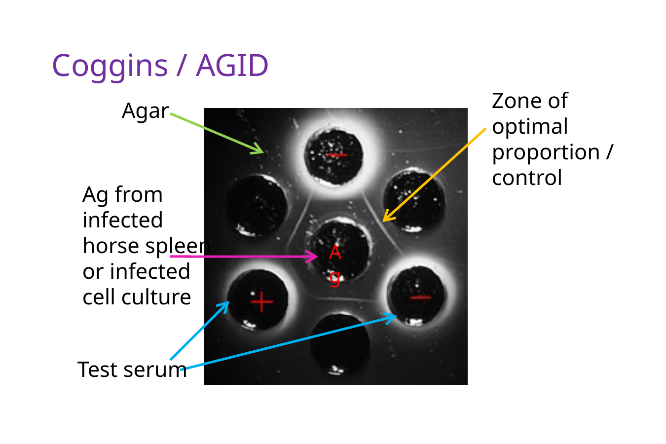
⭐ 31. Q: What does the Coggins test detect, and why is serum preferred?
The Coggins test is an AGID test used for equine infectious anemia (EIA).
AGID = agar gel immunodiffusion.
So it uses the same basic idea as the immunodiffusion card you just learned:
Antigen and antibody diffuse through agar → if they recognize each other and meet in the right proportions → a visible precipitation line forms.
For Coggins, the test is looking for antibodies against equine infectious anemia virus
So essentially:
“Does this horse have antibodies against EIA virus?”
If yes, that means the horse has been exposed/infected with the virus.
Why does the test use serum instead of plasma?
Remember:
Serum = blood fluid after clotting, no fibrinogen
Plasma = blood fluid before clotting, still contains fibrinogen/clotting proteins
The problem with plasma is that fibrin can form.
And fibrin can make its own cloudy/precipitated material in the agar and obscure the zones of optimal proportion
Another reason why Serum is preferred is because plasma contains anticoagulants, i.e. EDTA, which can dilute the blood sample
32. Q: What is agglutination?
Agglutination occurs when bivalent antibodies cross-link particulate antigens and produce visible clumping.
🔎 What this means:
Because each antibody can bind more than one target, it can physically link particles or cells together.
The lecture gives Coombs testing and MAT as examples involving agglutination.
⭐ 33. Q: How does the Coombs test work, and what does a positive test mean?
The Coombs test detects IgG, IgM, or complement attached to a patient’s RBCs, and agglutination after adding antiglobulin reagent indicates a positive result.
🔎 What this means:
The procedure shown uses washed patient RBCs.
Coombs antiglobulin reagent contains antibodies against:
IgG
IgM
and/or C3 complement
If those molecules are already attached to the RBCs, the reagent cross-links them and produces agglutination.
💡 Positive Coombs = patient RBCs are coated with antibody and/or complement.

🟡 LOWER PRIORITY 34. Q: What is the microscopic agglutination test used for?
The microscopic agglutination test is used to detect antibodies against various pathogens.
🔎 What this means:
MAT = microscopic agglutination test.
The professor notes that it is becoming less common because molecular methods such as PCR and sequencing have advanced.