Women's Health MT1
 Call Kai
 Learn
Learn Practice Test
 Spaced Repetition
 Match
Match Flashcards
 Knowt Play
1/61

There's no tags or description
Looks like no tags are added yet.
Name | Mastery | Learn | Test | Matching | Spaced | Call with Kai | Chat |
|---|
No analytics yet
Send a link to your students to track their progress
62 Terms
the HPG axis
female hypothalamus - pituitary - gonad axis
Hypothalamus releases pulsatile gonadotropin-releasing hormone (GnRH)
GnRH stimulates anterior pituitary to secrete follicle stimulating hormone (FSH) and luteinizing hormone (LH)
GnRH pulse frequency modulates gonadotropin secretion
Slow pulse → greater FSH
Fast pulse → greater LH
FSH + LH coordinate follicular development, ovulation, corpus luteum formation
Ovary produces estradiol e2, progesterone(p4), inhibin, → feed back to the hypothalamus and pituitary.
Note: males have much less variability, with LH pul,ses every 2-3 hours casing testosterone release (follows circadian rhythm)

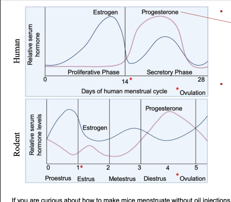
menstrual cycle diagram

Conditions that effect women exclusively/disproportionately/differently
exclusive: menopause, ovarian cancer
disproportionately: autoimmune diseases, Alzheimers disease
differently: cardiovascular diseases, lung cancer
Thalidomide tragedy & NIH Revitalization Act
In hopes of preventing another tragedy, after thalidomide exposure caused increased infant death, the FDA stopped including women of childbearing potential (almost all women) from clinical trials, resulting in a huge loss of data for how drugs work on women.
NIH:
This act ended the exclusion of women from these clinical trials mentioned above, and ensures that women and racial minorities were included in all clinical research.
Why are animal models insufficient for womens health sometimes
simultaneous menstruation is unusual, 98%% of mammals dont
placentation is much deeper in humans than other species
menopause only happens in whales and humans.
Endometriosis as an underfunded health burden
Endometriosis affects ~10% of women, causes substantial morbidity, and imposes high healthcare costs
(~3-fold higher than matched controls). Yet, the funding proportional to burden is low compared to other conditions. The study by Mirin et al. normalized funding to disability-adjusted life years (or DALY, a measure that considers prevalence, morbidity, and mortality in one number representing sum of years lost to disability) for various diseases. They found that endometriosis is one of the most underfunded conditions in the study. The ratio of actual funding compared to expected funding based on DALY was between 0.1-0.2 from 2015-2019, where a value of 1 would represent proportional funding
Fibrioids is another example
how do typical E2 concentrations in cell culture compare to peak menstrual cycle concentrations in vivo?
Cell culture concentrations are often very high! Cell culture E2 concentrations are often in the range of 10-100 nM, which is comparable to peak pregnancy but on the order of ~10-1000 times above the 0.2-2 nM range observed in menstruation. Importantly, high concentration does not just amplify effects, but can change them.
Menstrual vs Estrous cycle
Human: post ovulation, endometrium undergoes decidualization in response to p4
tissue remodels to support implantation
if no embryo —> corpus luteum demises, e2 p4 falls, menstruation starts
Rodent: 4 phases:
proestrus: estrogen increases
estrus: alongside ovulation, follows corpus luteum formation, shift from estrogen to progesterone dominant phase
decidualization doesn’t occur spontaneously, endometrium is remodeled not shed
continues until pregnancy or pseudopregnancy disrupts it and causes decidualization
Major hormones
Estrone (E1):
Predominant estrogen in post-menopausal women.
Estradiol (E2)
most studied
predominant in pre-menopasal women
made from estrone in ovaries/testes
can be synthesized from testosterone from hella diff cells.
Estetrol (E4):
Produced exclusively in human fetal liver.
Progesterone (P4):
primarily produced in ovary after ovulation or by placenta in pregnancy
produced by adrenal glands also in men and women in lower concentrations
metabolites help regulate menstrual cycle
can be converted to DHT and T
Testosterone (T):
produced in gonads, smaller amounts in adrenal cortex and skin
can be produced locally in brain and adipose tissues
concentration much higher in males than females
can be metabolized to DHT mostly in liver
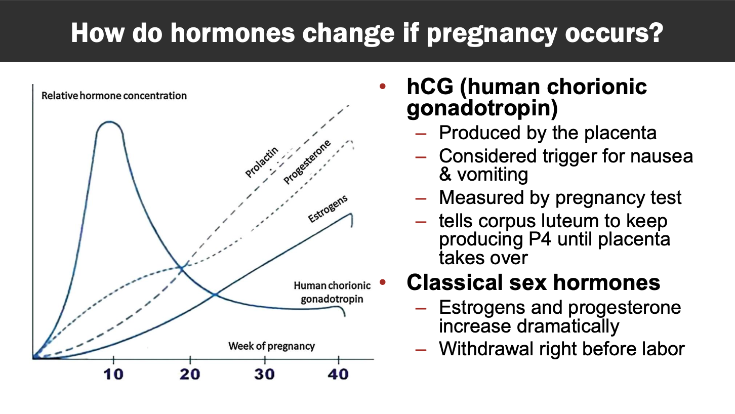
Hormone changes during pregnancy (E2 / P4)
E2 and P4 increase drastically throughout pregnancy instead of withdrawing (at end of menstrual cycle)
placenta is the predominant producer of P4 during pregnancy
corpus luteum does this in late menstruation and early pregnancy
In the days immediately preceding labor, P4 stays approximately the same in humans; receptor expression decrease is thought to contribute to functional withdrawal
In the days immediately preceding menstruation P4 and E2 decrease
hCG (human chorionic gonadotropin) has a sharp increase in early pregnancy, produced by placenta, causes nausea and vomiting, what a pregnancy test measures, tells corp lut to produce P4 until placenta takes over
Classical sex hormones: estrogens and progesterone increase dramatically, withdrawal right before labor
PBPK Models
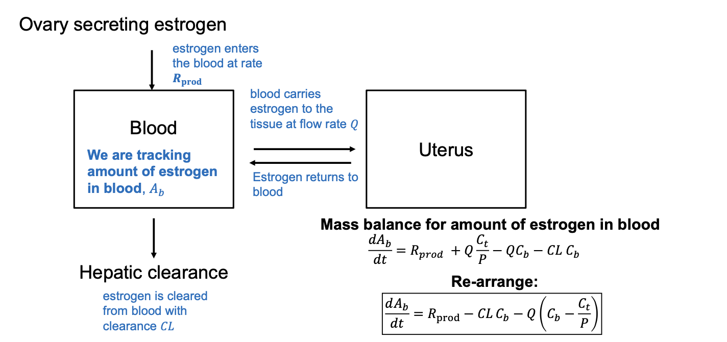
Pharmacokinetic model
standard perfusion limited PBPK assumption: tissue concentration is related to concentration in blood leaving the tissue by a partition coeffiient (P = Ct/Cvt)
Ct = tissue concentration, Cvt = concentration venous blood leaving tissue, P = partition coeff
Start by defining in vs out
substitute in any rate terms
multiply rates by their respective concentrations, remember to convert units as well
Ex: estrogen delivery to tissue = blood flow rate Q concentration of estrogen in blood (Cb) —> term is Q*CB
Autoimmune disease, estrogen, immune responses
Estrogen down-regulates autoimmune regulation
ARIE (Autoimmune regulator), transcription factor, makes sure t cells are destroyed if they react to self proteins
autoimmune diseases can worsen during periods of high hormonal fluctuation, or improve/worsen depending on disease and estrogen state.
estrogens DECREASE arie expression
androgens INCREASE arie expression
estrogen:
increases B cell antibody production
changes T cell regulation
enhances dendritic cell antigen presentation

x chromosome increases immune gene expression
2 X genes, if a gene escapes silencing it leads to double expression of certain genes:
TLR7 overexpression has been linked to lupus-like autoimmunity because excessive sensing of viral-like RNA can drive interferon production and chronic immune activation
Sex differences in microbiome and musosal immunity
Sex hormones and the microbiome regulate each other, so sex differences in microbiome and mucosal immunity may help explain why metabolic diseases like diabetes differ between males and females
What is SHBG and how would you expect endogenous levels of SHBG to impact estrogen uptake in rat and human tissues?
SHBG = sex hormone binding globulin. E2 binding to SHBG reduces free fraction of E2 in plasma, which is thought to be the active fraction, although bound E2 may be active to an extent via rapid dissociation. If E2 is bound to SHBG in the plasma, it should be less likely to enter the tissue as diffusion is limited by the carrier. SHBG is present in humans and not rats; however, E2 can bind to albumin in rats and humans with a similar effect.
factors that might influence tissue dose estimates for estrogen:
Uterine uptake of E2 depends on hormonal status because E2 may be impacting vascular permeability.
ER content and binding affinity are important determinants of how much E2 is deposited in tissues
Cardiovascular diseases present differently by sex

Main differences in cardiovascular system by sex
anatomy:
overall heart size, and ventricular architecture
coronary anatomy and microvasculature function
Hormones across lifespan lead to cardiovascular physiology changes
Disease phenotype
diff cellular/molecular response to injury
diff vascular remodeling and vascular biology
diff in calcific response
Key anatomical differences:
female heart 1/4th smaller
scaling for body size reduces but does not eliminate differences in cardiac mass!
female hearts are not just smaller versions of male hearts

How is it possible that female hearts exhibit lower cardiac output yet higher heart rate and ejection fraction?
Even though female hearts beat faster and have higher ejection fraction, male heart volumes are substantially larger volume leading to greater cardiac output
despite beating faster and pushing out more blood per beat, female hearts pump overall less blood
women also have higher contractility, and lower blood pressure

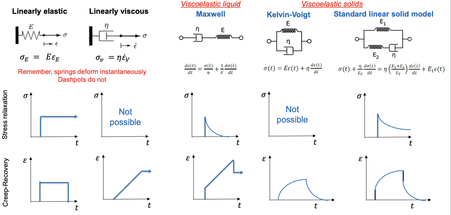
Stress vs Strain equations
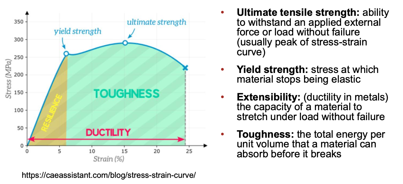
Stress: the internal resistive force per unit area that a material develops when subjected to an external load or force
Strain: the measure of how much a material deforms or changes shape when an external force is applied
linearly elastic solid (hookean) = stress depends on strain linearly (𝜎 = 𝐸𝜀)
Linearly viscous fluid (newtonian) = Stress linearly depends on strain rate (𝜎 = 𝐸𝜀) (𝜀 has dot on top of it for strain rate)
E = modulus, 𝜀 = strain, 𝜎 = stress

Viscoelastic solid vs fluid
solid: like memory foam, regains its shape —> acts like a solid
fluid: viscoscity eventually wins, keeps flowing: egg whites, dough
Stress vs strain curve
stress on Y axis, strain on X
when slope increasing —> area under curve = resilience
when slope mostly leveled off —> area under curve = toughness

Stress/strain/modulus in heart tissue
Female heart tissue is generally stiffer than male tissue
stiffness increases with age (esp in women)
Note: stiffness is a property of a structure, modulus describes properties of the material
hypertension mouse model:
when estrogen disrupted, female arteries were much less stiff, surpassed male
Young/LaPlace equation relation to the heart
YLP describes tension across a fluid interface, relates the pressure difference across a curved surface to the surface tension and the curvature of that surface.
note: the tissues we work with have thickness ofc
∆P = 𝜎𝜃ℎ / 𝑅
The stress in the wall goes up when:
the pressure is higher
the radius is larger
the wall is thinner

Heart adaptations to load

Sex diff in heart failure
HFrEF = can't squeeze (systolic, dilated, mostly male)
HFpEF = can't relax and fill (diastolic, stiff and thick, mostly female)

Cardiovascular diseases affect vasculature diff by sex
Female = vascular disease more likely in microvessels —> harder to detect
Male = epicardial coronary arteries more likely occluded —> easier to see
causes women to experience delays in diagnosis/treatment, less likely to be treated
While men often present with occlusion, women are more likely present with other dysfunctions such as diffuse atherosclerosis. Disease in the microvasculature is hard to detect with traditional imaging tools such as coronary angiography, highlighting the need for new diagnostics.
Hormones and cardiovascular function
estrogen is generally cardioprotective & for vasculature as well
vasculature —> estrogen increases nitric oxide (NO) release and vasodilation
NO + Estrogen = maintenance of vascular compliance
P4 and T protective against some arrhythmias, E2 can increase susceptibility
women at greater risk of drug induced arrhythmias
CAVD exhibits sex-specific patterns of extracellular matrix remodeling and calcification. Describe the predominant remodeling phenotype observed in males vs. females and explain how each contributes to valve stiffening.
CAVD increases stiffness of valves in males and females. However, calcification mediated by osteoblastic VICs is predominant in males, while fibrosis mediated by myofibroblastic VIC activation is more common in females. Fibrosis includes more collagen I/III activation and lower MMP activity.
Key sex specific differences in bones
obstetric selection: female skeleton must fascilitate childbirth without compromising bidpeal locomotion
energetic tradeoffs: female skeleton needs to adapt to pregnancy and gestation while balancing metabolic costs
sexual/ecological selection: males were selected for force generation, while females were selected for a compromise between mechanical function and reproductive demands.
Anatomy:
height, ribcage, skull, pelviship
Bone Density:
lower in females
regulated by sex hormones
decreases with age
Women’s pelvis shape and considerations
30% of maternal morbidity is due to obstructed labor
human infants have big ass heads in relation to maternal pelvis size
narrow pelvis good for bipedal walking/running, wide better for big head ass babies
sex differences in hip disorders / injuries
Hip fractures:
females have more fractures
males have higher death in response to a fracture
Hip dysplasia:
Females have less overall coverage of the femoral head and higher degree of acetabular anteversion angle (i.e. less anterior coverage) which is associated with greater joint instability and prevalence of dislocation.

Main Hip anatomy differences women vs men
Females:
less overall coverage of femoral head
higher degree of acetabular anteversion angle (less anterior coverage)
greater joint instability & prevalence of dislocation

Joint reaction force
Hip has hip abductor and body weight pushing down on it, and has femur pushing back up
we assume JRF acts as a pivot, points at an angle
JRF force generated within the hip joint to counteract forces acting on it
4x body weight when standing on one leg
running can get up to 5x body weight
contact stress = JRF / contact area
Bone internal structure
2 types: spongy & compact
have diff compressive strengths/strain
osteocytes sense and respond to bone mechanics
critical for bone remodeling feedback loop
dysregulation of this underlies multiple diseases
estrogen is a key modifier of osteocyte mechanotransduction
estrogen lowers threshold for osteocytes to respond to mechanical strain
broadly: hormones and activity substantially influence bone regulation
hormones are key regulatiors of bone
ex: maternal bone reabsorbed to meet calcium demands of lactation
Osteoporosis in women
80% people with it are women
menopause —> lower bone density —> osetoporosis —> fractures —> bad outcomes
major challenge today
Explain how E2, P4, PTHrP, and prolactin change during lactation relative to pregnancy and how they individually and collectively impact bone density
Rapid drop of E2 from pregnancy to lactation results in decreased bone formation and increased resorption. The rapid drop of P4 drop from pregnancy to lactation results in decreased bone formation and decreased osteoblast differentiation. At the same time, lactation maintains high PTHrP and prolactin levels established in pregnancy, which both generally promote resorption. Thus, the drop in E2 and P4 with little change in PTHrP and prolactin collectively shift toward more resorption and loss of bone mass.
Using the lecture data, calculate the approximate percent loss of bone mass from ages 40 to 60 years old in women and men. How might these differences in bone loss contribute to sex-specific patterns in hip replacement incidence later in life?

Explain one way in which estrogen may increase mechanosensitivity of osteocytes to exercise
Estrogen, relative to an estrogen depletion state like ovariectomy, binds to estrogen receptors on osteocytes leading to changes in abundance and organization of mechanosensing proteins. Collectively, this enables osteocytes to sense mechanical loading and respond by increasing bone formation more readily than without estrogen.
estrogen sensed, osteocytes more abundance and organized —> easier to sense mechanical loading —> increase bone formation
Uterus & myometrium expansion during pregnancy
uterus grows 500-1000x its original size
intrauterine volume expands from 10mL to 5mL
myometrium accommodates
predominantly hyperplasia (increase in cell #) of smooth muscles in early pregnancy
hypertrophy (increase in cell size) in late pregnancy, when biggest volume expansion occurs
stretch vs tough vs extensibility
uterine tissues ate strong and extensible (lots of stretch without failure)
Creep vs stress relaxation test
stress: think rubber band, hold it stretched, how the internal stress changes over time
control shape, measure force
creep: memory foam
control force, measure shape

Based on lecture, provide typical values (with units!) for the following along with a brief explanation of the functional importance of the property in pregnancy or parturition.

Stretch and mechanotransduction as a labor cue / Piezo 1
stretch suspected to be a key driver in labor
Explain or predict how might PIEZO1 contribute to the onset of labor and the
maintenance vs. lactation:
PIEZO1 is a stretch-activated ion channel that allows calcium to enter the cell cytoplasm under mechanical stretch, which may cause contraction. PIEZO1 channels are expressed abundantly in the human uterus near term. In rodent models, agonizing PIEZO1 can increase chance of labor while antagonization can reduce chance of labor in preterm models. In lactation, PIEZO1 is expressed but the functional significance is still unclear.
Blocking / knocking down Piezo1 lengthened pregnancy in preterm birth mouse model
why the myometrium and perimetrium are generally stiffer and more elastic than the endometrium (hypothesis)
The endometrium is unique in that it must be viscous and soft to support remodeling during embryo implantation,
whereas the myometrium and perimetrium must be stiff and elastic to resist excessive placental invasion while still being able to stretch and bear load as the fetus grows.

how collagen mechanics and remodeling contribute to maintaining pregnancy and enabling labor (Include toughness)
collagen is tough because it can break by slip and rupture
THIS IS HOW THE CERVIX STAYS TOUGH IN PREGNANCY
3 zones of collagen in the cervix, inner have longitudinal collagen fibers, middle zone has cirumferential collagen fibers
parturition requires cervical softening after being extremely sitff
10-100 fold softening
tissue remodeling and pressure during contraction concentrate stress on the cervix during labor
fibrous collagen is weak in compression, incredibly strong in tension
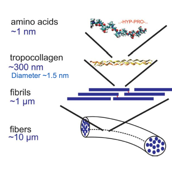
Tropocollagen length and staggered arrangement enable toughness (energy absorbed before failure)
Collagen gives tissues toughness through a hierarchical network of collagen fibrils and fibers that absorb energy during deformation. The staggered arrangement of collagen molecules and the long fibrillar structure facilitate energy dissipation through sliding of collagen molecules and fibrils, disruption of intermolecular bonds, and ultimately collagen rupture. In the cervix, highly organized and aligned collagen fibers help maintain mechanical integrity throughout pregnancy. Near labor, collagen remodeling, including degradation, reorganization, altered cross-linking, and increased hydration, reduces cervical stiffness and toughness. This softening allows the cervix to efface and dilate during labor contractions.

collagen structure
how does pushing help
thinner myometrium will increase value of pushing, thicker decreases

Post labor:
contract to expel placenta
if note —> postpartum hemmorhaging
uterine involution over first 4 weeks post partum
decrease in number and size of cells with vascular/collagen remodeling
uterus is permanently a bit bigger
Maternal-fetal placental interface
exhcnage: delivers O2 and nutrients to baby, removes CO2 and Waste
endorine: produces hormones to maintain pregnancy, prep for brirth/lactation
Estrogen/progesterone
hCG
immune: protects fetus while transferring maternal antibodies
barrier: controls maternal-fetal exchange and responds to O2, nutrients, inflammation
vascular remodeling is key to placenta formation:

uterine blood flow during pregnancy
changes dramatically to support fetus
increased blood supply to uterus = diverts blood from lower limbs
maternal blood volume / cardiac output increase by 30-50%
Maternal blood pressure falls
uterine arteries are controlled by muscle
contractions clamp vasculature causing hypoxia —> tissue death
necessary to shed placenta and stop post partum bleeding
shunting redirects blood flow
failed vascular remodeling / Why must spiral arteries remodel during normal pregnancy?
So blood enters the intervillous space gently instead of as a damaging high-velocity jet
Remodeling (loss of smooth muscle, widening) slows exit velocity so blood doesn't jet into and damage placental villous tissue.
flow rate when an artery DOES remodel and widen dramatically?:
Pressure drop shifts proximally; flow only increases modestly
small radius = blood jetting in
reynolds number predicts how smooth or rough the flow is

Placenta / amniotic membrane as a biomaterial
Amniotic membranes, valuable for wound healing due to their anti-inflammatory properties and because they are a source of human extracellular matrix, are currently processed for use in the clinical or tissue engineering. This usually involves decellularization with detergent and then lyophilization and reconstitution with other materials. The overarching limitation is variability. Sources of variability include the patient, what parts of the membranes were included, the method used for decellularization.
anatomy of mammay glands

Hormonal shifts drive mammary gland remodeling
signaling loop from HW

mechanical cues for maintaining lactation
infants apply cyclical stimulation that maintains lactation in lactogenesis III
prolactin = epithelial, ductal growth, milk synthesis
oxytocin release = myoepithelial contraction and “let-down”

Identify two mechanical cues provided by breast pumps and describe how they
mimic infant suckling.
cyclical stimulation of nipple, where the vacuum is applied, leads to oxytocin and prolactin release, much like cyclic suckling motion of infant. the earliest pumps were not cyclical
-vacuum from pump empties breast like an infant, reducing pressure and reducing expression of feedback inhibitor of lactation
Some key pregnancy adaptations and what can go wrong

Cardiovascular disease as a pregnancy complication / preeclampsia
40-50% increase in blood volume
30-50% increase in cardiac output
increased heart rate
Hypertension is the top complication
vascular dysfunction
Peripartum cardiomyopathy
reduced left ventricle ejection fraction (end of pregnancy)
Venous thromboembolism:
deep vein thrombosis or pulmonary embolism
normally, blood pressure drops slightly in pregnancy
Estrogen, relaxin, progesterone push the cardiovascular system toward vasodilation through distinct mechanisms, NO signaling, blunted myogenic tone, and reduced vasoconstrictor sensitivity, which together help accommodate the dramatic rise in blood volume and uteroplacental flow during pregnancy.
These hormones promote vascular compliance
preeclampsia can progress to eclampsia (mid to late pregnancy)
starts with abnormal placentation, sends systemic distress signals that exacerbate disease
stressed placenta releases sFlt-1 and soluble endoglin(sENG) into maternal circulation as damage control
sFlt-1 acts as a decoy receptor, sequesters VEGF and PIGF, sENG sequesters TGFb
systemic dysfunction: vasoconstriction, hypertension, capillary leak bc no dilation happening

Estrogen and relaxin impact on body
Relaxin acts systemically
important for pelvic adaptation, reproductive tissue stretch
receptors are lowk everywhere
promotes collagen remodeling (increased collegenase & MMP acivity)
implicated in joint instability & ligament injury risk

pregnancy impact on musculoskeletal mechanics
Pelvic floor:

models of viscoelastivity