BIOL 203 Lecture 12 - Forebrain
 Call Kai
 Learn
 Practice Test
 Spaced Repetition
 Match
 Flashcards
 Knowt Play
1/85

There's no tags or description
Looks like no tags are added yet.
86 Terms
prosencephalon
embryonic forebrain
cerebrum and diencephalon
what two major regions does the prosencephalon give rise to
cerebrum
higher cognitive functions, conscious perception, voluntary movement, memory, and language
diencephalon
comprised of the thalamus, hypothalamus, and epithalamus
diencephalon
sensory relay, homeostasis, autonomic and endocrine regulation
cerebrum
what is the top arrow pointing to?
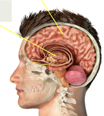
diencephalon
what is the bottom arrow point to?

scalp, skull, periosteal dura, meningeal dura, arachnoid, subarachnoid space, pia, brain
list the anatomical layers protecting the brain from superficial to deep
dura mater
the tough outer meningeal layer
arachnoid mater
middle meningeal layer (thin)
pia mater
thin, delicate meningeal layer; follows the contours of the gyri and sulci
subarachnoid space
actual CSF-filled space underneath arachnoid mater; trabeculae cross the space
falx cerebri
a fold of dura mater that separates the two cerebral hemispheres
gray matter
gray or white matter: processing
white matter
gray or white matter: communication
gray matter
contains many neuronal cell bodies, dendrites, synapses, and glial cells
gray matter
major site of information processing and integration
gray matter
in the cerebrum, forms the cerebral cortex
white matter
contains bundles of axons, many surrounded by myelin
white matter
carries signals between different CNS regions
white matter
in the cerebrum, lies mainly deep to the cerebral cortex
white matter
its lighter appearance is largely due to lipid-rich myelin
cerebral cortex (outer layer) and basal nuclei (deep gray matter structures)
what does gray matter form?
inner region containing nerve fiber tracts
what does white matter form?
pyramidal neurons
the principal excitatory projection neurons of the cerebral cortex
gray matter
central H-shaped region containing neuron cell bodies
white matter
outer region containing ascending and descending tracts
cerebral cortex, subcortical white matter, deep nuclei
three main components of the cerebrum
cerebral cortex
outer layer of gray matter; folded into gyri and sulci
subcortical white matter
axon tracts connecting different regions of the CNS
deep nuclei
collections of gray matter located within the white matter; include the basal nuclei
cerebral cortex
what is the top arrow pointing to?

subcortical white matter
what is the middle arrow pointing to?

deep nuclei
what is the bottom arrow pointing to?

primary area
functional area of cerebral cortex that is involved in processing specific sensory information or generating voluntary motor output
primary area
functional area of cerebral cortex that has the primary visual cortex, primary somatosensory cortex, and primary motor cortex
secondary area
functional area of cerebral cortex that performs more complex processing within a specific sensory or motor system
secondary area
functional area of cerebral cortex that has visual association areas and the premotor cortex
association area
functional area of cerebral cortex that integrates information from multiple cortical regions
association area
functional area of cerebral cortex that supports planning, reasoning, language, memory, attention, and decision-making
frontal, temporal, parietal, occipital, and insula
name the five lobes of the cerebrum
frontal lobe
what lobe is responsible for voluntary motor control, executive functions (planning, decision-making), and speech production (Broca’s area)
temporal lobe
what lobe is responsible for hearing (primary auditory cortex), language comprehension (Wernicke’s area), memory formation/processing, and olfactory processing
parietal lobe
what lobe is responsible for somatosensory processing: touch, pain, temperature, pressure, vibration, proprioception and sensory integration/spatial awareness
occipital lobe
what lobe is responsible for the primary visual cortex (visual processing and interpretation)
insula
what lobe is responsible for visceral sensory processing, taste perception (gustatory cortex), emotional awareness and salience processing, and interoception (internal body state)
blood-brain barrier (cerebral vasculature)
present in most brain capillaries; restricts uncontrolled entry of toxins, pathogens, and many large or hydrophilic molecules; allows selective passage of small lipid-soluble molecules (O2 and CO2), small nonpolar molecules, and essential substances (glucose, amino acids) via transport proteins
blood-CSF barrier (choroid plexus)
produces CSF; regulates movement of substances from blood into the CSF; helps maintain chemical stability of the CNS
brain-CSF barrier (pia arachnoid)
prevents uncontrolled exchange between blood and CSF
tight junctions
_____ _____ prevent paracellular diffusion in the BBB
capillary endothelial cells joined by tight junctions; basement membrane; pericytes; astrocyte end-feet
what are the four main components of the blood-brain barrier
choroid plexus
this contains the fenestrated capillaries covered by specialized epithelial cells; located within the lateral, third, and fourth ventricles; produces the most CSF through selective secretion of ions and water from the blood
blood-CSF barrier
formed primarily by tight junctions between choroid plexus epithelial cells; separates the blood/stroma from the CSF; the underlying capillaries are fenestrated and relatively permeable
blood flows through fenestrated capillaries in the choroid plexus, allowing substances to enter the surrounding stroma
what is the first step of the blood-CSF barrier
before reaching the CSF, substances must pass through choroid plexus epithelial cells, and these cells are connected by tight junctions, which control what can enter the CSF
what is the second step of the blood-CSF barrier
ventricular system
the _____ _____ is a network of interconnected cavities that contain CSF and provide a pathway for its movement
ependymal cells
_____ _____ line the ventricles and central canal; their cilia help move and mix CSF
lateral ventricles, third ventricle, and fourth ventricle
what are the main ventricles
between the thalami
where is the third ventricle located
between the brainstem and cerebellum
where is the fourth ventricle located
lateral ventricles
what is the light orange section called

third ventricle
what is the dark orange section called

fourth ventricle
what is the green section called

interventricular foramina (of Monro)
what is the connection between the lateral and third ventricle called (top arrow)

cerebral aqueduct (of Sylvius)
what is the connection between the lateral and fourth ventricle called (bottom arrow)

mechanical protection (shock absorber), helps maintain a stable chemical environment, nutrient transport and waste removal
what are the functions of CSF
lateral ventricles
first step of CSF flow pathway
interventricular foramina
second step of CSF flow pathway
third ventricle
third step of CSF flow pathway
cerebral aqueduct
fourth step of CSF flow pathway
fourth ventricle
fifth step of CSF flow pathway
median and lateral apertures
sixth step of CSF flow pathway
subarachnoid space around brain and spinal cord
seventh step of CSF flow pathway
passes through arachnoid granulations
eigth step of CSF flow pathway
drains into dural venous sinuses, mainly the superior sagittal sinus
ninth step of CSF flow pathway
arachnoid granulations
how is CSF reabsorbed into the venous system
dural venous sinuses
large venous channels within the dura mater; found between periosteal and meningeal layers of the dura mater; collect blood from cerebral veins and receive CSF; eventually drains into the internal jugular veins
association tracts
connect cortical areas within the same hemisphere
short
_____ fibers connect adjacent gyri (U-fibers)
long
_____ fibers connect more distant cortical regions, often in different lobes
commissural tracts
connect cortical regions across the two cerebral hemispheres, often corresponding regions; allow communication between the left and right sides of the brain
corpus callosum
largest commissural fiber bundle
projection tracts
connect the cerebral cortex with lower brain centers and the spinal cord; carry ascending sensory and descending motor signals
thalamus
lies superior to the brainstem and forms the lateral walls of the third ventricle; major relay station for most sensory information traveling to the cerebral cortex; relays motor information from the cerebellum and basal nuclei to motor areas of the cerebral cortex
epithalamus
contains the pineal gland
hypothalamus
inferior to the thalamus; forms the floor of the third ventricle; maintains homeostasis; regulates autonomic nervous system activity; regulates body temperature, hunger, thirst, and circadian rhythm; regulates endocrine function through the pituitary gland