BCMB 311- Quiz 5
 Call Kai
 Learn
 Practice Test
 Spaced Repetition
 Match
 Flashcards
 Knowt Play
1/127

There's no tags or description
Looks like no tags are added yet.
128 Terms
Why has yeast mating served as an early model for understanding how extracellular chemical signals can be perceived and produce intracellular changes?
Yeast cells “shmoo” in response to mating factors→ form mating projection
Define autocrine signaling.
Signals act on the same cell that released them.
Define endocrine signaling.
Hormones travel long distances, typically through blood stream
Define paracrine signaling.
Signals act on nearby cells
Define contact dependent signaling.
Requires direct physical contact between cells
Yeast releasing mating factor can be classified as which type of signal?
a. Autocrine
b. Endocrine
c. Paracrine
d. Contact dependent
c. Paracrine
A variety of biomolecules can act as signaling molecules. What are they?
Proteins
Peptides
Amino acids
Nucleotides
Steroid hormones
Fatty acid derivatives
Dissolved gases
What are hormones?
A molecule produces by one organ (gland) and released into the bloodstream
Binds to another organ (effector organ) to regulate physiology or behavior
Ultimately, hormones regulate gene expression
What are neurotransmitters?
A molecule released from a neuron and received by another neuron or effector organ (muscle, gland, etc.)
What is a water-soluble hormone?
Free (unbound) in the plasma
Usually peptides that bind to receptors on the membrane of the target cell→ stimulate the production of intracellular second messengers
UNABLE to penetrate the cell membrane
What is a lipid-soluble hormone?
Circulate bound to plasma proteins
Mechanism of action is directly mediated on the nuclear chromatin→ the formation of a hormone receptor complex→ activates RNA polymerase→ new mRNA→ specific protein
MUST cross the plasma and nuclear membranes
What two types of receptors do extracellular signal molecules bind to?
Cell-surface receptors
Intracellular receptors
What are cell-surface receptors?
Receptors on the plasma membrane that bind extracellular signal molecules and trigger the release of intracellular signaling molecules.
What are intracellular receptors?
Receptors inside the cell (inside nucleus) that bind small, hydrophobic signal molecules (usually steroid hormones)
What is signal transduction?
A stepwise progression of signaling stages: receptor→ transducers→ effectors
The receptor receives/perceives signal
Transducers relay signal
Effectors convert signal to an intracellular response
What are second messengers?
Small molecules that pass along a signal initiated by the binding of a ligand (“first messenger”) to its receptor
What are the three ways receptor signals are transduced?
Receptor-mediated activation of phosphorylation cascades that trigger the activation of various enzymes
Exchange of GDP for GTP be GTP-binding proteins (G proteins), which leads to generation of second messengers (cAMP, lipid breakdown products, Ca2+, etc.)
Conformational changes that open ion channels or recruit proteins into nuclear transcription complexes
The same signal molecule can induce different responses in different target cells. Describe this using acetylcholine.
Acts on heart pacemaker cells and decreases firing rate (less contractions)
Acts on salivary gland cells and increases secretion
Acts on skeletal muscle cells and increases contraction
How can extracellular signals act rapidly?
Can activate/deactivate an enzyme that is already present→ can be very fast
Phosphorylation/dephosphorylation
Ubiquitination
How can extracellular signals act slowly?
Can cause a protein to be synthesized by activating transcription→ much slower
Can be more energetically efficient because protein is only made if needed
What roles can intracellular signaling proteins play?
Relay signal
Amplify signal (transduce)
Detect from multiple sources and integrate
Distribute signal→ creating branches in info flow
Feedback→ positive or negative
What is a positive feedback loop?
Downstream effector proteins activate earlier steps→ often leads to substantial amplification
Allows pathway to exist in a stable “on” state in the absence of receptor activation or until acted upon by separate inactivation signal
What is a negative feedback loop?
Downstream effectors inhibit earlier steps
Pathway will shut itself off after activating to a certain level and when receptor is no longer active
New signal will be required to reactivate
How can some intracellular signaling proteins act as molecular switches?
Signaling by protein phosphorylation (activate/deactivate)
Signaling by GTP-binding proteins
What two types of regulatory proteins control the activity of monomeric GTPases?
Guanine nucleotide exchange factors (GEFs)
GTPase-activating proteins (GAPs)
What do GEFs do?
Promote the exchange of GDP for GTP→ switches protein on
What do GAPs do?
Stimulate the hydrolysis of GTP to GDP→ switches protein off
What are the three main classes of cell-surface receptors?
Ion channels
G-protein coupled receptors
Enzyme-coupled receptors
How do ion-channel-coupled receptors work?
Signal molecule binds receptor and causes a conformational change→ opens channel and allows ion flow
What are the two ways enzyme-coupled receptors work?
Signal molecule binds and causes inactive catalytic domains to form active dimer
Signal molecule binds and causes formation of dimer→ active associated enzyme can bind
All GPCRs have a similar structure. Describe this.
Seven transmembrane a helices
Extracellular side binds signaling molecules
Receptors can bind:
Small molecules (I.e., acetylcholine and epinephrine)
Proteins
Cytoplasmic side binds a G protein inside cell
G protein→ heterotrimeric GTPase
About 800 genes in humans encode GCPRs
What are the two types of GPCR responses?
Stimulatory→ Gs
Inhibitory→ Gi
What are the two main types of GTPases?
Small, monomeric GTPases (i.e., Ran)
G Proteins→ heterotrimeric GTPases containing a, B, and y subunits
a subunit→ binds GTP/GDP
By dimer
Both (a and y) are membrane-tethered by attachment to lipid head groups
What are the typical steps involved with activating a GPCR?
Ligand binds extracellularly
Cytosolic domain undergoes conformational change→ activates a G protein
Activated G protein exchanges GDP for GTP in a subunit
Often, a subunit dissociates and binds/activates other effector proteins to activate signal transduction
Free By dimer can also activate effectors

What does the activated a subunit do?
a subunit GTPase hydrolyzes its GTP to GDP (usually after a few seconds of activation)
Inactive a subunit releases target effector protein and rebinds the By dimer→ reforms inactive G protein
Effector protein inactivates once its no longer bound by the a subunit

What is Gi, a G protein, activation coupled to?
Gi receptor activation is directly coupled with the opening of K+ channels in the plasma membrane of heart pacemaker cells
Activated By subunit causes opening of ion channel
Enzymes activated by G proteins ____ the concentrations of small intracellular signaling molecules called ____
increase, second messengers
Second messengers diffuse to act on intracellular signaling proteins

Seven
Three
Two
GDP
GTP
GTPase
What is cAMP? How is it synthesized and degraded? What does it do?
cAMP is a common second messenger (released into cytosol)
Synthesized by adenylyl cyclase and degraded by cAMP phosphodiesterase
Can bind and activate downstream effectors
Synthesis/release is followed quickly by degradation/removal
What are the two main branches of the nervous system?
Somatic (conscious activities)
Autonomic (unconscious activites)
What are the two branches of the autonomic nervous system?
Parasympathetic (inhibitory)
Sympathetic (stimulatory)
What is the typical GPCR associated with the sympathetic nervous system?
Gs
Receptors: Adrenergic receptors
Ligand: Epinephrine
What is the typical GPCR associated with the parasympathetic nervous system?
Gi
Receptors: Muscarinic receptors
Ligand: Acetylcholine
How does Gi work in heart pacemaker cells?
Acetylcholine binds its GPCR
G protein activates
By complex opens K+ channel
Increases permeability to K+
Membrane is harder to activate→ slows heart rate
Why does epinephrine stimulate glycogen breakdown in skeletal muscle cells?
Muscle cells need to rapidly get energy to prepare for muscle contraction
Glycogen is a polymer of stored glucose and is present in muscle cells
We must get to breakdown mode to generate energy in the form of ATP
What are the steps of activation for B adrenergic receptor (Gs) in skeletal muscle cells?
Activation of epinephrine GPCR (B adrenergic receptor) activates adenylyl cyclase
cAMP is produced
cAMP activates protein kinase A (PKA)
PKA has many targets→ phosphorylation by PKA typically activates target
Phosphorylase kinase is activated
Phosphorylase kinase phosphorylates and activates glycogen phosphorylase→ breaks down glycogen, releasing glucose

A rise in intracellular cAMP can _____ gene transcription. Provide an example.
activate
Bacterial Lac operon→ expresses genes needed to catabolize lactose
How does cAMP activate PKA?
By removing inactivating “regulatory” subunits from catalytic subunits
What two signaling pathways are activated by phospholipase C?
IP3
DAG
Some GPCRs work by activating phospholipase C. Describe this in terms of the IP3 and DAG pathways.
Use G protein Gq
Both a and By are involved in activating phospholipase C
Phospholipase C hydrolyzes an inositol head group from a membrane lipid
Inositol 1,4,5-triphosphate (IP3) diffuses through cytosol and opens ER Ca2+ channels
Ca2+ floods into cytosol
Diacylglycerol (DAG) remains in plasma membrane and, together with Ca2+, helps activate protein kinase C (PKC)
PKC phosphorylates and activates downstream effectors to continue the signal

What is a second messenger?
Molecule/ligand is “first messenger”
Second messenger (Ca2+, cAMP, etc.) is released when ligand binds to extracellular receptor
Second messenger activates/inhibits processes in cytosol or nucleus (depending on if it’s Gs or Gi)
Briefly describe the activation of phospholipase C.
Hormone binds receptor
Gq protein is activated
Phospholipase C is activated
DAG and IP3 release from cell membrane
IP3 mobilizes calcium ions (has second messenger effects) from ER (ex. smooth muscle contraction)
How are IP3 and DAG produced?
Many second messengers are generated by breakdown of membrane phospholipids
Binding of some hormones to their receptors triggers activation of PLC
Results in the breakdown of inositol phospholipids to yield IP3 and DAG
Ca2+ signaling triggers many biological processes and is involved with egg fertilization. Describe this.
Fertilization of an egg by a sperm triggers an increase in cytosolic Ca2+ in the egg→ produces membrane changes that prevent fertilization by other sperm cells and initiates embryonic development
What does calmodulin do?
Calmodulin acts as an intermediary protein that senses calcium levels and relays signals to various calcium-sensitive enzymes
What does nitric oxide (NO) do?
NO triggers smooth muscle relaxation in blood vessel walls by bidning guanylyl cyclase and triggering the release of cyclic GMP
Enzyme in mitochondria (mitochondrial aldehyde dehydrogenase) converts nitroglycerine into nitric oxide
How do enzyme-coupled receptors work?
Act as enzymes themselves when activated (ex. receptor tyrosine kinase)
Binding of signal molecule activates recpetor’s enzymatic activity
Pass once through cell membrane (unlike GPCRs, which have 7 transmembrane configuration)
Binding of extracellular ligand activates tyrosine kinase activity
Results in phosphorylation of target protein in cytoplasm→ activates target protein
Usually involves phosphorylation cascade
All mediated by series of kinases
What three domains make up a single pass receptor tyrosine kinase and what do they do?
Extracellular domain→ responsible for ligand binding
A single transmembrane domain
Intracellular domain→ a tyrosine kinase: an enzyme that uses ATP to phosphorylate a tyrosine on a target protein
What is the role of kinases and phosphatases?
Phosphorylation typically occurs on serine, threonine, and tyrosine (side chains contain -OH group)
Phosphorylation cascade turned off by phosphatases→ remove phosphates
Phosphorylation cascade turned on by kinases→ add phosphates
Describe the activation of RTKs.
Ligand binding often causes receptor dimerization→ produces conformational change that activates intracellular kinase domains
Cytosolic domains of receptor are kinases that phosphorylate each other upon dimerization
Other proteins are recruited to phosphorylated sites and become activated→ assembly of intracellular signaling complex
Most RTKs activate the monomeric GTPase Ras. Describe this.
Ras is tethered to the membrane via lipid attachment
Activated RTK recruits an adaptor
Adaptor recruits Ras-GEF to membrane
Once enriched at the membrane, Ras-GEF activates Ras-GTP
Ras-GTP activates a signaling cascade (MAPK)
Ras GAP will eventually promote GTP hydrolysis to inactivate Ras until a new signaling event occurs. Phosphatases will eventually inactivate the kinases

Describe the MAP-kinase signaling module.
Ras-GTP activates MAP kinase kinase kinase
MAP kinase kinase kinase activates MAP kinase kinase
MAP kinase kinase activates MAP kinase
MAP kinase activates proteins X and Y (changes in protein activity) and transcription regulators A and B (changes in gene expression)
Mitogens are small proteins that promote mitosis
Each of these enzymes activates may targets, so each level serves as an amplification step
What is the relationship between some RTKs and PI-3 kinase?
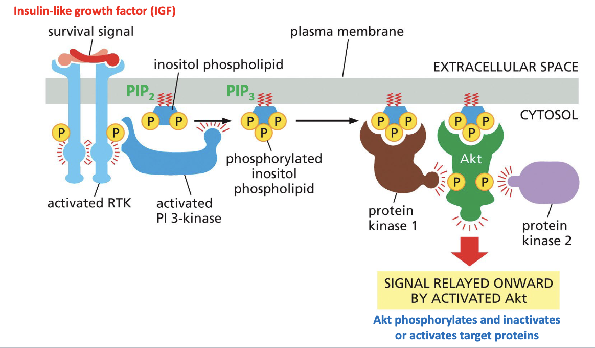
Some RTKs activate PI-3 kinase to produce lipid docking sites in the plasma membrane
One downstream target of PI-3 kinase is Akt kinase
The IGF signal promotes cell survival/groeth through Akt
Once the ligand dissociates, tyrosine phosphatases remove phosphates and inactivate RTK
Describe the effect of insulin-like growth factor (IGF) binding to RTK.
IGF binds and activates RTK
PI-3 kinase binds RTK and becomes activated
PI-3 kinase phosphorylates/activates PIP2 (located in membrane)
PIP2 phosphorylates/activates PIP3 (located in membrane)
PIP3 binds/activates PK1
PK1 and PK2 phosphorylate Akt
Akt relays signal onward
How does Akt promote cell survival?
Akt phosphorylates Bad of Bad/Bcl2 complex
Releases inactive Bad and active Bcl2
Bcl2 promotes cell survival by inhibition of apoptosis

How does the steroid hormone cortisol act?
By activating a transcription regulator
Steroid hormones bind nuclear hormone receptors
Nuclear hormone receptors are intracellular receptors that also act as transcription factors to activate transcription and specific genes
Cortisol crosses into cell through plasma membrane→ binds/activates nuclear receptor protein through conformational changes→ activated complex enters nucleus→ complex binds regulatory region of target gene→ activates transcription
How is a hormone response element activated and what does it do?
Lipid soluble molecules (ex. steroid hormones and vitamin D) are able to penetrate cell membrane
Bind with protein receptors in cytosol
Hormone-receptor complex binds to a regulatory sequence of the DNA called hormone response element
Transcription of specific gene or mRNA will be stimulated or inhibited by the hormone response element
Thyroid hormones have intranuclear receptors
How are estrogen receptors involved in breast tumors?
Immunohistochemical detection of estrogen receptor complex in nuclei of tumor cells
Breast tumor cells may or may not have estrogen receptors
If estrogen receptors are present, there is in advantage→ hormone therapy can control tumor growth
What is EGFR and what can mutations in it cause?
EGFR (epidermal growth factor receptor) is an RTK that normally responds to growth factors
In some lung cancers, mutations in EFGR gene change receptor’s structure
Mutated receptor can remain active even without growth factor binding
This continuously activates intracellular pathways that promote cell growth and survival
What is HER2 and how is it involved with breast cancer?
HER2 is an RTK found on cell membrane
Normally, HER2 works to activate signaling pathways that regulate cell growth and division
In some breast cancers, HER2 gene amplification produces too many HER2 receptors
Excessive receptor signaling stimulates pathways that promote uncontrolled cell proliferation
Clinical connection: HER2-targeted drugs (ex. Herceptin) can block HER2 signaling and help treat HER2-positive breast cancer
When adrenaline binds to adrenergic receptors on the surface of a muscle cell, it activates a G protein, initiating an intracellular signaling pathway in which the activated α subunit activates adenylyl cyclase, thereby increasing cAMP levels in the cell. The cAMP molecules then activate a cAMP-dependent kinase (PKA) that, in turn, activates enzymes that result in the breakdown of muscle glycogen, thus lowering glycogen levels. You obtain muscle cells that are defective in various components of the signaling pathway. Referring to the Figure provided, indicate how glycogen levels would be affected in the presence of adrenaline in the following cells. Would they be higher or lower than in normal cells treated with adrenaline?
A. cells that lack adenylyl cyclase
B. cells that lack the GPCR
C. cells that lack cAMP phosphodiesterase
D. cells that have an α subunit that cannot hydrolyze GTP
A: Higher
B: Higher
C: Lower
D: Lower
What are the three types of protein filaments? Order them from largest to smallest.
Microtubules> Intermediate filaments> Actin filaments
Describe the structure of microtubules.
Hollow tubes of 25 nm thickness
Made of a-tubulin and B-tubulin monomers, with B being a GTPase
Rigid but do not have high tensile strength
Has polarity→ plus and minus end
Minus end usually attaches to the microtubule organizing center (MTOC) in animal cells
What are the main functions of microtubules?
Organization within cytosol
Tracks for transport of vesicles and organelles
Movement of chromosomes during cell division
Serves as moving structural component within cilia and flagella
Provide structure for:
Mitotic spindle
Cilia and flagella
Centrioles
Neurons
What is the role of tubulin and protofilaments in microtubule structure?
a and B tubulin form a tubulin dimer
A linear strand of dimers is called a protofilament
A ring of protofilaments makes up a microtubule
All dimers and protofilaments assemble in the same orientation
Describe the polarity of microtubules.
a tubulin is exposed at the minus end
B tubulin is exposed at the plus end
Polarity is critical for directional transport of cargo along microtubules
Microtubules grow faster from their plus ends
Tubulin polymerizes from ____ sites on a _____. Can a and B tubulin spontaneously come together in vitro?
nucleation, centrosome
In vitro, a and B tubulin can spontaneously come together as rings of protofilaments, self-assembling into microtubules→ only at a concentration far higher than is present in the cell
What does it mean for microtubules to have a property known as dynamic instability?
Each microtubule grows and shrinks independently of its neighbors
What controls the dynamic instability of micro tubules? What favors assembly/disassembly? What does “catastrophe” refer to?
GTP hydrolysis:
B tubulin is a GTPase→ hydrolyzes its GTP over time
GTP-bound tubulin dimers favor assembly
GDP-bound tubulin dimers favor disassembly
“Catastrophe”→ loss of GTP tubulin cap and rapid depolymerization
How are microtubules stabilized? How do they behave when stabilized/unstabilized?
Can be stabilized by attachment to capping proteins
Unstabilized→ microtubules grow and shrink randomly
Stabilized→ microtubule plus ends that encounter a stabilizing capping protein will persist
Microtubules dynamically grow out from centrosomes in all directions and retract (disassemble), probing cell for attachment sites that will stabilize ends in places where microtubules are needed
How do microtubule-affecting drugs act?
Can bind and stabilize microtubules
Taxol→ anti-breast cancer
Can bind tubulin dimers and prevent polymerization
Colchicine, colcemid
Vinblastine, vincristine→ Hodgkin’s disease and Wilms tumor
What are the two motor proteins discussed? In what direction do they move and what are their functions?
Dynein
Retrograde: from + end to - end
Function: cilia motion
Kinesin
Anterograde: from - end to + end
Function: cell division and intracellular trafficking
Walk in a directional fashion
Require ATP
In a ciliated cell, how does a cilium “beat?” What is the role of basal bodies?
Cilium beats by performing a repetitive cycle of movements→ power stroke (whipping motion) followed by a recovery stroke
Basal bodies act as MTOC to stabilize minus ends
How do flagella propel a cell through fluid?
By using repetitive wavelike motion
What is the general structure of cilia and flagella? What is their role in the trachea, fallopian tubes, and sperm?
Microtubule-rich organelles
Structure: 9 + 2 arrangement of MT doublets
Trachea: moving mucus and dirt
Fallopian tubes: moving ovum from ovary to uterus
Sperm: motility

What is the result of dynein movement in isolated doublet microtubules vs. normal flagella? Where do immobilized dyneins try to “walk?”
Isolated doublet microtubules (no linking proteins)→ dynein produces microtubule sliding
Normal flagella (have linking proteins)→ dynein causes microtubule bending
Immobilized dynein→ try to walk toward minus end of other microtubule doublet

What causes Kartagener’s syndrome? What are the symptoms?
Mutation in dynein motor arm
Results in immotile cilia and sperm (flagella)
Symptoms:
Male and female infertility
Recurrent sinusitis
Bronchiectasis
May result in situs inversus→ inverse arrangement of internal organs
How are microtubules involved in transport along a nerve cell axon?
Microtubules guide the transport of organelles, vesicles, and macromolecules in both directions along a cell axon
Backward transport (to cell body)
Outward transport (to axon terminal)
Recall that centrosomes are usually near the nucleus such that the plus ends radiate out
What are the steps of kinesin movement?
Kinesins (with bound cargo) randomly bind to a microtubule
The leading head group exchanges its ADP for ATP→ triggers conformational change
Linker neck region of leading head group “zippers” onto head group→ throws lagging head group forward one “step”→ called power stroke
Trailing head group hydrolyzes ATP and linker region releases
Leading head group exchanges ADP for ATP→ linker neck region zippers→ process repeates

Describe the structure of actin filaments.
7 nm diameter (smaller than MTs 25 nm)
Twisted chain of identical actin monomers
Has polarity (+/- end)
Nucleation, dynamics, and branching heavily regulated by binding proteins
Most abundant near cell cortex
What are the main functions of actin filaments?
Vesicle and organelle trafficking
Help determine cell morphology and dynamics:
Microvilli
Phagocytosis
Cell constriction during cytokinesis
Cell motility by crawling along surfaces
Alongside bundles of myosin motor proteins→ make up contractile component of muscles
Provide mechanical strength to plasma membrane
What are 4 shapes/functions of actin filaments?
Microvilli (in intestinal cells)→ not motile like cilia, increase surface area for absorption
Contractile bundles→ generate contractile forces for cell shape, adhesion, migration, mechanical stress resistance, etc.
Filopodia→ protrusions used to reach out and adhere to surfaces as part of cell crawling
Contractile ring→ used during cytokinesis

What is the shape of actin filaments?
Two strands of identical actin monomers in a right-handed helix
What type of enzyme do actin monomers act as?
ATPases (B-tubulin of MTs=GTPase)
Where do actin monomers add to form the filament?
Actin monomers can add to either end, but addition to plus end is more favorable→ occurs at lower actin-ATP concentrations
Actin filaments can undergo treadmilling. Describe this.
Treadmilling→ occurs when monomers are added to the plus end and are eventually removed from the minus end

Can microtubules undergo treadmilling?
Yes→ tubulin is added to plus end and lost from minus end
What 3 drugs are discussed that act on actin filaments? How do they do this?
Phalloidin→ binds and stabilizes filaments
Cytochalasin→ Caps filament plus ends, preventing polymerization there
Latrunculin→ Binds actin monomers and prevents polymerization
________ ______ control the behavior of actin filaments in vertebrate cells.
Actin-binding proteins
What percent of cellular protein is actin? How much is in filaments vs. monomers?
About 5%
Half in filaments
Half are monomeric