Condensed Muscle List: Origins, Insertions, and Actions (click images to open)
 Call Kai
 Learn
 Practice Test
 Spaced Repetition
 Match
 Flashcards
 Knowt Play
1/63

Earn XP
Description and Tags
Comprehensive vocabulary flashcards detailing the anatomical origins, insertions, and primary actions for all 64 muscles from the condensed human muscular system reference.
64 Terms
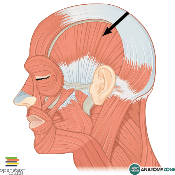
Temporalis
Origin: Temporal fossa and deep surface of temporal fascia.
Insertion: Coronoid process and anterior border of mandibular ramus.
Action: Elevates mandible; posterior fibers retract mandible.
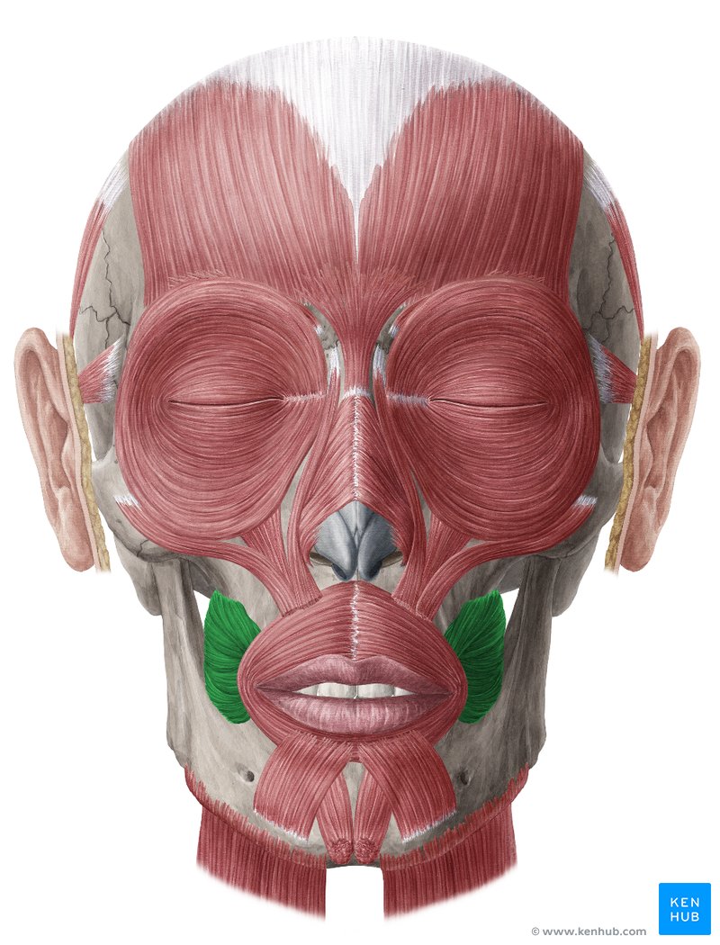
Masseter
Origin: Zygomatic arch.
Insertion: Lateral surface of mandibular ramus and angle; deep fibers also reach the coronoid process.
Action: Elevates mandible; superficial fibers assist protrusion.

Buccinator
Origin: Alveolar regions of maxilla and mandible opposite the molars; pterygomandibular raphe.
Insertion: Angle of mouth, blending with orbicularis oris.
Action: Compresses cheek against teeth; keeps food between chewing surfaces and assists blowing.
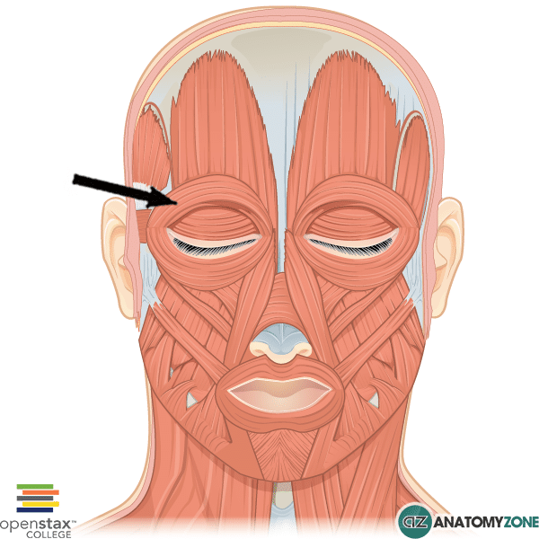
Orbicularis oculi
Origin: Medial orbital margin, medial palpebral ligament, and lacrimal bone.
Insertion: Skin around orbit, eyelids, and lateral palpebral raphe.
Action: Closes eyelids: gently for blinking and firmly for squinting.
Orbicularis oris
Origin: Maxilla, mandible, and interlacing fibers of surrounding facial muscles at the mouth angles.
Insertion: Skin and mucous membrane of lips.
Action: Closes, compresses, and protrudes lips.

Zygomaticus major
Origin: Lateral surface of zygomatic bone.
Insertion: Skin and muscle at angle of mouth (modiolus).
Action: Elevates and draws the mouth angle laterally, as in smiling.

Depressor anguli oris
Origin: Oblique line of mandible.
Insertion: Angle of mouth (modiolus).
Action: Depresses mouth angle.

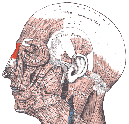
Procerus
Origin: Fascia over lower nasal bone and upper lateral nasal cartilage.
Insertion: Skin of lower forehead between eyebrows.
Action: Depresses medial eyebrows and wrinkles skin across the bridge of the nose.
Platysma
Origin: Superficial fascia over upper pectoralis major and deltoid.
Insertion: Inferior border of mandible and skin and muscles of lower face.
Action: Tenses neck skin; depresses lower lip and mouth angle; assists mandibular depression.

Sternocleidomastoid
Origin: Manubrium of sternum and medial third of clavicle.
Insertion: Mastoid process and lateral superior nuchal line.
Action:
a) Unilateral: bends neck to same side and rotates head to opposite side.
b) Bilateral: flexes neck; can extend head depending on posture.
Assists forced inspiration when head is fixed.


Levator scapulae
Origin: Transverse processes of C1-C4.
Insertion: Medial scapular border between superior angle and root of scapular spine.
Action: Elevates and downwardly rotates scapula; with scapula fixed, bends neck to same side.

Multifidus
Origin: Posterior sacrum, posterior superior iliac spine and sacroiliac ligaments; lumbar mammillary processes; thoracic transverse processes; cervical articular processes C4-C7.
Insertion: Spinous processes generally 2-4 vertebral levels above the origins.
Action: Stabilizes vertebrae; extends vertebral column bilaterally and rotates it to opposite side unilaterally.
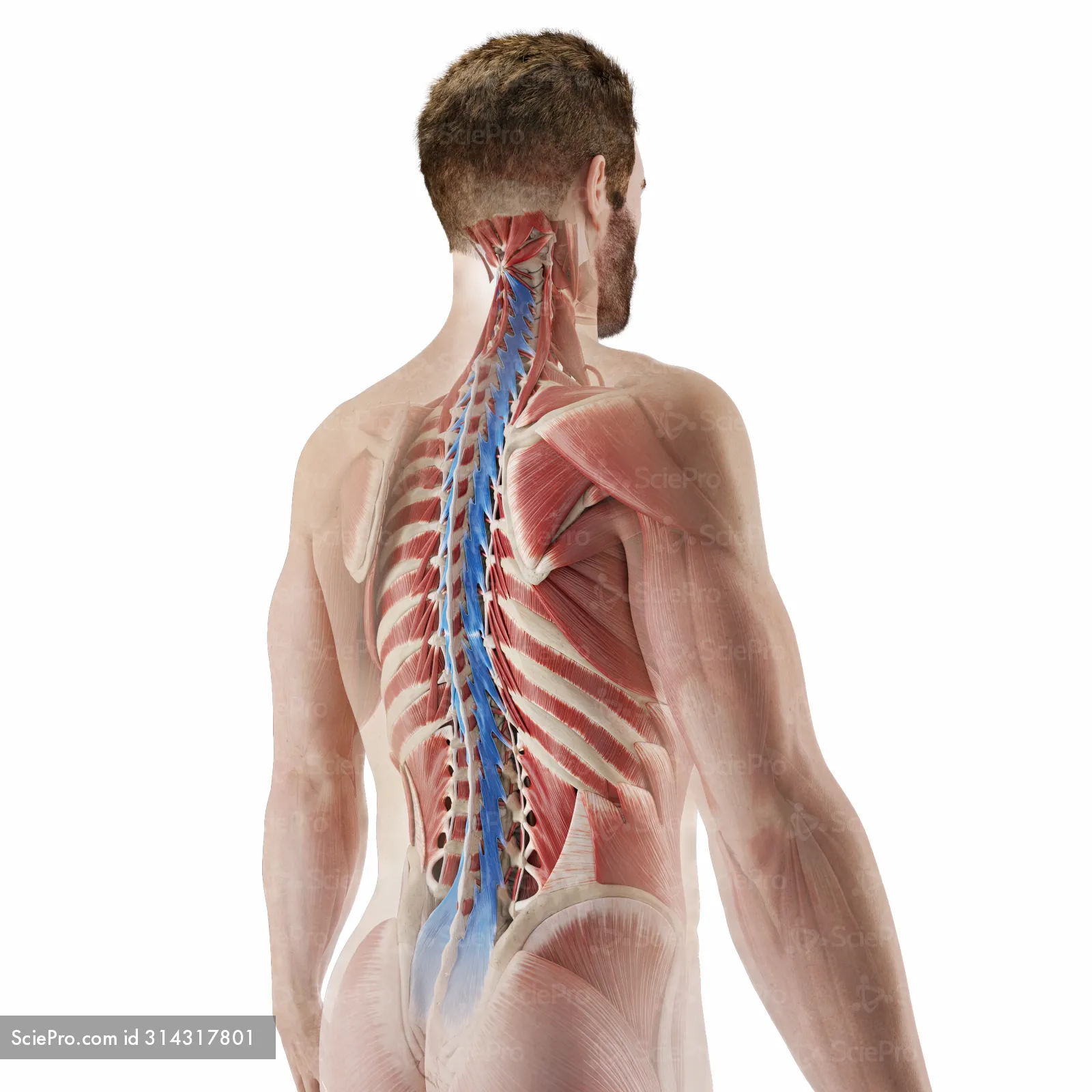
Erector spinae group
Origin: Common inferior tendon from posterior iliac crest, sacrum, and lower vertebral spinous processes; additional vertebral and rib attachments vary by subdivision.
Insertion: Ribs, vertebral transverse and spinous processes, and skull, depending on subdivision.
Action: Extends and laterally flexes vertebral column and head; maintains posture. Group comprises iliocostalis, longissimus, and spinalis.

Quadratus lumborum
Origin: Posterior iliac crest and iliolumbar ligament.
Insertion: Inferior border of rib 12 and transverse processes L1-L4.
Action: Laterally flexes trunk to same side; assists trunk extension and stabilizes rib 12 during respiration.

External intercostals
Origin: Inferior border of each rib, ribs 1-11.
Insertion: Superior border of rib immediately below, ribs 2-12.
Action: Elevate ribs during inspiration and support intercostal spaces.

Internal intercostals
Origin: Internal surface and inferior border of each upper rib and its costal cartilage.
Insertion: Superior border of rib immediately below.
Action: Interosseous parts depress ribs during forced expiration; interchondral (parasternal) parts elevate ribs during inspiration. Support intercostal spaces.

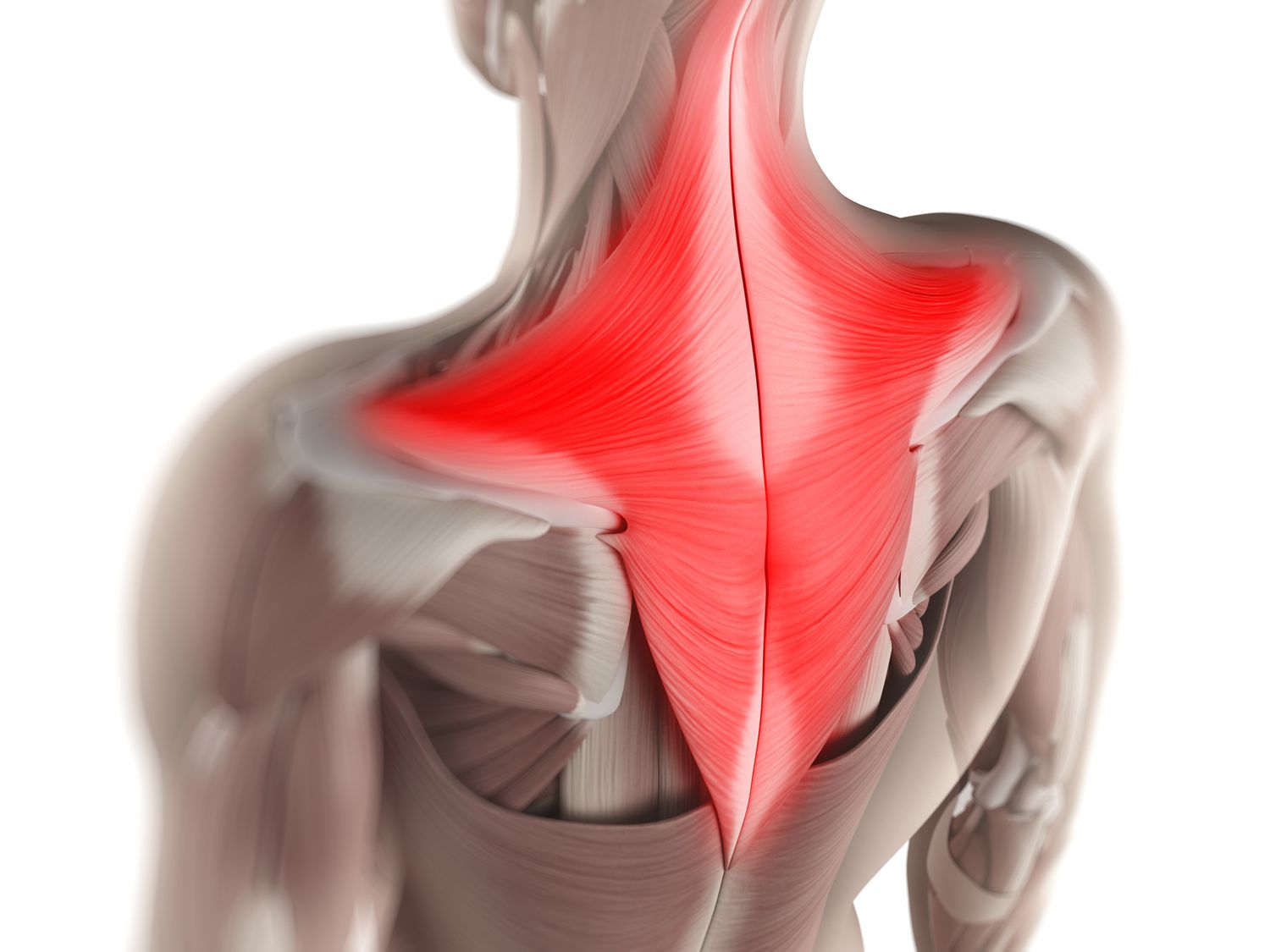
Trapezius
Origin: External occipital protuberance, medial superior nuchal line, nuchal ligament, and spinous processes C7-T12.
Insertion: Lateral third of clavicle, acromion, and scapular spine.
Action: Upper fibers elevate, middle fibers retract, and lower fibers depress scapula. Upper and lower fibers cooperate in upward rotation.
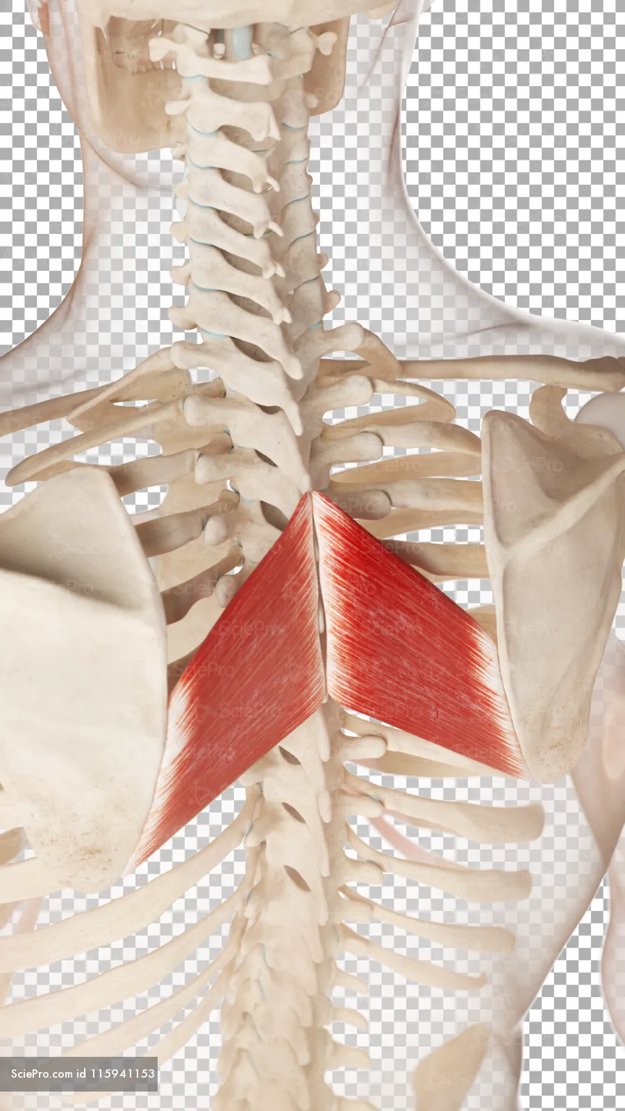
Rhomboid major
Origin: Spinous processes T2-T5 and associated supraspinous ligaments.
Insertion: Medial scapular border inferior to root of scapular spine.
Action: Retracts, elevates, and downwardly rotates scapula; holds it against thoracic wall.
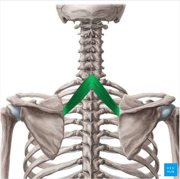
Rhomboid minor
Origin: Lower nuchal ligament and spinous processes C7-T1.
Insertion: Medial scapular border at root of scapular spine.
Action: Retracts, elevates, and downwardly rotates scapula; stabilizes it against thoracic wall.
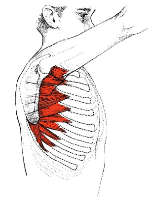
Serratus anterior
Origin: External surfaces of ribs 1-8 or 1-9.
Insertion: Anterior (costal) surface of medial scapular border.
Action: Protracts and upwardly rotates scapula; holds scapula against thoracic wall.


Pectoralis minor
Origin: Anterior surfaces of ribs 3-5 near costal cartilages.
Insertion: Medial border and superior surface of scapular coracoid process.
Action: Draws scapula anteriorly and inferiorly; stabilizes scapula. Assists inspiration when scapula is fixed.

Supraspinatus
Origin: Supraspinous fossa of scapula and overlying fascia.
Insertion: Superior facet of greater tubercle of humerus and shoulder joint capsule.
Action: Initiates and assists arm abduction; stabilizes humeral head in glenoid cavity.

Infraspinatus
Origin: Infraspinous fossa of scapula and overlying fascia.
Insertion: Middle facet of greater tubercle of humerus and shoulder joint capsule.
Action: Laterally rotates arm; stabilizes humeral head in glenoid cavity.

Teres minor
Origin: Upper two-thirds of posterior aspect of lateral scapular border.
Insertion: Inferior facet of greater tubercle of humerus and shoulder joint capsule.
Action: Laterally rotates arm, assists adduction, and stabilizes humeral head.

Deltoid
Origin: Lateral third of clavicle, acromion, and scapular spine.
Insertion: Deltoid tuberosity of humerus.
Action: Middle fibers abduct arm. Anterior fibers flex and medially rotate arm; posterior fibers extend and laterally rotate arm.

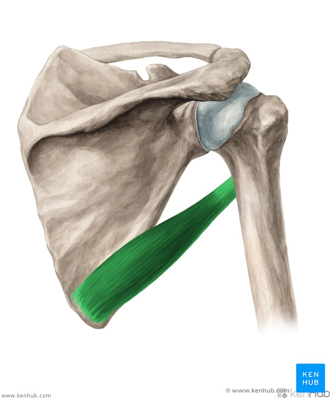
Teres major
Origin: Posterior surface of inferior scapular angle and adjacent lateral border.
Insertion: Medial lip of intertubercular sulcus of humerus (crest of lesser tubercle).
Action: Adducts, medially rotates, and extends arm.
Latissimus dorsi
Origin: Spinous processes T7-T12; thoracolumbar fascia, iliac crest, and lower 3-4 ribs.
Insertion: Floor of intertubercular sulcus of humerus.
Action: Extends, adducts, and medially rotates arm.

Pectoralis major
Origin: Medial half of clavicle, sternum, upper six costal cartilages, and external oblique aponeurosis.
Insertion: Lateral lip of intertubercular sulcus of humerus (crest of greater tubercle).
Action: Adducts and medially rotates arm. Clavicular part flexes arm; sternocostal part extends a flexed arm.

Biceps brachii
Origin: Long head: supraglenoid tubercle of scapula. Short head: coracoid process of scapula.
Insertion: Radial tuberosity and forearm fascia through bicipital aponeurosis.
Action: Supinates forearm and flexes elbow; assists shoulder flexion.

Triceps brachii
Origin: Long head: infraglenoid tubercle of scapula. Lateral head: posterior humerus superior to radial groove. Medial head: posterior humerus inferior to radial groove.
Insertion: Olecranon of ulna and forearm fascia.
Action: Extends elbow. Long head also assists shoulder extension and adduction.

Pronator teres
Origin: Humeral head: medial epicondyle of humerus. Ulnar head: coronoid process of ulna.
Insertion: Middle of lateral surface of radius.
Action: Pronates forearm; assists elbow flexion.


Palmaris longus
Origin: Medial epicondyle of humerus via common flexor tendon.
Insertion: Flexor retinaculum and palmar aponeurosis.
Action: Flexes wrist and tenses palmar aponeurosis.
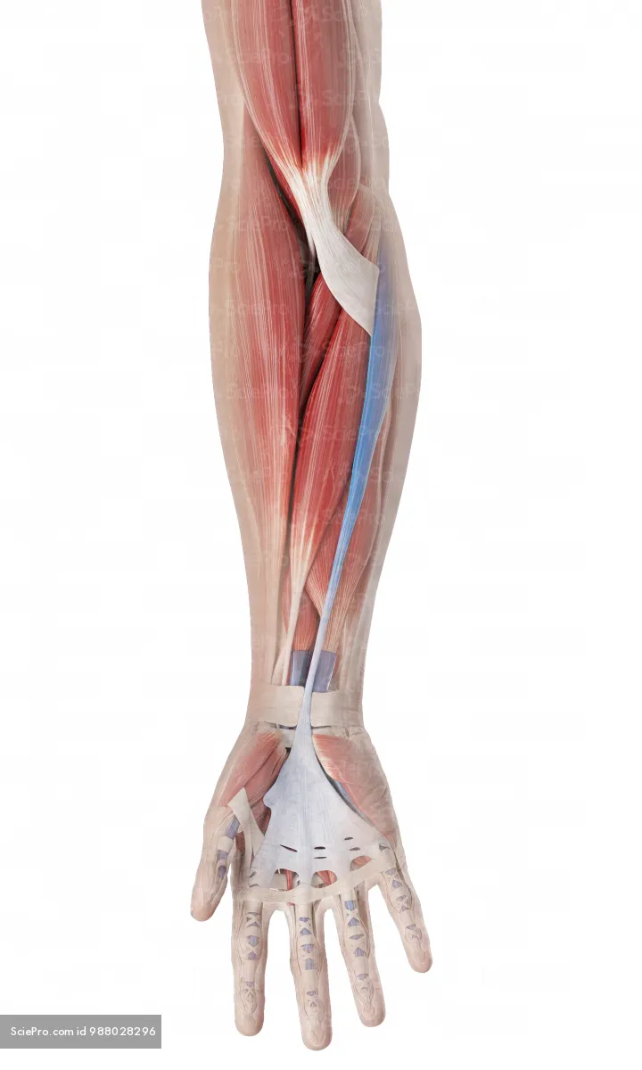
Flexor digitorum superficialis
Origin: Humeroulnar head: medial humeral epicondyle, ulnar collateral ligament, and coronoid process. Radial head: anterior border of radius.
Insertion: Sides of middle phalanges of digits 2-5; each tendon splits around the profundus tendon.
Action: Flexes proximal interphalangeal joints of digits 2-5; also assists metacarpophalangeal and wrist flexion.

Flexor pollicis longus
Origin: Anterior surface of radius and adjacent interosseous membrane.
Insertion: Palmar base of distal phalanx of thumb.
Action: Flexes thumb, especially at interphalangeal joint; assists wrist flexion.

Brachioradialis
Origin: Proximal two-thirds of lateral supracondylar ridge of humerus.
Insertion: Lateral distal radius at base of styloid process.
Action: Flexes elbow, especially with forearm in neutral position (thumb up); helps return forearm toward neutral from pronation or supination.

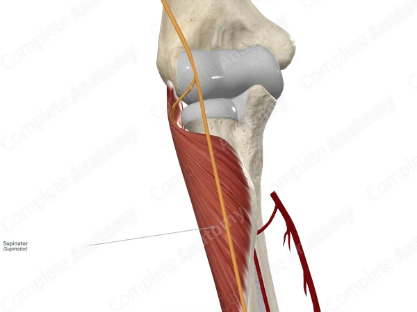
Supinator
Origin: Lateral humeral epicondyle, radial collateral and annular ligaments, and supinator crest and fossa of ulna.
Insertion: Anterior, lateral, and posterior surfaces of proximal third of radius.
Action: Supinates forearm.


Extensor digitorum
Origin: Lateral epicondyle of humerus via common extensor tendon.
Insertion: Extensor expansions of digits 2-5, reaching bases of middle and distal phalanges.
Action: Extends digits 2-5, primarily at metacarpophalangeal joints; assists interphalangeal and wrist extension.
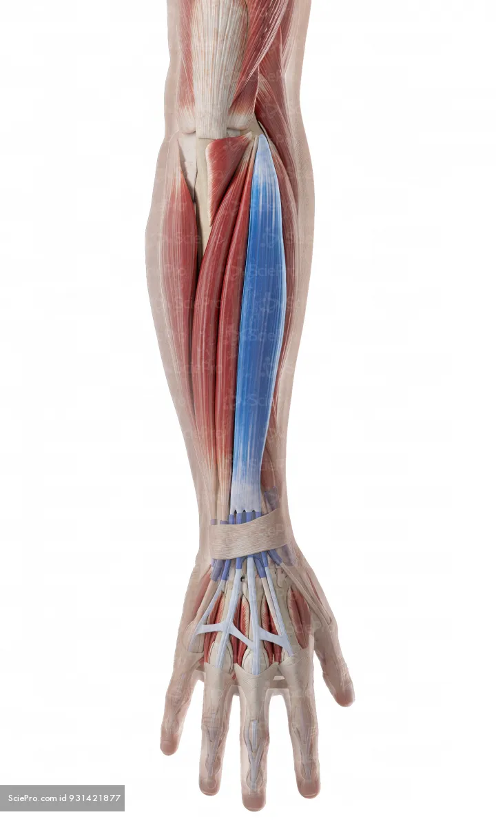
Extensor pollicis longus
Origin: Middle third of posterior ulna and adjacent interosseous membrane.
Insertion: Dorsal base of distal phalanx of thumb.
Action: Extends thumb, especially at interphalangeal joint; also assists extension at its other joints.


External oblique
Origin: External surfaces of ribs 5-12.
Insertion: Linea alba, pubic tubercle, and anterior half of iliac crest.
Action: Compresses abdominal contents and flexes trunk bilaterally. On one side, bends trunk to same side and rotates it to opposite side.


Internal oblique
Origin: Thoracolumbar fascia, iliac crest, and lateral inguinal ligament.
Insertion: Inferior borders of ribs 10-12, linea alba, and pubic crest and pecten pubis via conjoint tendon.
Action: Compresses abdominal contents and flexes trunk bilaterally. On one side, bends and rotates trunk to same side.
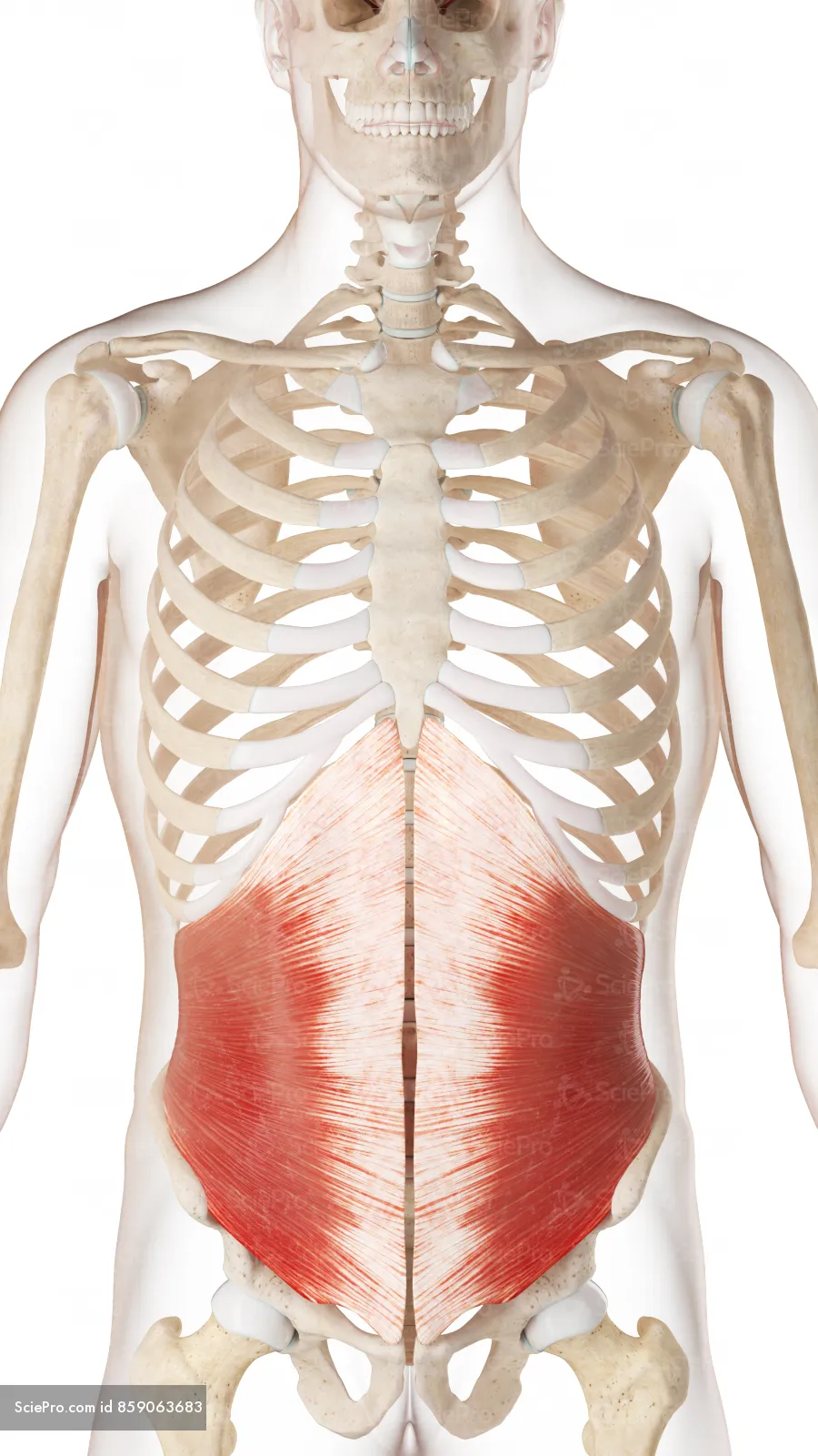

Transversus abdominis
Origin: Internal surfaces of costal cartilages 7-12, thoracolumbar fascia, iliac crest, and lateral third of inguinal ligament.
Insertion: Linea alba, pubic crest, and pecten pubis via conjoint tendon.
Action: Compresses and supports abdominal contents; increases intra-abdominal pressure and stabilizes trunk.

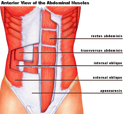
Rectus abdominis
Origin: Pubic crest and pubic symphysis.
Insertion: Xiphoid process and costal cartilages 5-7.
Action: Flexes trunk, compresses abdominal contents, and assists posterior pelvic tilt.

Gluteus maximus
Origin: Ilium posterior to posterior gluteal line, posterior sacrum and coccyx, and sacrotuberous ligament.
Insertion: Iliotibial tract and gluteal tuberosity of femur.
Action: Extends and laterally rotates thigh; helps stabilize hip and knee through iliotibial tract.

Gluteus medius
Origin: External ilium between anterior and posterior gluteal lines.
Insertion: Lateral surface of greater trochanter of femur.
Action: Abducts thigh and stabilizes pelvis during single-leg stance. Anterior fibers assist medial rotation.

Gluteus minimus
Origin: External ilium between anterior and inferior gluteal lines.
Insertion: Anterior surface of greater trochanter of femur and hip capsule.
Action: Abducts and medially rotates thigh; stabilizes pelvis during single-leg stance.

Iliopsoas group
Origin: Psoas major: bodies and discs T12-L5 and lumbar transverse processes. Iliacus: iliac fossa and adjacent iliac crest.
Insertion: Lesser trochanter of femur, chiefly through iliopsoas tendon.
Action: Flexes thigh at hip; with femur fixed, assists trunk flexion. Supports hip and lumbar stability.

Adductor longus
Origin: Body of pubis, inferior to pubic crest.
Insertion: Middle third of linea aspera of femur.
Action: Adducts thigh and assists hip flexion.

Gracilis
Origin: Body and inferior ramus of pubis.
Insertion: Superior medial tibia via pes anserinus.
Action: Adducts thigh; flexes knee and medially rotates leg when knee is flexed.

Rectus femoris
Origin: Anterior inferior iliac spine and ilium superior to acetabulum.
Insertion: Patella via quadriceps tendon; tibial tuberosity via patellar ligament.
Action: Extends knee and flexes thigh at hip.

Vastus lateralis
Origin: Greater trochanter, gluteal tuberosity, and lateral lip of linea aspera of femur.
Insertion: Patella via quadriceps tendon; tibial tuberosity via patellar ligament.
Action: Extends knee and helps stabilize patella.


Vastus medialis
Origin: Intertrochanteric line, medial lip of linea aspera, and medial supracondylar line of femur.
Insertion: Patella via quadriceps tendon; tibial tuberosity via patellar ligament.
Action: Extends knee and helps stabilize patella medially.

Sartorius
Origin: Anterior superior iliac spine and adjacent notch.
Insertion: Superior medial tibia via pes anserinus.
Action: Flexes, abducts, and laterally rotates thigh; flexes knee and medially rotates leg when knee is flexed.
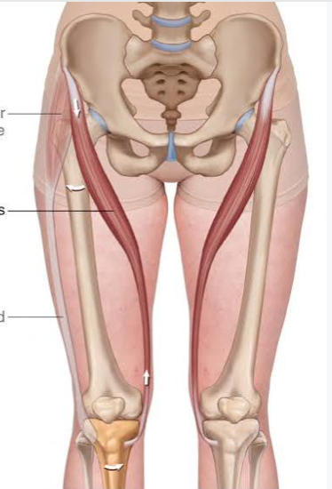

Biceps femoris
Origin: Long head: ischial tuberosity. Short head: lateral linea aspera and lateral supracondylar line of femur.
Insertion: Lateral side of fibular head.
Action: Flexes knee and laterally rotates flexed leg. Long head extends thigh at hip; short head does not cross hip.


Semitendinosus
Origin: Ischial tuberosity.
Insertion: Superior medial tibia via pes anserinus.
Action: Extends thigh, flexes knee, and medially rotates leg when knee is flexed.

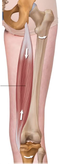
Semimembranosus
Origin: Superolateral impression of ischial tuberosity.
Insertion: Posterior surface of medial tibial condyle, with expansions including oblique popliteal ligament.
Action: Extends thigh, flexes knee, and medially rotates leg when knee is flexed.


Tibialis anterior
Origin: Lateral tibial condyle, proximal lateral tibia, and adjacent interosseous membrane. Insertion:
Medial and plantar surfaces of medial cuneiform and base of first metatarsal.
Action: Dorsiflexes ankle and inverts foot; supports medial longitudinal arch.

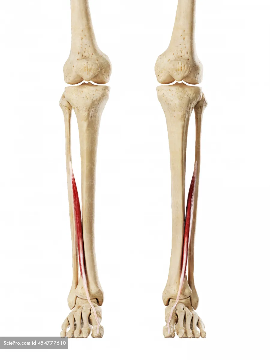
Extensor hallucis longus
Origin: Middle anterior fibula and adjacent interosseous membrane.
Insertion: Dorsal base of distal phalanx of great toe.
Action: Extends great toe and dorsiflexes ankle; assists inversion.

Extensor digitorum longus
Origin: Lateral tibial condyle, proximal anterior fibula, and interosseous membrane.
Insertion: Middle and distal phalanges of toes 2-5 through extensor expansions.
Action: Extends toes 2-5 and dorsiflexes ankle.

Gastrocnemius
Origin: Medial head: posterior femur superior to medial condyle. Lateral head: lateral femoral condyle and adjacent posterior femur.
Insertion: Posterior calcaneus through calcaneal (Achilles) tendon.
Action: Plantar flexes ankle and flexes knee.


Soleus
Origin: Posterior fibular head and proximal fibula, soleal line and medial border of tibia, and tendinous arch between tibia and fibula.
Insertion: Posterior calcaneus through calcaneal (Achilles) tendon.
Action: Plantar flexes ankle and helps maintain standing posture.

Flexor digitorum longus
Origin: Posterior tibia inferior to soleal line.
Insertion: Plantar bases of distal phalanges of toes 2-5.
Action: Flexes toes 2-5, especially distal interphalangeal joints; assists plantar flexion and inversion and supports longitudinal arches.

Flexor hallucis longus
Origin: Distal two-thirds of posterior fibula and adjacent interosseous membrane.
Insertion: Plantar base of distal phalanx of great toe.
Action: Flexes great toe; assists plantar flexion and supports medial longitudinal arch during push-off.

Fibularis longus (peroneus longus)
Origin: Head and proximal two-thirds of lateral fibula.
Insertion: Plantar surfaces of medial cuneiform and base of first metatarsal.
Action: Everts foot and assists ankle plantar flexion; supports transverse and longitudinal foot arches.

Dorsal interossei of foot
Origin: Adjacent sides of metatarsals 1-5; four bipennate muscles.
Insertion: Proximal phalangeal bases and extensor expansions: first to medial side of toe 2; remaining three to lateral sides of toes 2-4.
Action: Abduct toes 2-4 relative to axis of toe 2; flex metatarsophalangeal joints and assist interphalangeal extension.
