Critical care exam I
 Call Kai
 Learn
Learn Practice Test
 Spaced Repetition
 Match
Match Flashcards
 Knowt Play
1/29

There's no tags or description
Looks like no tags are added yet.
Name | Mastery | Learn | Test | Matching | Spaced | Call with Kai | Chat |
|---|
No analytics yet
Send a link to your students to track their progress
30 Terms
Normal Sinus Rhythm
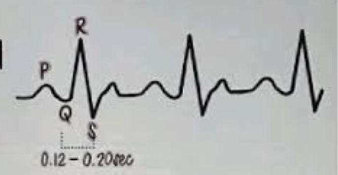
1st degree AV block
PR interval is longer than 0.20 secs, but P:QRS ratio 1:1
Usually regular
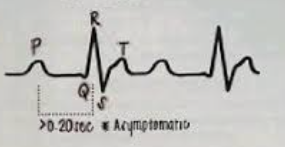
2nd degree AV block type I (Weckenbach)
PR interval gets longer & longer, than a QRS is dropped
Think: “longer, longer, drop”
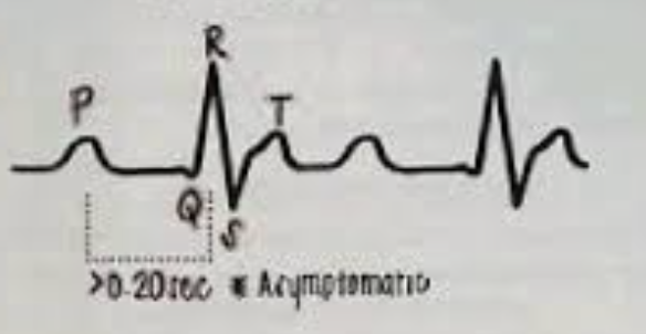
2nd degree AV block type II
PR interval stays constant, but QRS complexes are suddenly dropped
More serious; may require pacing

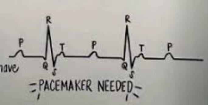
3rd degree/complete ❤ block
P waves & QRS complexes have no relationship to each other
Atria & ventricles beat independently
NSR - story and rhyme
The P & QRS are dating & never leave eachothers side 🙂

1st degree AV block - story and rhyme
The P & QRS got into an argument & are keeping their distance
Rhyme: “If the R is far from the P, then you have a 1st degree”

2nd degree AV block type I AKA weckenbach - story and rhyme
The P & QRS are in a cycle of breaking up & getting back together, repeat

Rhyme: “longer, longer, longer, DROP. Then you have a WECKENBACH!”
2nd degree AV block type II AKA mobitz II - story & rhyme
The P & QRS work it out but QRS is still upset & decides to go out
Rhyme: “If some QRS don’t get through, then you have a mobitz II”

3rd degree AV block - story & rhyme
The QRS catches P cheating so they separate & live single lives
Rhyme: “If P’s & Q’s don’t agree, then you have a 3rd degree”
PACEMAKER NEEDED

Asymptomatic bradycardia tx
Assess & monitor
Symptomatic / hemodynamically unstable bradycardia tx
May require atropine, pacing, or other interventions
Always assess the whole pt, not just the monitor
QT interval

Start of QRS —> end of T
The QT interval represents ventricular depolarization & depolarization
A prolonged QT interval increases the risk for dangerous/life threatening dysrhythmias
Atrial Flutter
Characteristic “sawtooth” pattern
Atrial rate is typically very fast
Ventricular rhythm may be regular or irregular

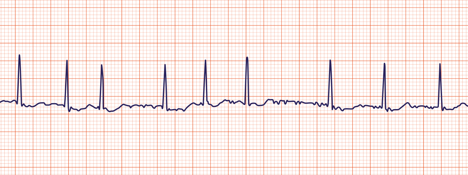
Atrial fibrillation
Irregularly irregular rhythm
No clearly identifiable normal P waves
High risk of thrombus / stroke
Afib tx concepts/common goals of tx
Rate control
Anticoagulation when indicated to decrease thromboembolic risk
Catheter Ablation
A procedure used to treat certain abnormal ❤ rhythms
A catheter is advanced through a blood vessel, commonly from the femoral area, toward the ❤
The provider identifies abnormal areas of cardiac electrical activity & uses
Heat (radio frequency)
OR freezing (cyroablation)
To destroy / isolate the abnormal tissue
Remember= Ablation = eliminate abnormal electric pathways
❤ failure common manifestations
Dyspnea
Fatigue
Fld retention
Peripheral edema
Pulmonary edema
Pulmonary crackles
Cough
Decreased appetite
Left sided HF
LEFT = LUNGS
Possible findings:
Dyspnea
Pulmonary edema
Crackles
Cough
Orthopnea
Right sided HF
RIGHT = Rest of body
Possible findings:
Peripheral edema
Increased abdominal girth
Ascites
Weight gain
JVD
Ejection Fraction
Ejection fraction measures the %age of blood pumped out of the ventricles with each contraction
Normal/preserved EF = 50—>70%
Low EF= reduced pumping function
HF diagnostics - Echocardiogram
Evaluates cardiac structure & function
Helps determine EF
HF diagnostics - BNP
Helps evaluate for HF
HF diagnostics - CXR
Can show pulmonary congestion/edema
HF diagnostics - ECG
Evaluates cardiac rhythm & electrical activity
Coronary Artery Disease (CAD) - Modifiable RF
HTN
Diabetes
Smoking
Obesity
Physical inactivity
Unhealthy diet
Increased cholesterol
Coronary Artery Disease (CAD) -- Non modifiable RF
Age
Family history/heredity
Atherosclerosis & thrombus formation
Adhesion
Platelets attach / bind to injured vessel wall
“I stick to the injury”
Activation
Platelets change shape & become activated
“Something is wrong! Get more platelets” Think like activation of an alarm to signal more platelets to come
Aggregation
Platelets clump together
“Everyone pile on!”
Easy memory trick:
A-A-A
Adhesion —> Activation —> Aggregation
Angiography & Angioplasty
The procedure may include:
Cardiac catheterization
Contrast dye
Angiography = visualization of coronary blood vessels
Angioplasty = the balloon fix
Stent placement (KEEP IT OPEN)
Stent = a small mesh tube used to help keep a coronary artery open
Important difference:
Angioplasty/stent is NOT the same as a CABG
CABG is open ❤ surgery using a blood vessel graft to create a new route
Post Angiography nursing care
Immediately following the procedure, monitor for:
Bleeding
Hematoma
Changes in cardiac rhythm
VS changes
Neurovascular status of the affected extremity
The pt may need bed rest & restrictions on movement